


Retropharyngeal abscesses, most common among young children, can cause sore throat, fever, neck stiffness, and stridor. Diagnosis requires lateral neck radiography or CT. Treatment is with endotracheal intubation, drainage, and antibiotics.
Retropharyngeal abscesses develop in the retropharyngeal lymph nodes at the back of the pharynx, adjacent to the vertebrae. They can be seeded by infection of the pharynx, sinuses, adenoids, or nose. Approximately 75% of cases occur in children younger than 5 years, with a median age around 3- to 4 years (1). Retropharyngeal abscesses beyond the age of 5 are less common because the retropharyngeal lymph nodes begin to regress thereafter (2). However, adults may develop infection after ingestion of a foreign body or after instrumentation. Common organisms include aerobic (Streptococcus and Staphylococcus species) and anaerobic (Bacteroides and Fusobacterium species) bacteria and, occasionally in adults and children, HIV or the bacteria that cause tuberculosis.
The most serious consequences include airway obstruction, septic shock, rupture of the abscess into the airway resulting in aspiration pneumonia or asphyxia, mediastinitis, carotid rupture, and suppurative thrombophlebitis of the internal jugular veins (Lemierre syndrome).
General references
1. Craig FW, Schunk JE. Retropharyngeal abscess in children: clinical presentation, utility of imaging, and current management. Pediatrics. 2003;111(6 Pt 1):1394-1398. doi:10.1542/peds.111.6.1394
2. Hajare PS, Bellad SA, Anand A, et al. Disparate Presentations of Retropharyngeal Abscess: An Experience From a Tertiary Care Centre. Indian J Otolaryngol Head Neck Surg. 2022;74(Suppl 3):5984-5989. doi:10.1007/s12070-021-02655-0
Symptoms and Signs of Retropharyngeal Abscess
Symptoms and signs of a retropharyngeal abscess are usually preceded in children by an acute upper respiratory infection and in adults by foreign body ingestion or instrumentation. Children may have odynophagia, dysphagia, fever, cervical lymphadenopathy, nuchal rigidity, stridor, dyspnea, snoring or noisy breathing, and torticollis.
Adults may have severe neck pain but less often have stridor.
The posterior pharyngeal wall may bulge to one side on physical examination.
Diagnosis of Retropharyngeal Abscess
Radiography
CT
The diagnosis of retropharyngeal abscess is made on the basis of imaging. A retropharyngeal abscess is suspected in patients with severe, unexplained sore throat and neck stiffness, stridor, or noisy breathing.
Lateral soft-tissue radiographs of the neck, taken in the maximum possible hyperextension, during inspiration, and without rotation, may show focal widening of the prevertebral soft tissues, reversal of normal cervical lordosis, air in the prevertebral soft tissues, or erosion of the adjacent vertebral body.

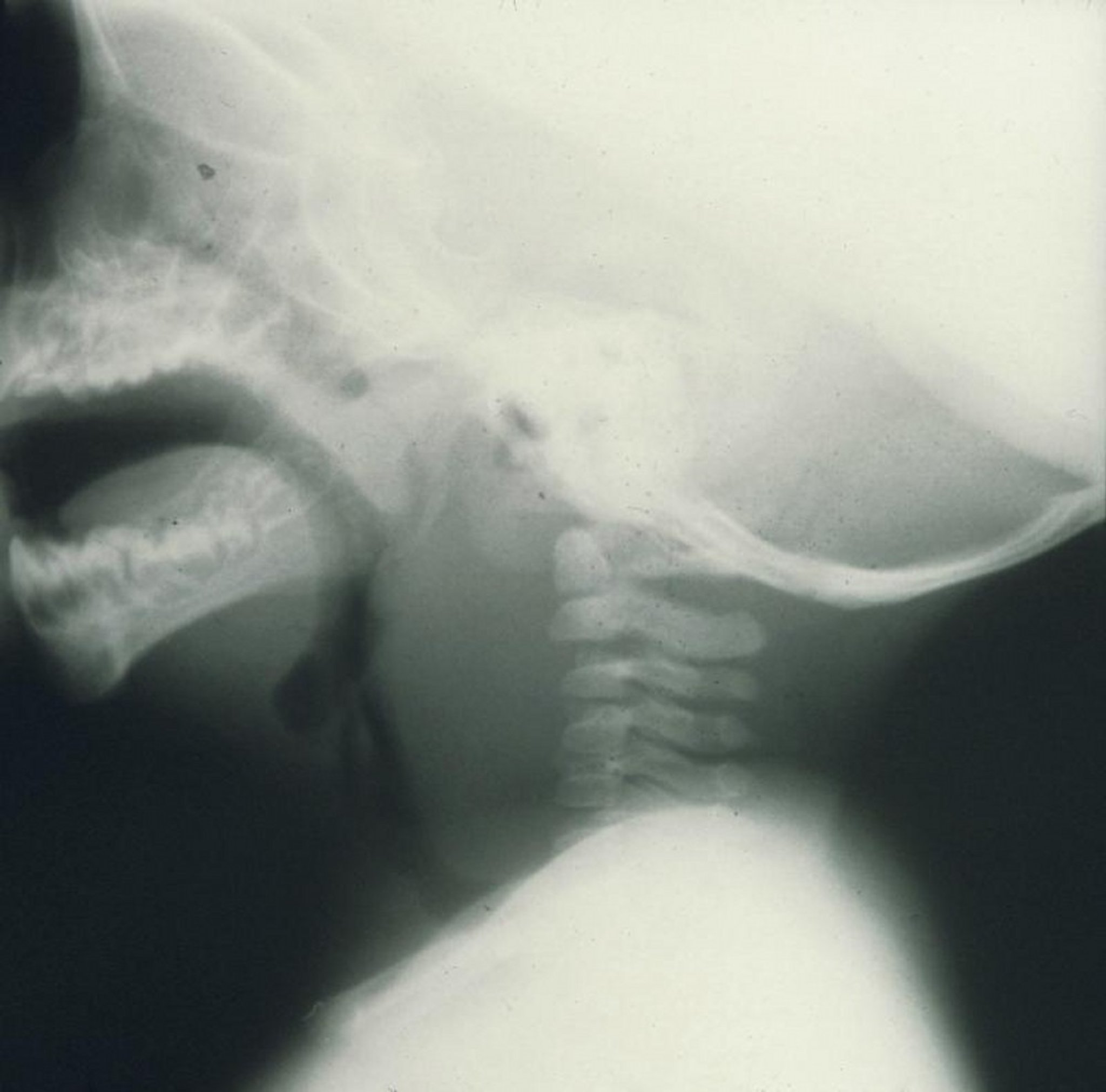
This lateral radiograph of a child shows marked swelling anterior to the cervical vertebrae caused by a retropharyngeal abscess.
Image provided by Clarence T. Sasaki, MD.
CT can help diagnose questionable cases, help differentiate cellulitis from an abscess, and assess the extent of an abscess.
Treatment of Retropharyngeal Abscess
Antibiotics (eg, ceftriaxone, clindamycin)
Adjunctive glucocorticoids
Usually surgical drainage
The treatment of retropharyngeal abscess involves airway stabilization and medical management including broad-spectrum, typically intravenous antibiotics or additional surgical management, depending on patient age, abscess characteristics, and clinical response. Approaches are based mainly on observational data (1).
Antibiotics, such as a broad-spectrum cephalosporin (eg, ceftriaxone 50 to 75 mg/kg IV once/day) or clindamycin, may occasionally be sufficient for children with small abscesses administered for approximately 3 weeks (2). Adjunctive glucocorticoids are increasingly used in pediatric patients to reduce symptom duration and appear to improve clinical outcomes including length of hospital stay (3).
Surgical drainage is reserved for patients with airway compromise, lack of response to antibiotic therapy, abscess size > 3 cm, immunocompromised status, or complications. The preferred approach is incision and drainage (ie, drainage through an incision in the posterior pharyngeal wall). Endotracheal intubation is performed preoperatively and maintained for 24 to 48 hours.
Treatment references
1. Cheng J, Elden L. Children with deep space neck infections: Our experience with 178 children. Otolaryngol Head Neck Surg. 2013;148 (6):1037–1042. doi: 10.1177/0194599813482292
2. Fitzsimons M, Boyle S, Colreavy M, et al. Retrospective review of 33 cases of paediatric retropharyngeal and parapharyngeal infections and a proposed algorithm for managing future cases. J Laryngol Otol. Published online July 14, 2025. doi:10.1017/S0022215125102703
3. Villanueva-Fernández E, Casanueva-Muruáis R, Vivanco-Allende A, et al. Role of steroids in conservative treatment of parapharyngeal and retropharyngeal abscess in children. Eur Arch Otorhinolaryngol. 2022 Nov;279(11):5331-5338. doi: 10.1007/s00405-022-07423-6. Epub 2022 Jun 29. PMID: 35767057; PMCID: PMC9519669
Drug Information for the Topic







