


Chromoblastomycosis is a specific type of cutaneous infection caused by one of several species of dematiaceous (pigmented) fungi. Symptoms are ulcerating nodules on exposed body parts. Diagnosis is by appearance, histopathology, and culture. Treatment is with itraconazole, another mold-active triazole, or terbinafine and surgical excision.
Chromoblastomycosis is a chronic, progressive cutaneous infection affecting immunocompetent people, mostly in tropical or subtropical areas; it is characterized by formation of papillomatous nodules that tend to ulcerate.
Chromoblastomycosis is caused by traumatic implantation of dematiaceous (pigmented) fungi. Common species include Fonsecaea pedrosoi, Cladophialophora carrionii, and Phialophora verrucosa. This infection occurs particularly in farmers and other agricultural workers without adequate protective footwear and clothing. It can lead to granulomatous and fibrotic inflammation, dysregulated T-helper cell and humoral immune responses, and cause the formation of muriform (sclerotic or Medlar) bodies in tissue, which are pathognomonic of disease (1).
(See also Overview of Fungal Infections.)
General reference
1. Queiroz-Telles F, de Hoog S, Santos DW, et al. Chromoblastomycosis. Clin Microbiol Rev. 2017;30(1):233-276. doi:10.1128/CMR.00032-16
Symptoms and Signs of Chromoblastomycosis
Usually, chromoblastomycosis begins on the foot or leg, but other exposed body parts may be infected, especially at sites of epidermal disruption.
Early, small, itchy, enlarging papules may resemble dermatophytosis (ringworm). These papules extend to form dull red or violaceous, sharply demarcated patches with indurated bases. Several weeks or months later, new lesions, projecting 1 to 2 mm above the skin, may appear along paths of lymphatic drainage. Hard, dull red or grayish cauliflower-shaped nodular projections may develop in the center of patches and, if the infection is untreated, gradually extend to cover extremities over the course of many years.
Lymphatics may be obstructed, itching may persist, and secondary bacterial superinfections may develop, causing ulcerations and occasionally septicemia.

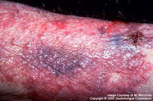
In chromoblastomycosis, small papules initially extend to form dull red or violaceous, sharply demarcated patches with indurated bases.
In chromoblastomycosis, small papules initially extend to form dull red or violaceous, sharply demarcated patches with
Image courtesy of www.doctorfungus.org © 2005.

In chronic chromoblastomycosis, hard, dull, red or grayish, cauliflower-shaped nodular projections develop over years and extend to cover the extremities.
In chronic chromoblastomycosis, hard, dull, red or grayish, cauliflower-shaped nodular projections develop over years a
Image courtesy of www.doctorfungus.org © 2005.

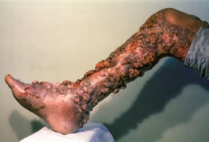
This photo shows extensive chromoblastomycosis involving the right lower extremity. Multiple verrucous and tumorous lesions can be seen.
This photo shows extensive chromoblastomycosis involving the right lower extremity. Multiple verrucous and tumorous les
Image courtesy of Karen McKoy, MD.


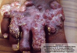
This photo shows thick, verrucous, and lichenified plaques of micaceous scale on the right leg of a patient with chromoblastomycosis.
This photo shows thick, verrucous, and lichenified plaques of micaceous scale on the right leg of a patient with chromo
SCIENCE PHOTO LIBRARY
In chromoblastomycosis, small papules initially extend to form dull red or violaceous, sharply demarcated patches with indurated bases.
In chromoblastomycosis, small papules initially extend to form dull red or violaceous, sharply demarcated patches with
Image courtesy of www.doctorfungus.org © 2005.
In chronic chromoblastomycosis, hard, dull, red or grayish, cauliflower-shaped nodular projections develop over years and extend to cover the extremities.
In chronic chromoblastomycosis, hard, dull, red or grayish, cauliflower-shaped nodular projections develop over years a
Image courtesy of www.doctorfungus.org © 2005.
This photo shows extensive chromoblastomycosis involving the right lower extremity. Multiple verrucous and tumorous lesions can be seen.
This photo shows extensive chromoblastomycosis involving the right lower extremity. Multiple verrucous and tumorous les
Image courtesy of Karen McKoy, MD.
This photo shows thick, verrucous, and lichenified plaques of micaceous scale on the right leg of a patient with chromoblastomycosis.
This photo shows thick, verrucous, and lichenified plaques of micaceous scale on the right leg of a patient with chromo
SCIENCE PHOTO LIBRARY
Diagnosis of Chromoblastomycosis
Histopathology
Culture
The diagnosis of chromoblastomycosis is confirmed by the observation of muriform bodies in tissue and by the isolation and identification of the causal species in culture (1).
Fontana-Masson staining for melanin helps confirm the presence of the muriform (sclerotic or Medlar) bodies, which are pathognomonic.
Late chromoblastomycosis lesions have a characteristic appearance, but early lesions may be mistaken for dermatophytoses.
Diagnosis reference
1. Queiroz-Telles F, Esterre P, Perez-Blanco M, Vitale RG, Salgado CG, Bonifaz A. Chromoblastomycosis: an overview of clinical manifestations, diagnosis and treatment. Med Mycol. 2009;47(1):3-15. doi:10.1080/13693780802538001
Treatment of Chromoblastomycosis
Itraconazole
Posaconazole, voriconazole, or terbinafine
Often surgery or cryotherapy
(See also Antifungal Medications.)
Itraconazole is the most effective medication for chromoblastomycosis, although not all patients respond. Flucytosine is sometimes added to prevent relapse. Flucytosine should not be used as monotherapy.
Anecdotal reports suggest that posaconazole, voriconazole, or terbinafine may also be effective.
Amphotericin B is ineffective (1).
For localized lesions, surgical excision may be curative.
Adjunctive therapies such as cryotherapy are often helpful, although response is slow.
Treatment reference
1. Brito AC, Bittencourt MJS. Chromoblastomycosis: an etiological, epidemiological, clinical, diagnostic, and treatment update. An Bras Dermatol. 2018;93(4):495-506. doi:10.1590/abd1806-4841.20187321
Drug Information for the Topic


