


Mycetoma is a chronic, progressive, local infection caused by fungi or bacteria involving the feet, upper extremities, or back. Symptoms include tumefaction and formation of sinus tracts. Diagnosis is clinical, confirmed by microscopic examination of exudates and culture. Treatment includes antimicrobials, surgical debridement, and sometimes amputation.
(See also Overview of Fungal Infections.)
Mycetomas are local infections caused by fungi or bacteria (1). They are classically divided into actinomycetoma (bacterial, most commonly due to Nocardia, Streptomyces, and Actinomadura) and eumycetoma (fungal, most commonly due to Madurella, Pseudallescheria, Scedosporium, and Fusarium species) (2).
Mycetoma occurs mainly in tropical or subtropical areas, including the southern United States. It occurs in equatorial regions of Africa, Latin America, and Asia known as the “mycetoma belt.” Eumycetomas are the most common type in Africa. Actinomycetomas cause most cases in South and Central America and in some Asian countries.
Mycetoma is acquired when organisms enter through sites of penetrating local trauma on bare skin of the feet or on the extremities or backs of workers carrying contaminated vegetation or other objects. Men aged 20 to 40 are most often affected, presumably because of trauma incurred while working outdoors.
Infections spread through contiguous subcutaneous areas, resulting in tumefaction and formation of multiple draining sinuses that exude characteristic grains of clumped organisms. Microscopic tissue reactions may be primarily suppurative or granulomatous depending on the specific causative agent. As the infection progresses, bacterial superinfections can develop.
General references
1. van de Sande WWJ, Fahal AH. An updated list of eumycetoma causative agents and their differences in grain formation and treatment response. Clin Microbiol Rev. 2024;37(2):e0003423. doi:10.1128/cmr.00034-23
2. Verma P, Jha A. Mycetoma: reviewing a neglected disease. Clin Exp Dermatol. 2019;44(2):123-129. doi:10.1111/ced.13642
Symptoms and Signs of Mycetoma
The initial lesion of mycetoma may variably be a papule, a fixed subcutaneous nodule, a vesicle with an indurated base, or a subcutaneous abscess that ruptures to form a fistula to the skin surface. Fibrosis is common in and around early lesions. Tenderness is minimal or absent unless acute suppurative bacterial superinfection is present.
Infection progresses slowly over months or years, gradually extending to and destroying contiguous muscles, tendons, fascia, and bones. The disease remains confined to the affected limb and does not disseminate. Eventually, muscle wasting, deformity, and tissue destruction prevent use of the affected limb.
In advanced infections, involved extremities appear markedly edematous, forming a club-shaped mass of cystic areas. The multiple draining and intercommunicating sinus tracts and fistulas in these areas discharge thick or serosanguineous exudates containing characteristic grains, which may be white or black.

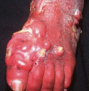
This image shows multiple, infiltrated nodules on the foot of a patient with mycetoma.
This image shows multiple, infiltrated nodules on the foot of a patient with mycetoma.
© Springer Science+Business Media


This posterior view of the left ankle shows eumycetoma, also called Madura foot.
This posterior view of the left ankle shows eumycetoma, also called Madura foot.
MEDICIMAGE / SCIENCE PHOTO LIBRARY

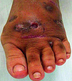
This image shows multiple subcutaneous nodules and rupture to the skin surface.
This image shows multiple subcutaneous nodules and rupture to the skin surface.
© Springer Science+Business Media

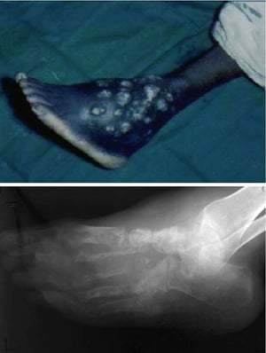
This photo shows chronic, nonhealing wounds with recurring episodes of drainage and ulceration (top) and advanced destruction of the entire bony architecture of the foot (bottom).
This photo shows chronic, nonhealing wounds with recurring episodes of drainage and ulceration (top) and advanced destr
© Springer Science+Business Media
This image shows multiple, infiltrated nodules on the foot of a patient with mycetoma.
This image shows multiple, infiltrated nodules on the foot of a patient with mycetoma.
© Springer Science+Business Media
This posterior view of the left ankle shows eumycetoma, also called Madura foot.
This posterior view of the left ankle shows eumycetoma, also called Madura foot.
MEDICIMAGE / SCIENCE PHOTO LIBRARY
This image shows multiple subcutaneous nodules and rupture to the skin surface.
This image shows multiple subcutaneous nodules and rupture to the skin surface.
© Springer Science+Business Media
This photo shows chronic, nonhealing wounds with recurring episodes of drainage and ulceration (top) and advanced destruction of the entire bony architecture of the foot (bottom).
This photo shows chronic, nonhealing wounds with recurring episodes of drainage and ulceration (top) and advanced destr
© Springer Science+Business Media
Diagnosis of Mycetoma
Examination and culture of exudates
Causative microorganisms can be identified presumptively by gross and microscopic examination of grains from lesional exudates, which contain pathognomonic, irregularly shaped, variably colored, 0.5- to 2-mm granules. Crushing and culture of these granules provides definitive identification. Exudate specimens may yield multiple bacteria and fungi, some of which are potential causes of superinfections.
Treatment of Mycetoma
Antibacterials or antifungals (depending on etiology)
Surgery in refractory cases
(See also Antifungal Medications.)
Treatment depends on the causative microorganism and the extent of the disease. The clinician must distinguish between actinomycetoma and eumycetoma.
In infections caused by actinomycetoma, including Nocardia, sulfonamides and certain other antibacterials, sometimes in combination, are used.
For eumycetoma, the treatment of choice is prolonged antifungal therapy, most commonly with oral itraconazole, sometimes in combination with surgical debridement or excision of the lesion as appropriate. Cure rates remain suboptimal. Triazoles (eg, voriconazole, posaconazole) may be considered in refractory cases, but evidence is limited. Antifungal treatment should be continued for at least 12 months.
Surgical debridement is occasionally necessary. Repeated debridement of the diseased tissue, including bone, may be required. Limb amputation to prevent potentially fatal severe secondary bacterial infections may be needed in advanced cases.
More Information
The following English-language resource may be useful. Please note that The Manual is not responsible for the content of this resource.
Mycetoma Research Center (World Health Organization Collaborating Center on Mycetoma): Mycetoma Management Guidelines
Drug Information for the Topic


