


Paracoccidioidomycosis is a progressive mycosis of the lungs, skin, mucous membranes, lymph nodes, and internal organs caused by Paracoccidioides brasiliensis. Symptoms include skin ulcers, adenitis, and pain due to abdominal organ involvement. Diagnosis is clinical and microscopic, confirmed by culture. Treatment is with azoles (eg, itraconazole), amphotericin B, or sulfonamides.
(See also Overview of Fungal Infections.)
Paracoccidioidomycosis occurs mostly in discrete foci in South and Central America, most often in men aged 40 to 50, especially agricultural workers in rural endemic regions, especially Brazil (1). It is rare in the United States.
Although a relatively unusual opportunistic infection, paracoccidioidomycosis sometimes occurs in immunocompromised patients but infrequently occurs in those with advanced HIV infection.
Although specific environmental sources for Paracoccidioides brasiliensis remain undefined, it is presumed to exist in soil as a mold, with infection resulting from inhalation of conidia (spores produced by the mycelial form of the fungus). Conidia convert to invasive yeasts in the lungs and are assumed to spread to other sites via blood and lymphatics.
General reference
1. Blotta MH, Mamoni RL, Oliveira SJ, et al. Endemic regions of paracoccidioidomycosis in Brazil: a clinical and epidemiologic study of 584 cases in the southeast region. Am J Trop Med Hyg. 1999;61(3):390-394. doi:10.4269/ajtmh.1999.61.390
Symptoms and Signs of Paracoccidioidomycosis
Most people who inhale conidia of P. brasiliensis develop an asymptomatic pulmonary infection.
Symptomatic disease usually manifests as acute pneumonia, which may spontaneously resolve. Clinically apparent infections can become chronic and progressive but are not usually fatal. There are 3 patterns:
Mucocutaneous: Infections most often involve the face, especially at the nasal and oral mucocutaneous borders. Yeasts are usually abundantly present within pinpoint lesions throughout granular bases of slowly expanding ulcers. Regional lymph nodes enlarge, become necrotic, and discharge necrotic material through the skin.
Lymphatic: Cervical, supraclavicular, or axillary nodes enlarge but are painless.
Visceral: Typically, focal lesions cause enlargement mainly of the liver, spleen, and abdominal lymph nodes, sometimes causing abdominal pain.
Infections may be mixed, involving combinations of all 3 patterns.
Paracoccidioidomycosis may manifest as:
An acute/subacute form that usually affects patients < 30 years of age and manifests as disseminated disease (involving the lymph nodes, liver, spleen, and bone marrow)
A chronic form that mostly affects patients > 30 years of age due to reactivation that can cause chronic pulmonary disease with pulmonary fibrosis, emphysema, and lung bullae

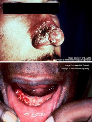
Paracoccidioides brasiliensis infection becomes clinically apparent as mucocutaneous ulcers, especially around the nose (top) and mouth, including the tongue, pharynx, and gums (bottom).
Paracoccidioides brasiliensis infection becomes clinically apparent as mucocutaneous ulcers, especially around the nose
Images courtesy of www.doctorfungus.org © 2005.

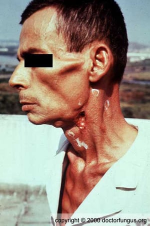
In Paracoccidioides brasiliensis infection, regional lymph nodes may become necrotic.
In Paracoccidioides brasiliensis infection, regional lymph nodes may become necrotic.
Image courtesy of www.doctorfungus.org © 2005.

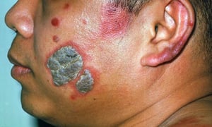
The person in this photo has left facial and auricular involvement. This photo shows 2 gray verrucous plaques on an erythematous base, erythematous nodules, and violaceous patches.
The person in this photo has left facial and auricular involvement. This photo shows 2 gray verrucous plaques on an ery
Image courtesy of Karen McKoy, MD.
Paracoccidioides brasiliensis infection becomes clinically apparent as mucocutaneous ulcers, especially around the nose (top) and mouth, including the tongue, pharynx, and gums (bottom).
Paracoccidioides brasiliensis infection becomes clinically apparent as mucocutaneous ulcers, especially around the nose
Images courtesy of www.doctorfungus.org © 2005.
In Paracoccidioides brasiliensis infection, regional lymph nodes may become necrotic.
In Paracoccidioides brasiliensis infection, regional lymph nodes may become necrotic.
Image courtesy of www.doctorfungus.org © 2005.
The person in this photo has left facial and auricular involvement. This photo shows 2 gray verrucous plaques on an erythematous base, erythematous nodules, and violaceous patches.
The person in this photo has left facial and auricular involvement. This photo shows 2 gray verrucous plaques on an ery
Image courtesy of Karen McKoy, MD.
Diagnosis of Paracoccidioidomycosis
Culture and/or histopathology
Serologic methods
Imaging studies
Clinical findings suggest the diagnosis of paracoccidioidomycosis. The diagnosis is established by direct identification of Paracoccidioides species in clinical specimens through microscopy, histopathology, or culture. Imaging studies can detect deeper tissue involvement.
Culture is diagnostic, although observation of large (often > 15 micrometers) yeasts that form characteristic multiple buds (pilot wheel) in specimens provides strong presumptive evidence. Serologic testing, particularly double immunodiffusion, is a valuable adjunct, offering 90% sensitivity and 100% specificity (1).
Because culturing P. brasiliensis can pose a severe biohazard to laboratory personnel, the laboratory should be notified of the suspected diagnosis.
Imaging tests, such as chest radiographs or chest CT scans, can identify lung involvement.
Diagnosis reference
1. Moreto TC, Marques ME, de Oliveira ML, Moris DV, de Carvalho LR, Mendes RP. Accuracy of routine diagnostic tests used in paracoccidioidomycosis patients at a university hospital. Trans R Soc Trop Med Hyg. 2011;105(8):473-478. doi:10.1016/j.trstmh.2011.03.001
Treatment of Paracoccidioidomycosis
Itraconazole
(See also Antifungal Medications.)
Azoles are highly effective. Oral itraconazole is generally considered the medication of choice, primarily because it costs less than other azoles that are available in endemic areas.
IV amphotericin B can also eliminate the infection and is often used in very severe cases.
Sulfonamides (eg, sulfamethoxazole/trimethoprim), which are widely used in some countries because they are inexpensive, can suppress growth of Paracoccidioides and cause lesions to regress. They may need to be given for long periods of time, often exceeding 2 years (1).
Treatment reference
1. Nery AF, Crepaldi NP, Rossi SBRS, et al. Therapeutic Response in Adult Patients with Nonsevere Chronic Paracoccidioidomycosis Treated with Sulfamethoxazole-Trimethoprim: A Retrospective Study. Am J Trop Med Hyg. 2017;97(2):556-562. doi:10.4269/ajtmh.16-0255
Drug Information for the Topic


