

Sporotrichosis is a cutaneous infection caused by the saprophytic molds of Sporothrix species. Pulmonary and hematogenous involvement is uncommon. Symptoms are cutaneous nodules that spread via lymphatics and break down into abscesses and ulcers. The diagnosis is made by culture. Treatment is with itraconazole or amphotericin B.
(See also Overview of Fungal Infections.)
Sporotrichosis is a cutaneous mycosis caused by infection with dimorphic fungi of the genus Sporothrix, most commonly Sporothrix schenckii and related species such as S. brasiliensis and S. globosa. Sporothrix species are dimorphic fungi that grow as a yeast in tissue and in culture at 37° C (98.6° F) but as a filamentous fungus at 30° C (86° F).
Sporothrix species reside on rose or barberry bushes, in sphagnum moss, in hay, and in other mulches (1). Horticulturists, gardeners, farm laborers, and timber workers are most often infected, typically after minor trauma involving contaminated material. In contrast to the other dimorphic fungi, Sporothrix species are not usually inhaled but enter the body through erosions and abrasions in the skin.
Cats play a central role in the zoonotic transmission of Sporothrix infection, particularly in Latin America (2). Transmission occurs primarily through scratches, bites, or contact with exudates from skin lesions of infected cats. Outbreaks in Brazil and other parts of South America have been driven by zoonotic transmission, and S. brasiliensis is the predominantly involved species. (See also Sporotrichosis in Animals.)
General references
1. Centers for Disease Control and Prevention (CDC). Sporotrichosis Basics. April 24, 2024.
2. Rodrigues AM, Della Terra PP, Gremião ID, Pereira SA, Orofino-Costa R, de Camargo ZP. The threat of emerging and re-emerging pathogenic Sporothrix species. Mycopathologia. 2020;185(5):813-842. doi:10.1007/s11046-020-00425-0
Symptoms and Signs of Sporotrichosis
Lymphocutaneous infections are most common. They characteristically involve one hand and arm, but they can occur anywhere on the body; primary lesions may occur on exposed surfaces of the feet or face.
A primary lesion may appear as a small, nontender papule or, occasionally, as a slowly expanding subcutaneous nodule that eventually becomes necrotic and sometimes ulcerates. Scratches or bites may become infected after contact with infected cats. Other lesions may become infected after contact with exudates from ulcerated lesions present on infected cats.
Typically, a few days or weeks later, a chain of lymph nodes that drains the affected area begins to enlarge slowly but progressively, forming movable subcutaneous nodules. This spread along lymphatic drainage lines is called sporotrichoid spread. Without treatment, the overlying skin becomes erythematous and may later necrose, sometimes causing an abscess and ulceration; bacterial superinfection may result. Systemic symptoms and signs of infection are notably absent.

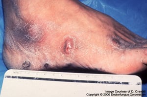
Sporotrichosis starts as a small papule or subcutaneous nodule that may ulcerate or necrose.
Sporotrichosis starts as a small papule or subcutaneous nodule that may ulcerate or necrose.
Image courtesy of www.doctorfungus.org © 2005.
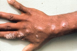
This photo shows cutaneous nodules in a lymphangitic (sporotrichoid) distribution over the dorsum of the right hand.
This photo shows cutaneous nodules in a lymphangitic (sporotrichoid) distribution over the dorsum of the right hand.
Image courtesy of Karen McKoy, MD.

Sporotrichosis starts as a small papule or subcutaneous nodule that may ulcerate or necrose. The infection spreads lymphatically.
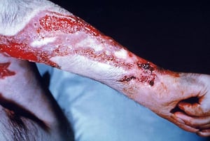
If untreated, diffuse erythema, ulcers, and abscesses may develop as is seen in this example of severe bacterial superinfection.
Sporotrichosis starts as a small papule or subcutaneous nodule that may ulcerate or necrose. The infection spreads lymp
CDC/ Dr. Lucille K. Georg
Sporotrichosis starts as a small papule or subcutaneous nodule that may ulcerate or necrose.
Sporotrichosis starts as a small papule or subcutaneous nodule that may ulcerate or necrose.
Image courtesy of www.doctorfungus.org © 2005.
This photo shows cutaneous nodules in a lymphangitic (sporotrichoid) distribution over the dorsum of the right hand.
This photo shows cutaneous nodules in a lymphangitic (sporotrichoid) distribution over the dorsum of the right hand.
Image courtesy of Karen McKoy, MD.
Sporotrichosis starts as a small papule or subcutaneous nodule that may ulcerate or necrose. The infection spreads lymphatically.
If untreated, diffuse erythema, ulcers, and abscesses may develop as is seen in this example of severe bacterial superinfection.
Sporotrichosis starts as a small papule or subcutaneous nodule that may ulcerate or necrose. The infection spreads lymp
CDC/ Dr. Lucille K. Georg
Lymphocutaneous sporotrichosis is chronic and indolent; it is potentially fatal, but only if a bacterial superinfection causes sepsis.
Rarely, in patients without primary lymphocutaneous lesions, hematogenous spread leads to indolent infections of multiple peripheral joints, sometimes bones, and, less often, genitals, liver, spleen, kidneys, or meninges. These infections are more common among patients with immunocompromise secondary to another disorder (eg, alcohol use disorder). Equally rare is chronic pneumonia caused by inhaling spores and manifested by localized infiltrates or cavities, which occurs most often in patients with preexisting chronic lung disease.
Diagnosis of Sporotrichosis
Culture and histopathologic tissue examination
Molecular methods
The diagnosis of sporotrichosis is suspected based on clinical presentation in a patient with a history of possible exposure to the fungus (eg, gardener, landscaper, forester).
The characteristic nodular lymphangitis/cutaneous clinical presentation of sporotrichosis can also be caused by other pathogens, including Mycobacterium tuberculosis, nontuberculous mycobacteria, Nocardia, Francisella tularensis, and Leishmania brasiliensis. A microbiologic diagnosis can usually be established when appropriate histologic stains and cultures of biopsied tissues are obtained (1). During the early, nondisseminated stage, the primary lesion may be misdiagnosed as a spider bite.
Culture of tissue from the active infection site provides the definitive diagnosis. Sporothrix species yeasts can be seen only rarely in fixed-tissue specimens, even with special staining. Serologic tests are not available.
Polymerase chain reaction (PCR)–based assays provide rapid, highly sensitive and specific detection of Sporothrix DNA in tissue or clinical specimens (2). Molecular methods such as PCR can help confirm the diagnosis when cultures are negative or not feasible.
Diagnosis references
1. Tobin EH, Jih WW. Sporotrichoid lymphocutaneous infections: Etiology, diagnosis and therapy. Am Fam Physician. 2001;63(2):326–332.
2. Zhang M, Li F, Li R, Gong J, Zhao F. Fast diagnosis of sporotrichosis caused by Sporothrix globosa, Sporothrix schenckii, and Sporothrix brasiliensis based on multiplex real-time PCR. PLoS Negl Trop Dis. 2019;13(2):e0007219. Published 2019 Feb 28. doi:10.1371/journal.pntd.0007219
Treatment of Sporotrichosis
For lymphocutaneous disease, itraconazole
For severe, disseminated disease, amphotericin B
Itraconazole is the treatment of choice for lymphocutaneous disease (Itraconazole is the treatment of choice for lymphocutaneous disease (1). Oral itraconazole should be continued for 2 to 4 weeks after all lesions have resolved (typically for a total of 3 to 6 months). Posaconazole has shown in vitro activity and may be an alternative treatment option.). Oral itraconazole should be continued for 2 to 4 weeks after all lesions have resolved (typically for a total of 3 to 6 months). Posaconazole has shown in vitro activity and may be an alternative treatment option.
Severe, disseminated disease requires a lipid formulation of IV amphotericin B; after a favorable response, treatment is switched to oral itraconazole for a total of 12 months of treatment. Patients with advanced HIV infection may require lifelong maintenance therapy with Severe, disseminated disease requires a lipid formulation of IV amphotericin B; after a favorable response, treatment is switched to oral itraconazole for a total of 12 months of treatment. Patients with advanced HIV infection may require lifelong maintenance therapy withitraconazole for meningeal and disseminated infections.
Treatment reference
1. Kauffman CA, Bustamante B, Chapman SW, Pappas PG; Infectious Diseases Society of America. Clinical practice guidelines for the management of sporotrichosis: 2007 update by the Infectious Diseases Society of America. Clin Infect Dis. 2007;45(10):1255-1265. doi:10.1086/522765
Drug Information for the Topic


