


Frostbite is injury due to freezing of tissue. Initial manifestations may be deceptively benign. Skin may appear white or blistered and is numb; rewarming causes substantial pain. Gangrene may develop. Severely damaged tissue may autoamputate. Treatment is rewarming in warm (37 to 39° C) water, local care, and pain management. Surgical amputation may occasionally be indicated. However, surgical intervention, often guided by imaging results, is usually delayed until definitive demarcation of necrotic tissue occurs.
Frostbite usually occurs in extreme cold, especially at high altitude, and is aggravated by hypothermia. Distal extremities and exposed skin such as cheeks, ears, and nose are affected most often (1). (See also Overview of Cold Injury.)

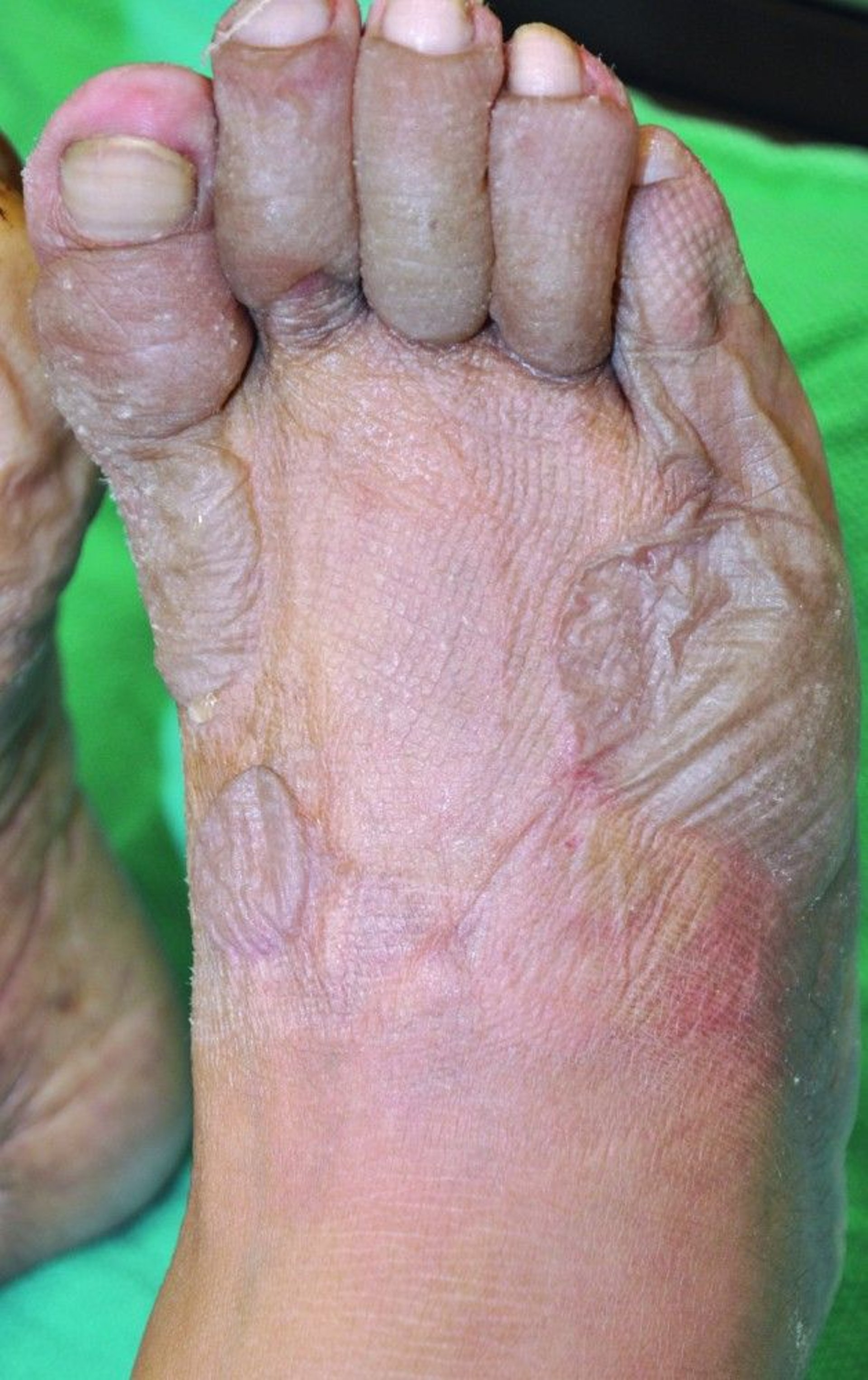
This photo shows pale nail beds, edematous gray toes, and blisters on the foot. The absence of blood in blisters suggests that frostbite is superficial (here, second degree) and that residual tissue loss is unlikely after healing.
© Springer Science+Business Media

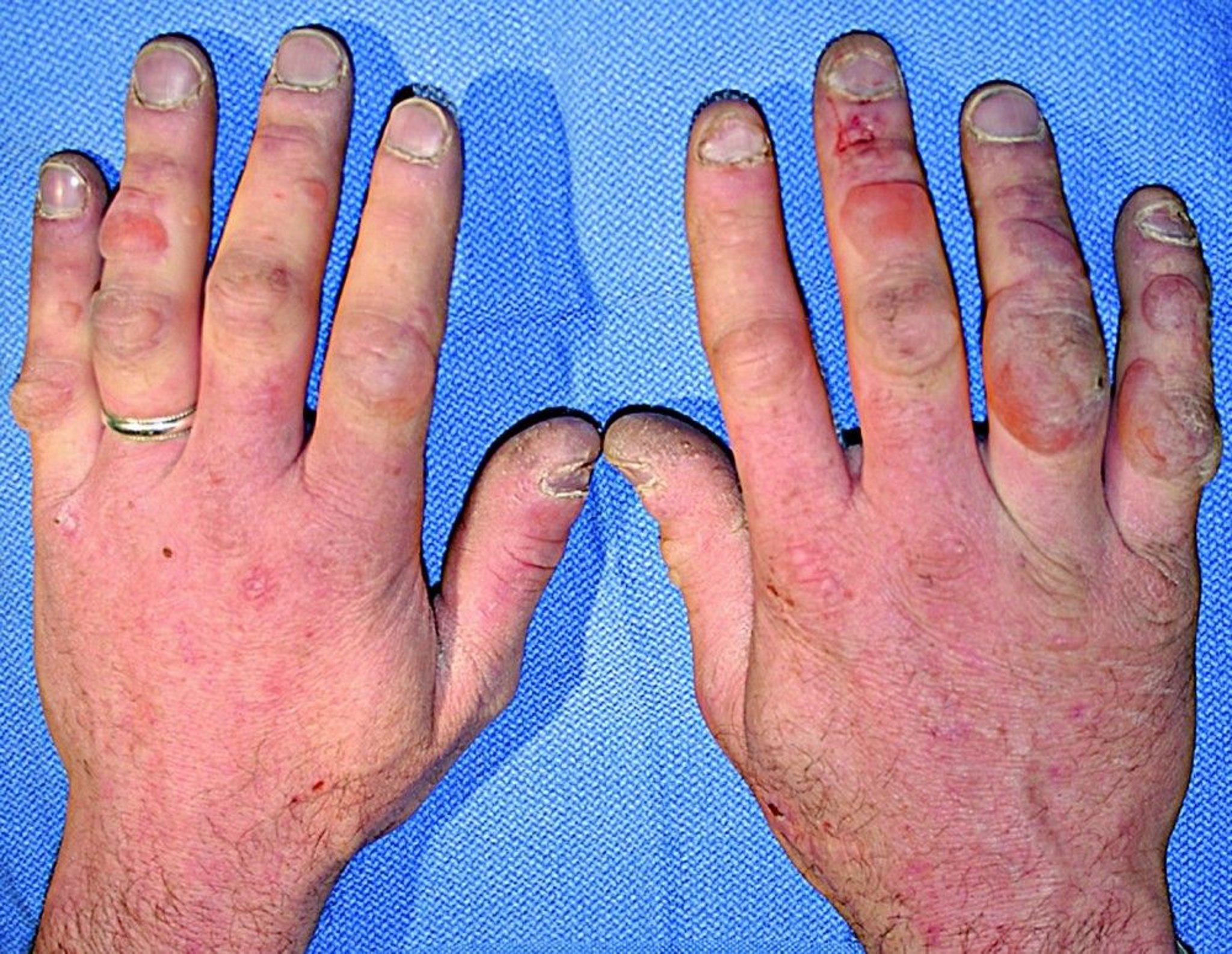
This photo shows frostbite (here, third-degree) of the hands. Hemorrhagic blisters (ring and small fingers) and cyanotic fingertips may portend necrosis. The ring should be removed before swelling occurs.
© Springer Science+Business Media
Ice crystals form within or between tissue cells, essentially freezing the tissue and causing cell death. Adjacent unfrozen areas are at risk because local vasoconstriction and thrombosis can cause endothelial and ischemic damage. With reperfusion during rewarming, inflammatory cytokines (eg, thromboxanes, prostaglandins) are released, exacerbating tissue injury. The depth of tissue loss depends on the duration and depth of freezing.
General reference
1. McIntosh SE, Freer L, Grissom CK, et al. Wilderness Medical Society Clinical Practice Guidelines for the Prevention and Treatment of Frostbite: 2024 Update. Wilderness Environ Med. 2024;35(2):183-197. doi:10.1177/10806032231222359
Symptoms and Signs of Frostbite
The affected area is cold, hard, white, and numb. When warmed, the area becomes blotchy red, swollen, and painful. Blisters form within 4 to 6 hours, but the full extent of injury may not be apparent for several days.
Frostnip is a nonfreezing tissue injury, often a precursor to frostbite, involving numbness and pallor of exposed skin.
Superficial frostbite does not involve permanent tissue loss (1):
First-degree frostbite demonstrates mild edema or plaque on rewarming.
Second-degree frostbite involves blisters that are filled with serous (clear or milky) fluid on rewarming and are surrounded by edema and erythema.
Deep frostbite involves the dermis or deeper tissue layers and involves permanent tissue loss (1):
Third-degree frostbite involves hemorrhagic blisters and edema.
Fourth-degree frostbite has cyanotic and minimally edematous tissue on rewarming, with necrosis and eventual mummification or gangrene.
Gangrene, when it occurs, can be dry or wet. Dry gangrene, due to freezing of deep tissue, results in a hard black carapace over healthy tissue. Wet gangrene is gray, edematous, and soft and is less common. Wet gangrene is characterized by infection; dry gangrene is less likely to become infected.

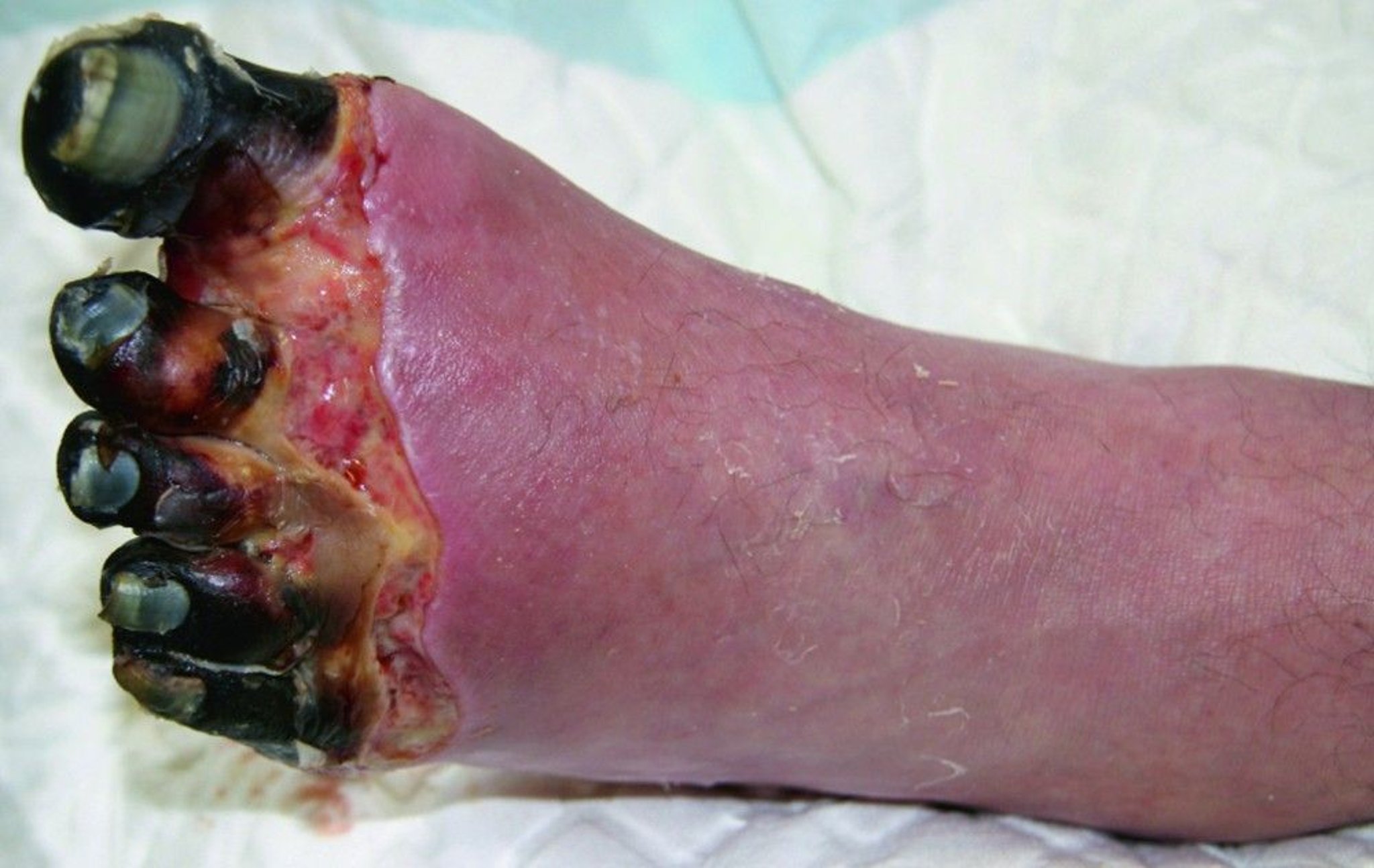
This photo shows fourth-degree frostbite with necrosis of affected tissue.
© Springer Science+Business Media
Severely damaged tissue may autoamputate. Compartment syndrome may develop. All degrees of frostbite may cause faulty nail growth and long-term neuropathic symptoms: sensitivity to cold, excessive sweating, and numbness (symptoms resembling those of complex regional pain syndrome) (2).
Symptoms and signs references
1. McIntosh SE, Freer L, Grissom CK, et al. Wilderness Medical Society Clinical Practice Guidelines for the Prevention and Treatment of Frostbite: 2024 Update. Wilderness Environ Med. 2024;35(2):183-197. doi:10.1177/10806032231222359
2. Regli IB, Strapazzon G, Falla M, Oberhammer R, Brugger H. Long-Term Sequelae of Frostbite-A Scoping Review. Int J Environ Res Public Health. 2021;18(18):9655. doi:10.3390/ijerph18189655
Diagnosis of Frostbite
History and physical examination
Diagnosis is based on clinical findings after rewarming. Many early characteristics of frostbite (eg, coldness, numbness, white or red color) are also characteristic of nonfreezing cold injuries; thus, the definitive diagnosis of frostbite may require repeated observation until more specific characteristics (eg, hemorrhagic blisters, black carapace) develop.
Treatment of Frostbite
Treatment of hypothermia
Rewarming in warm (37 to 39° C) water
Ibuprofen
Iloprost or thrombolytic therapy when available
Tetanus prophylaxis
Supportive measures, including analgesia
Local wound care
Sometimes surgery
Prehospital care
In the field, frostbitten extremities should be rewarmed rapidly by totally immersing the affected area in water that is tolerably warm to the touch (37 to 39° C) (1). Rewarming with an uncontrolled dry heat source (eg, fire, heating pad) should be avoided because the affected area is numb and at risk of burns. Rubbing, which may further damage tissue, should also be avoided.
The longer an area remains frozen, the greater the ultimate damage may be. However, if a patient must walk any distance to receive care, it may be necessary to delay thawing the feet. Thawed tissue is particularly sensitive to the trauma of walking and, if refrozen, will be more severely damaged than if left frozen (1). If thawing must be delayed, the frozen area is gently cleansed, air dried, and protected in sterile and dry bulky compresses. Patients are given analgesics, if available, and the whole body is kept warm.
If available, ibuprofen is recommended based on the potential for prostaglandin inhibition to reduce vasoconstriction and tissue ischemia (1).
Acute care
Once the patient is in the hospital, core temperature is stabilized (ie, hypothermia is treated) and extremities are rapidly rewarmed in large containers of circulating water kept at approximately 37 to 39° C; 15 to 30 minutes is usually adequate (1). All jewelry must be removed. Pain may be severe during rewarming. Parenteral analgesics, including opioids, are often required.
Patients are encouraged to move the affected part gently during thawing. Large, clear blisters generally are left intact or aspirated using sterile technique. Hemorrhagic blisters are left intact to avoid secondary desiccation of deep dermal layers. Broken vesicles are debrided.
For severe injury presenting within 48 to 72 hours, infusion of a prostacyclin analog, such as iloprost, is indicated after rewarming (1, 2, 3). Iloprost is used to treat severe frostbite. Studies have shown reduction in risk of digit amputation by improving microvascular perfusion and inhibiting platelet aggregation (4). If the injury is deep (at a distal interphalangeal joint or more proximally) and there is a risk of amputation, intra-arterial or intravenous thrombolytic (fibrinolytic) therapy should be considered within the first 24 hours of the injury (5). IV low molecular weight dextran may be considered as an alternative if iloprost or thrombolytics are unavailable.
Although other vasodilators such as phenoxybenzamine, papaverine, nicardipine, or nitroglycerin have also been used as adjunctive therapy, strong evidence to support their use is lacking (1). Anticoagulants (eg, heparin), IV low molecular weight dextran, and intra-arterial vasodilators (eg, reserpine, tolazoline) have no proven clinical benefit and should not be used.
Anti-inflammatory measures (eg, ibuprofen and topical aloe vera) are helpful. Affected areas are left open to warm air, and extremities are elevated to decrease edema. There are insufficient data to support hyperbaric oxygen therapy (1, 6).
Empiric prophylaxis is not indicated unless there is gross contamination, crush injury, or wet gangrene. If wet gangrene is present, broad-spectrum antibiotics are administered. Tetanus prophylaxis is administered based on current guidelines. If tissue damage is severe, tissue pressure is monitored for indications of compartment syndrome (see Compartment Syndrome: Diagnosis, How To Measure Compartment Pressure in a Forearm, and How To Measure Compartment Pressure in the Lower Leg).
Ongoing care
Adequate nutrition is important to sustain metabolic heat production. Consider using Doppler ultrasound to assess pulses and tissue status.
Other imaging tests include radionuclide scanning, MRI, microwave thermography, and laser-Doppler flowmetry to help assess circulation, determine tissue viability, and thus guide treatment. MRI, and magnetic resonance angiography in particular, as well as technetium-99 bone scanning, may establish the line of demarcation of viable tissue before clinical demarcation and thus make earlier surgical debridement or amputation possible (1). However, whether earlier surgery improves long-term outcome is unclear. Usually, surgery is delayed as long as possible because the black carapace is often shed, leaving viable tissue. Advise patients with severe frostbite hat many weeks of observation may be required before demarcation and the extent of tissue loss become apparent.
Long-term management may also include whirlpool baths at 37 to 39° C 1 to 2 times a day followed by gentle drying, rest, and time. No totally effective treatment for the long-lasting symptoms of frostbite (eg, numbness, hypersensitivity to cold) is known. Chemical or surgical sympathectomy for late neuropathic symptoms is not advised.
Treatment references
1. McIntosh SE, Freer L, Grissom CK, et al. Wilderness Medical Society Clinical Practice Guidelines for the Prevention and Treatment of Frostbite: 2024 Update. Wilderness Environ Med. 2024;35(2):183-197. doi:10.1177/10806032231222359
2. Iloprost (Aurlumyn) for frostbite. Med Lett Drugs Ther. 2024;66(1707):114. doi:10.58347/tml.2024.1707b
3. Vampola C-L, Fahrbach K, Davis C. Risk of Amputation in Severe Frostbite - A Systematic Literature Review and Meta-analysis to Evaluate Iloprost and Standard of Care. Wilderness Environ Med. 2023;34(4):e4. https://doi.org/10.1016/j.wem.2023.08.016
4. Harris E. FDA Approves Iloprost as First Drug to Treat Severe Frostbite. JAMA. 2024;331(11):907. doi:10.1001/jama.2024.1116
5. Hickey S, Whitson A, Jones L, et al. Guidelines for Thrombolytic Therapy for Frostbite. J Burn Care Res. 2020;41(1):176-183. doi:10.1093/jbcr/irz148
6. Lorentzen AK, Davis C, Penninga L. Interventions for frostbite injuries. Cochrane Database Syst Rev. 2020;12(12):CD012980. doi:10.1002/14651858.CD012980.pub2
Key Points
Depth of injury is difficult to recognize initially, although blood-filled blisters indicate deep damage.
Thaw frostbitten tissue as soon as possible using water that is tolerably warm to the touch (37 to 39° C); analgesia is usually required.
Avoid thawing and refreezing.
Keep affected areas uncovered, clean, dry, and elevated.
Black tissue may represent a black carapace that will be shed or gangrene that will require amputation; surgery is usually delayed until the demarcation is clear.
Neuropathic symptoms (eg, sensitivity to cold, numbness) may persist indefinitely.
Drug Information for the Topic







