


Fractures of the nasal bones or cartilaginous injury may result in swelling, point tenderness, hypermobility, crepitus, epistaxis, and periorbital bruising. Diagnosis is usually clinical. Treatment may include reduction, stabilization through internal packing, and splinting. A septal hematoma should be drained immediately.
The nasal bones are commonly fractured facial bones because of their central location and protrusion. Depending on the mechanism of injury, fractures of the maxilla, orbit, or cribriform plate and injury to the nasolacrimal ducts may occur concurrently.
Complications of nasal fractures include cosmetic deformity and functional obstruction. Septal hematomas are subperichondrial blood collections that may lead to avascular or septic necrosis of the cartilage with resultant deformity (saddle nose). Cribriform plate fracture may cause a cerebrospinal fluid (CSF) leak, with increased risk of meningitis or brain abscess. Fortunately, this complication is rare (1).

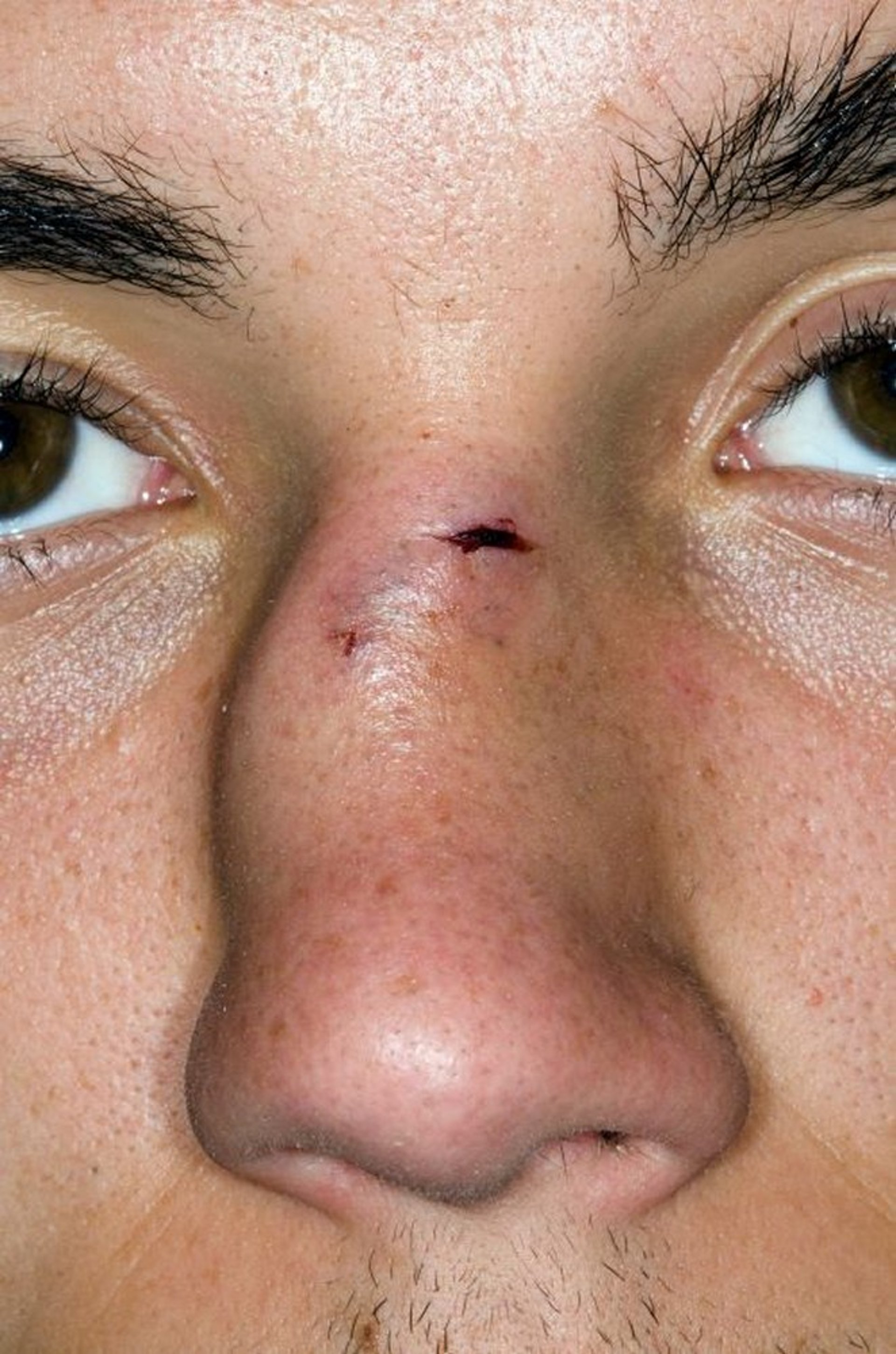
Ecchymosis, deviation of the nasal bones, and a laceration characterize this open nasal fracture.
DR P. MARAZZI/SCIENCE PHOTO LIBRARY
General reference
1. Umana GE, Pucci R, Palmisciano P, et al. Cerebrospinal Fluid Leaks After Anterior Skull Base Trauma: A Systematic Review of the Literature. World Neurosurg. 2022;157:193-206.e2. doi:10.1016/j.wneu.2021.10.065
Symptoms and Signs of Nasal Fractures
Facial trauma resulting in epistaxis may indicate a nasal fracture. Cerebrospinal fluid (CSF) rhinorrhea appears as clear drainage but may be mixed with blood, making it difficult to identify.
Physical examination finding may include obvious or subtle nasal deformity, swelling, point tenderness, crepitus, and instability. Lacerations, ecchymosis (nasal and periorbital), septal deviation, and nasal obstruction may be present. Septal hematoma appears as a purplish bulge on the septum.
Diagnosis of Nasal Fractures
History and physical examination alone
Diagnosis is usually based on history and physical examination alone.
Plain radiographs of an uncomplicated nasal fracture are not helpful because their sensitivity and specificity are poor. If other facial fractures or complications are suspected, CT of facial bones is performed.
If CSF rhinorrhea is suspected, testing nasal discharge for beta-2-transferrin is an effective test for detecting CSF (1). Bedside glucose test strips do not accurately identify CSF rhinorrhea and are not recommended.
Diagnosis reference
1. Mantur M, Łukaszewicz-Zając M, Mroczko B, et al. Cerebrospinal fluid leakage--reliable diagnostic methods. Clin Chim Acta. 2011;412(11-12):837-840. doi:10.1016/j.cca.2011.02.017
Treatment of Nasal Fractures
Symptomatic care
For septal hematomas, immediate drainage
For deformities, delayed reduction
Immediate treatment includes symptomatic control with ice and analgesics. Septal hematomas must be immediately incised and drained to prevent avascular or septic necrosis of the septal cartilage.
Reduction is needed only for fractures causing clinically visible deformity or nasal airway obstruction. The end-point of reduction is determined by clinical appearance or decreased obstruction of the nasal cavity. Reduction is usually deferred for 3 to 5 days after injury to allow swelling to subside but should take place within 2 weeks of the injury, before bony callus formation. Nasal fractures in adults may be reduced after a local anesthetic is given; children may require general anesthesia.
For reduction, a blunt nasal fracture elevator instrument is passed through the nares and placed under the depressed nasal bone, which is lifted anteriorly and laterally while pressure is applied to the other side of the nose to bring the nasal dorsum to the midline (1). The nose may be stabilized with internal packing (consisting of antibiotic-impregnated strip gauze, silicone elastomer sponges, or similar products) placed high within the nasal vestibule, as well as with external splinting. Internal packing is left in place for 4 to 7 days; external splinting is left for 7 to 14 days. Antibiotic prophylaxis effective against staphylococci is required for the duration of nasal packing to decrease the risk of toxic shock syndrome.
Cartilaginous injuries often do not require reduction. In the rare circumstance that a deformity persists after swelling subsides, a reduction and splinting after a local anesthetic is given are usually sufficient.
Septal fractures are difficult to hold in position and often require septal surgery later.
Cribriform plate fractures with cerebrospinal fluid (CSF) leak initially require hospital admission with bed rest and head elevation (2). Placement of a lumbar drain may be required for a persistent CSF leak. Drain management and need for antibiotics vary by institution. If the CSF leak does not resolve, surgical repair of the skull base may be required.
Treatment references
1. Trujillo O, Lee C. Nasal Fractures: Acute, Subacute, and Delayed Management. Otolaryngol Clin North Am. 2023;56(6):1089-1099. doi:10.1016/j.otc.2023.05.004
2. Bell RB, Dierks EJ, Homer L, et al. Management of cerebrospinal fluid leak associated with craniomaxillofacial trauma. J Oral Maxillofac Surg. 2004;62(6):676-684. doi:10.1016/j.joms.2003.08.032
Key Points
The main concerns with nasal fractures are septal hematoma, epistaxis, nasal obstruction, cosmetic problems, and rare cribriform plate fractures.
Nasal radiographs are unnecessary.
Immediately drain septal hematomas.
Delay reduction and some other treatments for 3 to 5 days to allow edema to resolve.


