

Achilles tendinopathy is a condition characterized by local pain, swelling, and impaired function of portions of the Achilles tendon. Symptoms include posterior heel pain that worsens with activity. Diagnosis is clinical, but may be supported by imaging (ultrasound or magnetic resonance imaging). Treatment includes resistance exercise and other measures (eg, orthoses, NSAIDs).
Achilles tendinopathy is very common among running athletes. The calf muscles attach to the calcaneus via the Achilles tendon. During running, the calf muscles help with the lift-off phase of gait. Repetitive forces from running combined with insufficient recovery time can lead to a failed healing response to repetitive microtrauma, resulting in degenerative changes of the tendon matrix (eg, disorganization of collagen fibers, neovascularization) (1). These changes can cause the tendon thickening. Non-resolving inflammation is also recognized as a feature of chronic tendinopathy. Fluoroquinolone antibiotics can also increase the risk of Achilles tendinopathy and complete tendon rupture (2).
A complete tear of the Achilles tendon is a serious injury, usually resulting from sudden, forceful stress. Tendon tears can occur with minimal exertion in people who have taken fluoroquinolone antibiotics.
Achilles tendinopathy may be associated with a Haglund deformity, which is a posterior superior calcaneal prominence located adjacent to the Achilles tendon. The presence of a Haglund deformity does not correlate directly with the patient's symptoms (3).
General references
1. Traweger A, Scott A, Kjaer M, et al. Achilles tendinopathy. Nat Rev Dis Primers. 2025;11(1):20. Published 2025 Mar 27. doi:10.1038/s41572-025-00602-9
2. Shu Y, Zhang Q, He X, Liu Y, Wu P, Chen L. Fluoroquinolone-associated suspected tendonitis and tendon rupture: A pharmacovigilance analysis from 2016 to 2021 based on the FAERS database. Front Pharmacol. 2022;13:990241. doi:10.3389/fphar.2022.990241
3. Kang S, Thordarson DB, Charlton TP. Insertional Achilles tendinitis and Haglund's deformity. Foot Ankle Int. 2012 Jun;33(6):487-491. doi: 10.3113/FAI.2012.0487
Symptoms and Signs of Achilles Tendinopathy
The primary symptom of Achilles tendon inflammation is pain in the back of the heel, which initially increases when exercise is begun and often lessens as exercise continues.
A complete tear of the Achilles tendon typically occurs with a sudden forceful plantarflexion movement, particularly among athletes who do not regularly engage in high-intensity recreational activities (eg, "weekend warriors") such as jumping or playing basketball. Patients relate a sensation of having been struck in the back of the ankle and calf, accompanied by a popping or snapping feeling in the back of the leg. This sensation is immediately followed by the inability to bear weight or push off of the affected limb.
Diagnosis of Achilles Tendinopathy
Primarily history and physical examination
The diagnosis is largely based on clinical findings. Pain is observed at the tendon insertion at the calcaneus and along the tendon, particularly in the watershed area 2 to 6 cm from the insertion site, during resisted muscle strength testing in plantarflexion as well as with passive stretching in dorsiflexion.
On examination, an inflamed or partially torn Achilles tendon is tender when squeezed between the fingers. Complete tears are differentiated by:
Sudden, severe pain and inability to bear weight on the extremity
A palpable defect along the course of the tendon as well as loss of ankle plantarflexion strength
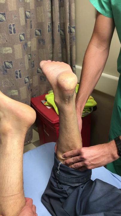
A positive Thompson test (while the patient lies prone on the examination table and foot is hanging off the edge of the table, the examiner squeezes the calf muscle; this maneuver by the examiner does not cause the normally expected plantarflexion of the foot)

Treatment of Achilles Tendinopathy
Ice, nonsteroidal anti-inflammatory drugs (NSAIDs), and stretches
Modification of activities
Heel lifts and walking boot
Exercise program for persistent symptoms
Tendon inflammation should initially be treated with ice, gentle calf muscle stretching, and use of NSAIDs.
A heel lift can be temporarily placed in the shoes or within a walking boot to take tension off the tendon by creating plantarflexion at the ankle. The heel lift should be removed after the initial pain subsides, and the patient should begin a dedicated stretching routine with a progressive increase in duration and frequency. Athletes should be instructed to avoid uphill and downhill running until the tendon is not painful and to engage in cross-training aerobic conditioning.
For symptoms lasting longer than 3 months, an exercise program is generally recommended. Exercises, especially eccentric or resistance training, have been shown to decrease pain and improve function (1, 2). Exercise protocols typically involve 3 months of structured exercises involving calf muscle loading, with gradual improvement expected over several weeks to months.
Most clinicians treat complete tears of the Achilles tendon with surgical repair. However, some studies (3, 4, 5) have shown that nonoperative management (including immediate immobilization of the ankle in plantarflexion then highly structured rehabilitation activities) gives long-term results similar to those of surgical repair in terms of ankle strength, range of motion in the ankle, re-rupture rates of the tendon, and ability to return to pre-injury activity.

1. Stand facing or next to wall with hands on the wall.
2. Bend knee on involved side to place top of involved foot dow
Courtesy of Tomah Memorial Hospital, Department of Physical Therapy, Tomah, WI; Elizabeth C.K. Bender, MSPT, ATC, CSCS; and Whitney Gnewikow, DPT, ATC.

1. Sit in chair.
2. Bend knee on involved side to place top of involved foot down toward the floor with toes pointing d
Courtesy of Tomah Memorial Hospital, Department of Physical Therapy, Tomah, WI; Elizabeth C.K. Bender, MSPT, ATC, CSCS; and Whitney Gnewikow, DPT, ATC.

1. Stand facing or next to wall with hands on the wall for support.
2. Place uninvolved leg forward.
3. Keep rear leg s
Courtesy of Tomah Memorial Hospital, Department of Physical Therapy, Tomah, WI; Elizabeth C.K. Bender, MSPT, ATC, CSCS; and Whitney Gnewikow, DPT, ATC.

1. Stand facing or next to wall with hands on the wall for support.
2. Place uninvolved leg forward.
3. Keep heels on t
Courtesy of Tomah Memorial Hospital, Department of Physical Therapy, Tomah, WI; Elizabeth C.K. Bender, MSPT, ATC, CSCS; and Whitney Gnewikow, DPT, ATC.

1. Stand on balls of feet with heels off the floor.
2. Walk on balls of feet while keeping knees straight.
3. Walk as f
Courtesy of Tomah Memorial Hospital, Department of Physical Therapy, Tomah, WI; Elizabeth C.K. Bender, MSPT, ATC, CSCS; and Whitney Gnewikow, DPT, ATC.

1. Stand on heels with balls of feet off the floor.
2. Walk on heels while keeping knees straight.
3. Walk as far as ab
Courtesy of Tomah Memorial Hospital, Department of Physical Therapy, Tomah, WI; Elizabeth C.K. Bender, MSPT, ATC, CSCS; and Whitney Gnewikow, DPT, ATC.

1. Stand with both feet on step with heels off edge of step. Hold on for support.
2. Raise up on balls of feet.
3. Focu
Courtesy of Tomah Memorial Hospital, Department of Physical Therapy, Tomah, WI; Elizabeth C.K. Bender, MSPT, ATC, CSCS; and Whitney Gnewikow, DPT, ATC.


1. Stand facing or next to wall with hands on the wall.
2. Bend knee on involved side to place top of involved foot down toward the floor with toes pointing down.
3. Bend knee on uninvolved side and slowly lower body until stretch is felt over the top of the foot and ankle.
4. Hold exercise for 30 second.
5. Perform 1 set of 4 repetitions, 3 times a day.
6. Special Instructions
a. Perform plantarflexion stretching while either sitting or standing, whichever is most comfortable.
Courtesy of Tomah Memorial Hospital, Department of Physical Therapy, Tomah, WI; Elizabeth C.K. Bender, MSPT, ATC, CSCS; and Whitney Gnewikow, DPT, ATC.

1. Sit in chair.
2. Bend knee on involved side to place top of involved foot down toward the floor with toes pointing down.
3. Slowly sit forward in the chair and push foot into plantarflexion until a stretch is felt over the top of the foot and ankle.
4. Hold exercise for 30 seconds.
5. Perform 1 set of 4 repetitions, 3 times a day.
6. Special Instructions
a. Perform plantarflexion stretching while either sitting or standing, whichever is most comfortable.
Courtesy of Tomah Memorial Hospital, Department of Physical Therapy, Tomah, WI; Elizabeth C.K. Bender, MSPT, ATC, CSCS; and Whitney Gnewikow, DPT, ATC.

1. Stand facing or next to wall with hands on the wall for support.
2. Place uninvolved leg forward.
3. Keep rear leg straight with knees and toes pointing toward the wall, keep rear heel on the floor.
4. Bend knee on uninvolved leg and lean hips toward the wall to feel a stretch along the calf of the rear leg.
5. Hold exercise for 30 seconds.
6. Perform 1 set of 4 repetitions, 3 times a day.
(It is important to maintain a straight back posture with the heel firmly planted [not lifted] during the stretch.)
Courtesy of Tomah Memorial Hospital, Department of Physical Therapy, Tomah, WI; Elizabeth C.K. Bender, MSPT, ATC, CSCS; and Whitney Gnewikow, DPT, ATC.

1. Stand facing or next to wall with hands on the wall for support.
2. Place uninvolved leg forward.
3. Keep heels on the floor and slowly bend both knees to feel stretch along the calf of the rear leg.
4. Hold exercise for 30 seconds.
5. Perform 1 set of 4 repetitions, 3 times a day.
Courtesy of Tomah Memorial Hospital, Department of Physical Therapy, Tomah, WI; Elizabeth C.K. Bender, MSPT, ATC, CSCS; and Whitney Gnewikow, DPT, ATC.

1. Stand on balls of feet with heels off the floor.
2. Walk on balls of feet while keeping knees straight.
3. Walk as far as able during given time frame, stop at point of fatigue.
4. Perform 3 sets of 1 minute, 1 time a day.
Courtesy of Tomah Memorial Hospital, Department of Physical Therapy, Tomah, WI; Elizabeth C.K. Bender, MSPT, ATC, CSCS; and Whitney Gnewikow, DPT, ATC.

1. Stand on heels with balls of feet off the floor.
2. Walk on heels while keeping knees straight.
3. Walk as far as able during given time frame, stop at point of fatigue.
4. Perform 3 sets of 1 minute, 1 time a day.
Courtesy of Tomah Memorial Hospital, Department of Physical Therapy, Tomah, WI; Elizabeth C.K. Bender, MSPT, ATC, CSCS; and Whitney Gnewikow, DPT, ATC.

1. Stand with both feet on step with heels off edge of step. Hold on for support.
2. Raise up on balls of feet.
3. Focus on slowly lowering heels to below the height of the step.
4. Return to starting position and repeat.
5. Perform 3 sets of 10 repetitions, 1 time a day.
Courtesy of Tomah Memorial Hospital, Department of Physical Therapy, Tomah, WI; Elizabeth C.K. Bender, MSPT, ATC, CSCS; and Whitney Gnewikow, DPT, ATC.
Treatment references
1. Irby A, Gutierrez J, Chamberlin C, Thomas SJ, Rosen AB: Clinical management of tendinopathy: A systematic review of systematic reviews evaluating the effectiveness of tendinopathy treatments. Scand J Med Sci Sports 30(10):1810-1826, 2020. doi:10.1111/sms.13734
2. Wilson F, Walshe M, O'Dwyer T, Bennett K, Mockler D, Bleakley C: Exercise, orthoses and splinting for treating Achilles tendinopathy: a systematic review with meta-analysis. Br J Sports Med 52(24):1564-1574, 2018. doi:10.1136/bjsports-2017-098913
3. Lantto L, Heikkinen J, Flinkkila T, et al: A prospective randomized trial comparing surgical and nonsurgical treatments of acute Achilles tendon ruptures. Am J Sports Med 44(9):2406-2414, 2016. doi: 10.1177/0363546516651060
4. Parisien RL, Dodson CC, Trofa DP, et al: Face off: Surgical versus nonsurgical treatment of acute Achilles tendon ruptures. AAOS Now July 2016, cover.
5. Myhrvold SB, Brouwer EF, Andresen TKM, et al: Nonoperative or surgical treatment of acute Achilles' tendon rupture. N Engl J Med 386(15):1409-1420, 2022. doi: 10.1056/NEJMoa2108447


