Rubella is a viral infection that may cause adenopathy, rash, and sometimes constitutional symptoms, which are usually mild and brief. Infection during early pregnancy can cause fetal demise or congenital rubella syndrome. Diagnosis is usually clinical. Treatment is supportive. Vaccination is effective for prevention.
Rubella is the most common cause worldwide of vaccine-preventable congenital anomalies. Globally, an estimated 100,000 infants are born with congenital rubella syndrome annually (1). The risk of congenital rubella infection, and of congenital anomalies due to this infection, is highest in the first 12 weeks of gestation (2).
The Rubella virus is an RNA virus, which is spread by respiratory droplets through close contact or through the air. Rubella is less contagious than measles. Rash is a characteristic clinical manifestation. Infected people can transmit rubella from 7 days before a rash appears until 7 days after onset of the rash (3); the period of greatest risk is from a few days before the rash appears to 7 days after onset of the rash. Some patients are asymptomatic, but they can transmit the virus.
Congenitally infected infants may shed rubella virus for many months after birth.
Immunity appears to be lifelong after natural infection (4). However, in unvaccinated populations, 10 to 15% of young adults have not had childhood infection and are susceptible.
In 2004, rubella was declared eliminated in the United States, and the last endemic rubella cases in the Americas were diagnosed in 2009 (2, 4, 5). In the United States, among the few people (< 10) who develop rubella each year, most acquired the infection while living or traveling outside of the United States.
Postnatally acquired rubella infection is discussed here; see Congenital Rubella for information about prenatal rubella infection.
General references
1. World Health Organization (WHO). Rubella. May 14, 2024. Accessed February 6, 2026.
2. McLean HQ, Fiebelkorn AP, Temte JL, Wallace GS; Centers for Disease Control and Prevention. Prevention of measles, rubella, congenital rubella syndrome, and mumps, 2013: summary recommendations of the Advisory Committee on Immunization Practices (ACIP). MMWR Recomm Rep. 2013;62(RR-04):1-34.
3. Committee on Infectious Diseases, American Academy of Pediatrics. Rubella in Red Book: 2024–2027 Report of the Committee on Infectious Diseases, ed. 33, edited by Kimberlin DW, Banerjee R, Barnett ED, Lynfield R, and Sawyer MH. Itasca, American Academy of Pediatrics, 2024.
4. Pan American Health Organization (PAHO). Rubella. Accessed December 17, 2025.
5. Centers for Disease Control and Prevention (CDC). Impact of U.S. MMR Vaccination Program. Rubella Elimination: A Public Health Success Story. January 17, 2025. Accessed December 17, 2025.
Symptoms and Signs of Rubella
The average incubation period is 17 days, but can be as short as 12 days or as long as 23 days. In adults, there is typically a 1- to 5-day prodrome, usually consisting of low-grade fever, malaise, conjunctivitis, lymphadenopathy, headache, stiff joints, transient arthritis, and mild rhinitis; tender swelling of the suboccipital, postauricular, and posterior cervical nodes is characteristic (1). Up to half of adults, however, are asymptomatic (1).
Prodromal symptoms may be minimal or absent in children; they may include malaise and arthralgias. Symptoms are mild in many cases.
On examination, there is pharyngeal injection at the onset.

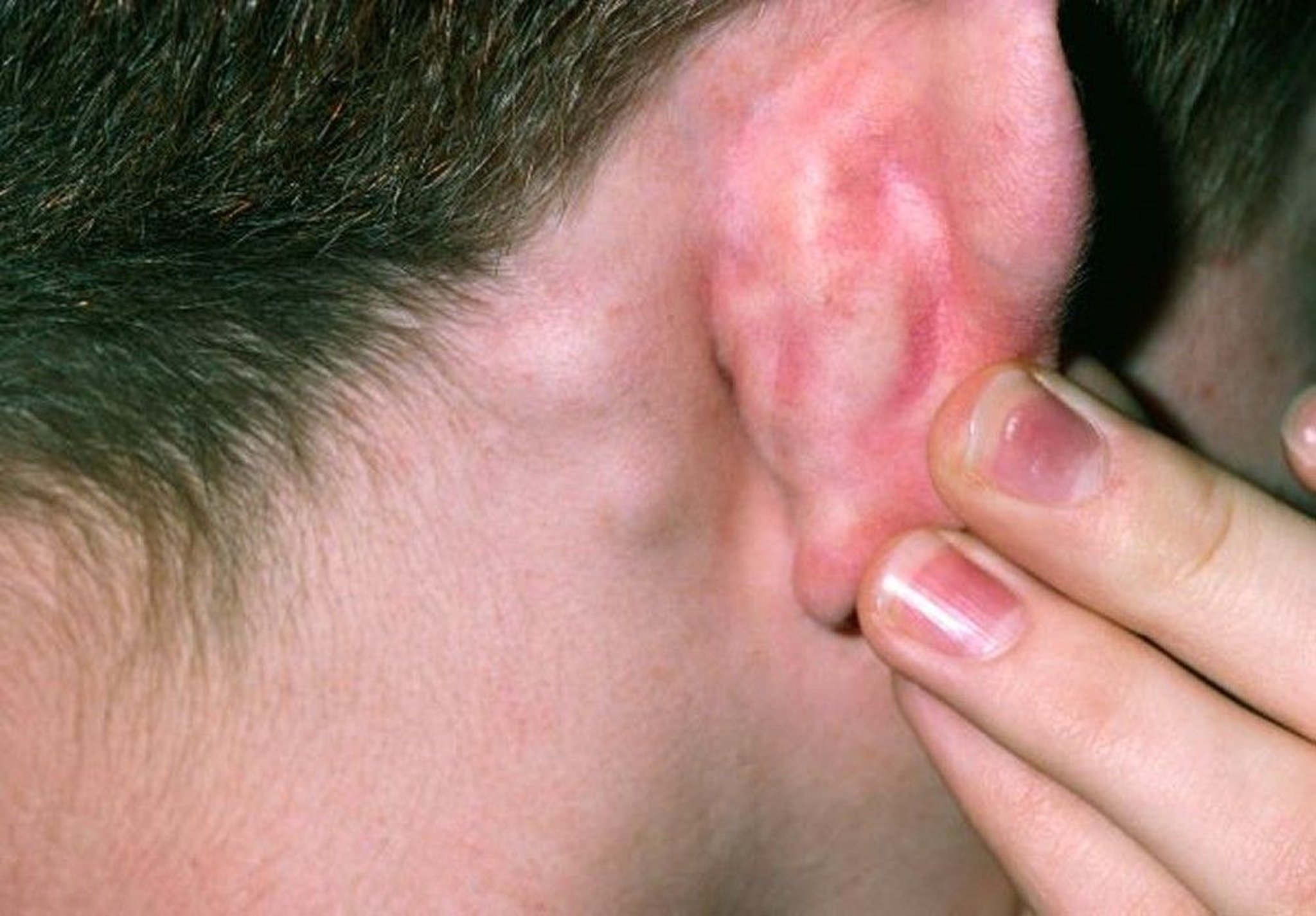
This image shows enlarged posterior auricular lymph nodes, which are commonly seen in rubella infection.
DR P. MARAZZI/SCIENCE PHOTO LIBRARY
After the prodrome, a maculopapular rash that appears similar to that of measles but is less extensive and more evanescent develops. Rash is often the first sign in children. It begins on the face and neck and quickly spreads to the trunk and extremities. At onset, a blanching, macular erythema may appear, particularly on the face. On the 2nd day, the rash often becomes more scarlatiniform (pinpoint) with a reddish flush. Petechiae form on the soft palate (Forchheimer spots), later coalescing into a red blush. The rash lasts 3 to 5 days.

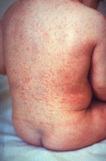
The rash of rubella is similar in distribution to that of measles (rubeola) but it is less intensely red and confluent.
Encephalitis occurs rarely (1 in 6000 cases) (1, 2). Complete resolution is usual, but encephalitis can cause seizures and can be fatal (3).
Thrombocytopenic purpura and otitis media occur rarely.
Symptoms and signs references
1. Committee on Infectious Diseases, American Academy of Pediatrics. Rubella in Red Book: 2024–2027 Report of the Committee on Infectious Diseases, ed. 33, edited by Kimberlin DW, Banerjee R, Barnett ED, Lynfield R, and Sawyer MH. Itasca, American Academy of Pediatrics, 2024.
2. Winter AK, Moss WJ. Rubella. Lancet. 2022;399(10332):1336-1346. doi:10.1016/S0140-6736(21)02691-X
3. Chaari A, Bahloul M, Berrajah L, et al. Childhood rubella encephalitis: diagnosis, management, and outcome. J Child Neurol. 2014;29(1):49-53. doi:10.1177/0883073812469443
Diagnosis of Rubella
History and physical examination
Serologic or reverse transcription–polymerase chain reaction (RT-PCR) testing
Rubella is suspected in patients with characteristic adenopathy and rash and with epidemiologic risk factors (eg, travel to an endemic area). Diagnosis is often based solely on history and physical examination.
Laboratory diagnosis is necessary for pregnant patients, neonates, and patients with encephalitis. Also, laboratory evaluation is strongly encouraged for all suspected cases of rubella for public health purposes.
For laboratory diagnosis, detection of viral RNA by RT-PCR testing of throat, nasal, or urine specimens is the most sensitive and specific way to confirm the diagnosis (1); genotype analysis is useful in epidemiologic investigations. Serologic testing is also commonly used to aid in the diagnosis (2) and evaluate a child's serologic response. A ≥ 4-fold rise between acute and convalescent (at least 10 days after the first sample, often 4 to 8 weeks) IgG antibody titers confirms the diagnosis. IgM antibody testing can also be used but is subject to false-positive and false-negative results.
(See also Diagnosis of Congenital Rubella for more information about diagnosis in pregnant patients, fetuses, and neonates.)
Differential diagnosis
Other causes of similar clinical manifestations include measles, erythema infectiosum (parvovirus B19 infection), enterovirus infections (eg, echovirus, coxsackievirus), infectious mononucleosis, scarlet fever, secondary syphilis, and drug rashes (see table ). Infections with enteroviruses and erythema infectiosum may be clinically indistinguishable.
Some of these conditions can be distinguished from rubella as follows:
Measles: Rubella is differentiated from measles by the milder, more evanescent rash, milder and briefer constitutional symptoms, and absence of Koplik spots, photophobia, and cough.
Infectious mononucleosis: Infectious mononucleosis can be differentiated by its more severe pharyngitis, more prolonged malaise, and atypical lymphocytosis and with Epstein-Barr virus antibody testing.
Scarlet fever: Within a day of onset, scarlet fever usually causes more severe constitutional symptoms and pharyngitis than does rubella.
Secondary syphilis: In secondary syphilis, adenopathy is not tender, and the rash is usually prominent on the palms and soles. Also, laboratory diagnosis of syphilis is usually readily available.
Diagnosis references
1. Committee on Infectious Diseases, American Academy of Pediatrics. Rubella in Red Book: 2024–2027 Report of the Committee on Infectious Diseases, ed. 33, edited by Kimberlin DW, Banerjee R, Barnett ED, Lynfield R, and Sawyer MH. Itasca, American Academy of Pediatrics, 2024.
2. Winter AK, Moss WJ. Rubella. Lancet. 2022;399(10332):1336-1346. doi:10.1016/S0140-6736(21)02691-X
Treatment of Rubella
Supportive care
Treatment of rubella is symptomatic.
No specific therapy for encephalitis is available; antiseizure medications are given if seizures occur (1).
Treatment reference
1. Winter AK, Moss WJ. Rubella. Lancet. 2022;399(10332):1336-1346. doi:10.1016/S0140-6736(21)02691-X
Prevention of Rubella
Vaccination with live-attenuated virus vaccine containing measles, mumps, and rubella (MMR) is administered routinely to children in most nations that have a robust health care system. In the United States, the MMR vaccine is recommended for children and is administered in 2 doses: 1 at age 12 to 15 months and 1 at age 4 to 6 years.
MMR vaccination generally provides lasting immunity (1). The effectiveness of the MMR vaccine in preventing rubella in children was 93 to 97% after 1 dose and 100% after 2 doses, based on observational studies (2).
MMR is a live vaccine and is contraindicated during pregnancy. To prevent congenital rubella infection, unvaccinated patients who could become pregnant should receive 1 dose of the MMR vaccine and then wait 4 weeks before trying to conceive. Ideally, patients planning to become pregnant should be tested to confirm immunity to rubella at a preconceptual visit. Some people do not develop immunity after childhood vaccination but are likely to become immune with an additional dose of MMR vaccine (3).
MMR vaccine is also contraindicated in certain immunocompromised patients and in patients who have had an anaphylactic reaction to a vaccine component or previous vaccine dose (4).
See MMR Vaccine for more information, including indications, contraindications and precautions, dosing and administration, and adverse effects.
Postexposure vaccination or treatment with immune globulin has not been found to be effective (5).
Patients with rubella should be isolated, staying home from work, school, or child care, for 7 days after rash onset (5, 6). Caregivers of a hospitalized patient should observe droplet precautions for the same period of time.
(See also Prevention of Congenital Rubella for more information about preventing rubella infection in pregnant patients, fetuses, and neonates.)
Prevention references
1. McLean HQ, Fiebelkorn AP, Temte JL, Wallace GS; Centers for Disease Control and Prevention. Prevention of measles, rubella, congenital rubella syndrome, and mumps, 2013: Summary recommendations of the Advisory Committee on Immunization Practices (ACIP). MMWR Recomm Rep. 2013;62(RR-04):1–34.
2. Di Pietrantonj C, Rivetti A, Marchione P, et al. Vaccines for measles, mumps, rubella, and varicella in children. Cochrane Database Syst Rev. 2020;4(4):CD004407. doi:10.1002/14651858.CD004407.pub4
3. Haas DM, Flowers CA, Congdon CL. Rubella, rubeola, and mumps in pregnant women: susceptibilities and strategies for testing and vaccinating. Obstet Gynecol. 2005;106(2):295-300. doi:10.1097/01.AOG.0000171110.49973.e3
4. National Center for Immunization and Respiratory Diseases. General recommendations on immunization --- recommendations of the Advisory Committee on Immunization Practices (ACIP). MMWR Recomm Rep. 2011;60(2):1-64.
5. Committee on Infectious Diseases, American Academy of Pediatrics. Rubella in Red Book: 2024–2027 Report of the Committee on Infectious Diseases, ed. 33, edited by Kimberlin DW, Banerjee R, Barnett ED, Lynfield R, and Sawyer MH. Itasca, American Academy of Pediatrics, 2024.
6. CDC. Clinical Overview of Rubella. July 15, 2024. Accessed February 6, 2026.
Progressive Rubella Panencephalitis
Progressive rubella panencephalitis is a neurologic disorder occurring in children with congenital rubella. It is presumably due to persistence or reactivation of rubella virus infection.
Progressive rubella panencephalitis is a usually fatal neurodegenerative disorder that can develop months to years after rubella infection (1). The primary infection is usually congenital rubella. The first manifestation may be neurologic deficits or symptoms that develop during early adolescence.
The diagnosis of progressive rubella panencephalitis is considered when a child with congenital rubella develops progressive spasticity, ataxia, mental deterioration, and seizures. Testing involves at least cerebrospinal fluid (CSF) examination and serologic testing. CSF total protein and globulin and rubella antibody titers in CSF and serum are elevated. An elevated level of IgG antibodies to rubella in CSF is diagnostic (1).
A CT scan or autopsy may show ventricular enlargement due to cerebellar atrophy and white matter disease (2). Brain biopsy may be necessary to exclude other causes of encephalitis or encephalopathy.
Rubella virus usually cannot be recovered by viral culture or immunohistologic testing.
No specific treatment for progressive rubella panencephalitis exists. Symptoms (eg, seizures, muscle spasticity, weakness) are managed as appropriate.
Progressive rubella panencephalitis references
1. Winter AK, Moss WJ. Rubella. Lancet. 2022;399(10332):1336-1346. doi:10.1016/S0140-6736(21)02691-X
2. Townsend JJ, Baringer JR, Wolinsky JS, et al. Progressive rubella panencephalitis. Late onset after congenital rubella. N Engl J Med. 1975;292(19):990-993. doi:10.1056/NEJM197505082921902
Key Points
Rubella causes a scarlatiniform rash and often low-grade fever, malaise, conjunctivitis, and lymphadenopathy (characteristically involving the suboccipital, postauricular, and posterior cervical nodes).
Most cases of postnatal rubella infection are mild and complications are few except for rare cases of encephalitis and the risk during early pregnancy that infection can cause spontaneous abortion, stillbirth, or congenital defects.
Laboratory diagnosis is strongly encouraged for all suspected cases for public health purposes; serologic or reverse transcription–polymerase chain reaction testing can be performed.
Rubella is preventable by vaccination; vaccination is contraindicated during pregnancy.