

Coal worker pneumoconiosis results from the inhalation of dust generated by the drilling, blasting, or crushing of coal and by the equipment and processes used to extract coal. Exposures in coal mining are heterogenous, which contributes to a wide spectrum of disease in coal miners. The clinical manifestations of coal worker pneumoconiosis range from minimal symptoms to progressive massive fibrosis with impaired lung function. Chronic obstructive pulmonary disease and bronchitis are common in coal miners. Diagnosis is based on history, chest x-ray findings, and pulmonary function test results. Treatment is generally supportive.
(See also Overview of Environmental and Occupational Pulmonary Disease.)
Coal worker pneumoconiosis is a nodular interstitial lung disease that results from exposure to coal mining dust. Coal worker pneumoconiosis can present as
Simple pneumonoconiosis
Complicated pneumonoconiosis, also known as progressive massive fibrosis
Simple coal worker pneumoconiosis is characterized by upper lobe predominance of small rounded nodular opacities and typically is not associated with symptoms or pulmonary dysfunction.
In complicated coal worker pneumoconiosis or progressive massive fibrosis, nodules coalesce to form larger parenchymal masses, usually in the upper posterior lung fields, and symptoms are prominent.
Following the enactment of the Coal Mine Health and Safety Act in 1969, there was a decline in coal worker pneumoconiosis in the United States. However, since the late 1990s there has been a resurgence of coal worker pneumoconiosis, especially severe progressive disease (1). This resurgence is most likely due to greater silica exposure (1). Possible explanations for the increased silica exposure include disregard for health and safety regulations, greater silica content in the coal mining dust, less accessible coal seams that require cutting through more rock, and changes in work practices, such as the use of high-powered equipment that can produce more dust with finer particles.
General reference
1. Cohen RA, Rose CS, Go LHT, et al. Pathology and Mineralogy Demonstrate Respirable Crystalline Silica Is a Major Cause of Severe Pneumoconiosis in U.S. Coal Miners. Ann Am Thorac Soc 2022; 19(9), 1469-1478. doi:10.1513/AnnalsATS.202109-1064OC
Etiology of Coal Worker Pneumoconiosis
Coal worker pneumoconiosis is caused by chronic inhalation of coal mining dust, typically for ≥ 10 years. Cumulative dust exposure is the most important risk factor in the development of coal worker pneumoconiosis. The quantity of respirable crystalline silica in coal mining dust is also an important risk factor for progressive disease. Miners working in underground mines, closer to the extraction point, and those involved in cutting or drilling are at greater risk of coal worker pneumoconiosis.
Pathophysiology of Coal Worker Pneumoconiosis
Coal mining dust contains variable amounts of silica in addition to other components. In coal worker pneumoconiosis, alveolar macrophages engulf coal dust particles, which results in cell death and activation of inflammatory and fibrotic pathways. Effector cells promote inflammation and fibrosis around the coal dust particles, leading to the development of coal macules and nodules, which may be surrounded by emphysematous destruction of alveoli.
Nodules can coalesce into larger lesions, which is characteristic of progressive massive fibrosis. The risk of progressive massive fibrosis increases with greater exposure to silica. Progressive massive fibrosis can develop and continue to expand even after exposure to coal dust has ceased (1).
Pathophysiology reference
1. Hall NB, Blackley DJ, Markle T, et al. Postexposure progression of pneumoconiosis among former Appalachian coal miners. Am J Ind Med 2022; 65(12), 953-958. doi:10.1002/ajim.23431
Symptoms and Signs of Coal Worker Pneumoconiosis
The clinical presentation of coal worker pneumoconiosis is variable, ranging from limited pneumoconiosis to progressive massive fibrosis. Symptoms commonly include dyspnea, cough, and sputum production. Onset is typically insidious, and disease can progress even after exposure ceases. Progressive massive fibrosis can progress to end-stage lung disease.
Complications
Coal dust can cause chronic obstructive pulmonary disease (COPD) independent of smoking. Obstructive lung disease occurs in coal miners even in the absence of coal worker pneumoconiosis. Chronic bronchitis with symptoms of cough and sputum production is also common. The decline in lung function increases with greater cumulative dust exposure.
Coal miners are also at increased risk of developing rheumatoid arthritis. Rheumatoid arthritis can develop before or after lung disease manifests. Rheumatoid arthritis can have several pulmonary manifestations, including the development of rheumatoid nodules (called rheumatoid pneumoconiosis or Caplan syndrome). Clinically, it is important to distinguish rheumatoid nodules from cancer and infection.
Lung cancer risk is elevated in workers exposed to coal dust. The mining environment contains multiple contributors to lung cancer risk, including silica and diesel exhaust fumes. Because coal dust contains varying amounts of silica, the risk of tuberculosis is increased in coal worker pneumoconiosis.
Diagnosis of Coal Worker Pneumoconiosis
History of coal mining exposure
Chest CT or x-ray
Diagnosis of coal worker pneumoconiosis is based on a history of coal mining exposure and chest x-ray or CT appearance consistent with coal worker pneumoconiosis whether or not the patient is symptomatic.
Chest CT or x-ray reveals diffuse, small, rounded opacities or nodules in patients with simple coal worker pneumoconiosis. Chest CT is more sensitive and specific than chest x-ray for detecting coalescing nodules, early progressive massive fibrosis, emphysema, and cancer.

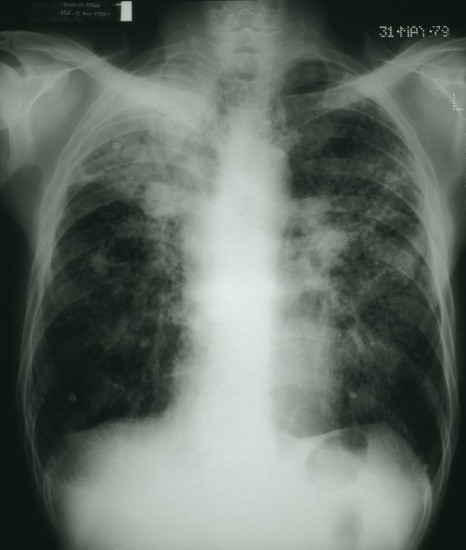
STEVE ALLEN/SCIENCE PHOTO LIBRARY
Pulmonary function tests can show obstructive, restrictive, or mixed abnormalities. Impairment of diffusing capacity for carbon monoxide (DLCO) is common. Abnormalities can be present even without radiographic findings (1).
Diagnosis reference
1. Go LHT, Almberg KS, Rose CS, et al. Prevalence and severity of abnormal lung function among US former coal miners with and without radiographic coal workers' pneumoconiosis. Occup Environ Med 2022; 79(8), 527-532. doi:10.1136/oemed-2021-107872
Treatment of Coal Worker Pneumoconiosis
Reduction of further exposure
Supportive treatment
Workers with more advanced coal worker pneumoconiosis, especially those with progressive massive fibrosis, should be restricted from further exposure. In early and mild disease, the impacts of job loss should be taken into account when considering removal from exposure.
Treatment is directed toward the clinical manifestations of coal worker pneumoconiosis. Treatment is not indicated in early and asymptomatic coal worker pneumoconiosis. Workers with obstructive lung disease may benefit from treatment for COPD.
Patients with pulmonary hypertension, hypoxemia, or both are given supplemental oxygen therapy. Pulmonary rehabilitation can help more severely affected workers carry out activities of daily living.
Lung transplantation should be considered in patients with progressive respiratory failure.
Progression is common even after exposure ceases, so monitoring should continue in former coal miners. Medical surveillance can identify new or progressive radiologic findings that develop after exposure to coal dust ends.
Smoking cessation and surveillance for tuberculosis are recommended for all exposed workers. Patients with coal worker pneumoconiosis should stay up to date with recommended vaccinations, including those against pneumococci, COVID, and influenza.
Prevention of Coal Worker Pneumoconiosis
Preventive measures begin with eliminating or reducing exposure. The most effective primary prevention is implementation of engineering controls to limit respirable mine dust. Despite long-standing regulations, exposures continue to occur in the mining trade, resulting in persistent occurrence of disease, including severe forms.
Respiratory masks provide only limited protection and should be used in conjunction with a comprehensive exposure control program.
Medical surveillance of current miners can hep identify disease at an earlier stage.
Key Points
Chronic inhalation of coal mining dust (which also contains variable amounts of silica) causes coal worker pneumoconiosis and COPD, and can progress to end-stage lung disease.
Diagnosis is based on history of exposure, chest imaging, and pulmonary function testing.
Treatment is supportive, including reduction of further exposure.
Surveillance of coal workers enables early diagnosis and institution of preventive measures, including exposure reduction, smoking cessation, tuberculosis screening, and routine vaccinations.
More Information
The following English-language resources may be useful. Please note that THE MANUAL is not responsible for the content of these resources.
GoLHT, Almberg KS, Rose CS, et al. Prevalence and severity of abnormal lung function among US former coal miners with and without radiographic coal workers' pneumoconiosis. Occup Environ Med 2022; 79(8), 527-532. doi:10.1136/oemed-2021-107872
Weissman DN. Progressive massive fibrosis: An overview of the recent literature. Pharmacol Ther 2022; 240:108232. doi:10.1016/j.pharmthera.2022.108232


