

Wrist fractures may involve the lower end of one or both of the forearm bones (radius or ulna) or, less often, a bone in the base of the hand.
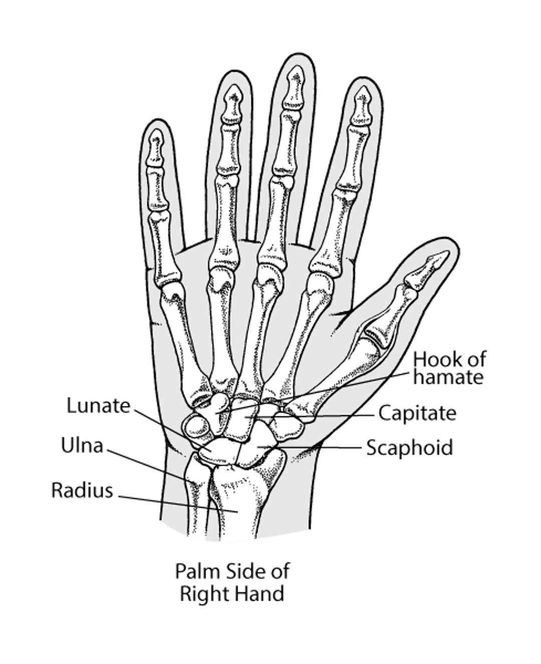
The wrist consists of:
The two long bones in the forearm (radius and ulna)
Eight small bones at the base of the hand (called carpal bones)
The carpal bones are located between the forearm bones and the bones of the hand.
(See also Overview of Fractures.)
Fractures of the Lower Forearm
(Colles Fracture; Smith Fracture)
Fractures usually involve the larger lower forearm bone (radius). Sometimes the smaller one (ulna) is also broken. These fractures are considered wrist fractures.
Wrist fractures usually result from a fall on an outstretched hand.
The fractured wrist is painful, swollen, and tender and sometimes becomes stiff and persistently painful.
Doctors can usually identify these fractures on x-rays, but occasionally, computed tomography is needed.
Often, the bone fragments can be put back in place (reduced) by manipulation, then held in place by a splint, but sometimes surgery is required.
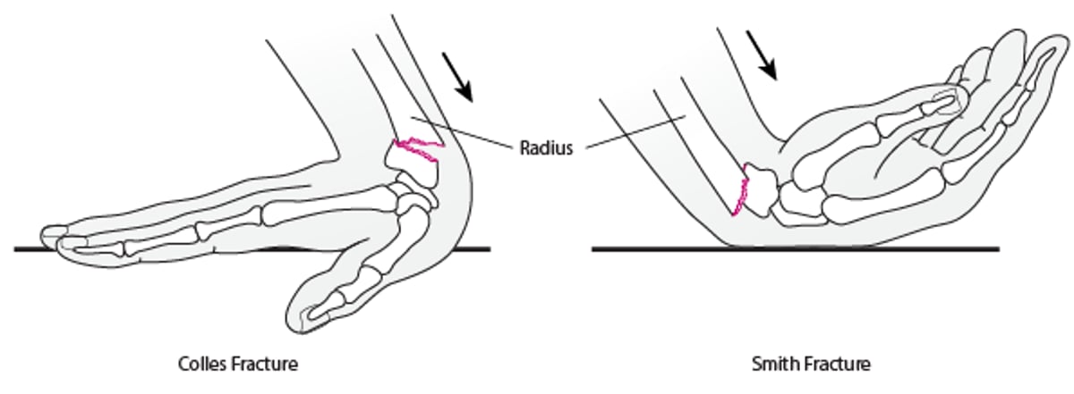
Usually, the wrist is fractured when people fall on an outstretched hand (to break the fall), with the wrist bent back. As a result, the radius breaks near the wrist, and the broken end of the radius is displaced up, toward the back of the hand. This type of fracture is called a Colles fracture. Sometimes the wrist is obviously out of position. If the force of the impact is great, the two ends of the broken radius are jammed into each other, and the bone appears shortened. Often, part of the ulna is also fractured. Colles fractures are common among older adults, especially if they have osteoporosis, which weakens bone.
Less often, the wrist is fractured when people fall with the hand bent forward or when the back of the wrist is hit—for example, with a hockey or lacrosse stick. The broken end of the radius is displaced down, toward the palm side of the wrist. This type of fracture is called a Smith fracture.
Wrist Fractures: Colles and Smith
Wrist fractures include
|

Fractures of the radius may extend into the wrist joint.
The median nerve—the nerve involved in carpal tunnel syndrome—may be damaged. This nerve travels through the wrist to the palm and some fingers.
Symptoms of Lower Forearm Fractures
The wrist is painful, swollen, and tender. If the median nerve is damaged, the tip of the index finger is numb, and people may have difficulty pinching their thumb and little finger together.
A wrist fracture may result in stiffness, persistent pain, and/or osteoarthritis, particularly if the fracture extends into the wrist joint or the broken pieces are out of place.
Diagnosis of Lower Forearm Fractures
X-rays
Occasionally computed tomography
(See also Diagnosis of Fractures.)
If people think they may have fractured their wrist, they should see a doctor.
Doctors can usually identify wrist fractures on x-rays. Occasionally, computed tomography (CT) is necessary to identify joint fractures. CT combines x-rays with computer technology to produce a more detailed, three-dimensional image of the injured area.
Treatment of Lower Forearm Fractures
Realignment of the broken bones done with or without surgery
A cast or a metal frame with pins
For many wrist fractures, doctors realign (reduce) the broken pieces without surgery (closed reduction). Before realigning the fracture, doctors may do one of the following to prevent people from feeling pain:
People may be given an opioid pain reliever and/or a sedative.
A hematoma block may be used.
For a hematoma block, a needle is inserted through the skin into the fracture where blood has collected (a hematoma is an accumulation of blood), and lidocaine (an anesthetic) is injected, numbing the fractured bones before the bones are realigned. For a hematoma block, a needle is inserted through the skin into the fracture where blood has collected (a hematoma is an accumulation of blood), and lidocaine (an anesthetic) is injected, numbing the fractured bones before the bones are realigned.
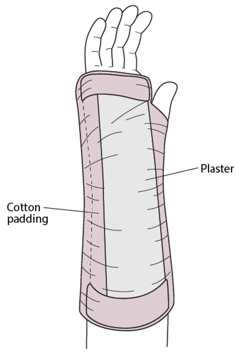
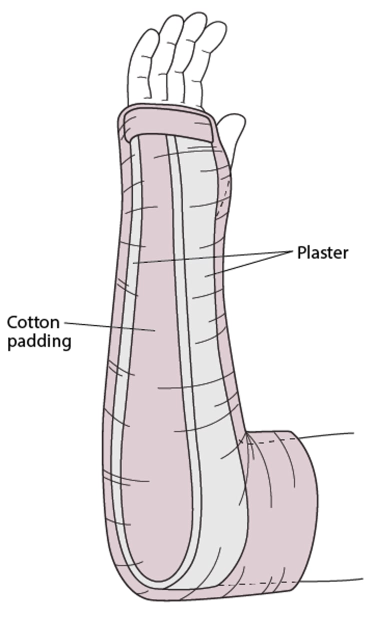
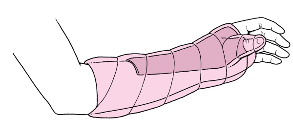
After the bones are realigned, a splint (such as a volar or sugar tong splint) is applied. The person is then referred to an orthopedic surgeon for follow-up. Usually, the splint is removed, and a cast is applied. The cast is worn for about 4 to 6 weeks.
Volar Splint

Sugar Tong Splint

If the joint is affected or the broken bones are jammed close together, surgery (open reduction with internal fixation, or ORIF) may be done to realign and immobilize the broken pieces, especially in active adults who need to be able to fully use their wrist.
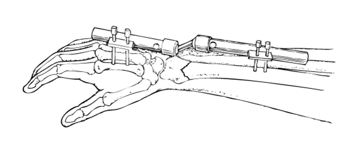
The bone may be immobilized by attaching a plate directly to it during surgery. Or doctors may attach a frame of rods to the outside of the wrist with stainless steel pins inserted through the skin into the bone (called an external fixator). An external fixator is usually temporary. It is used until surgery to insert more permanent hardware devices can be done.
People are advised to move their fingers, elbow (if it is not immobilized), and shoulder every day to prevent stiffness. Elevating the hand helps control swelling. The comfort, flexibility, and strength of the wrist can continue to improve for 6 to 12 months after the fracture.
External Fixator

Scaphoid Fractures
Scaphoid fractures involve a bone in the wrist near the base of the thumb (scaphoid bone).
Scaphoid fractures usually result from a fall on an outstretched hand.
These fractures can disrupt blood flow to the scaphoid bone, causing osteoarthritis and loss of function.
The thumb side of the wrist is tender and swollen, and rotating the wrist is particularly painful.
X-rays are taken, but because scaphoid fractures may be hard to see soon after the injury, other imaging tests may be needed, or a splint may be placed and x-rays repeated after 7 to 10 days.
A thumb spica splint is often effective, but sometimes surgery is needed to put the broken pieces of bone in place and hold them in place.
The scaphoid bone is the most commonly injured of the eight wrist (carpal) bones, located between the bones of the forearm and those of the hand.
Bones in the Wrist

Scaphoid fractures usually result from a fall on an outstretched hand. These fractures often cause problems because they often disrupt the blood supply to the scaphoid bone. Without its blood supply, the following may occur:
The bone may not heal and may then deteriorate and collapse.
Tissue may die (called osteonecrosis), resulting in osteoarthritis and disability.
The bones may not grow back together (called nonunion).
These problems occur infrequently and can occur even when treatment is optimal.
Symptoms of Scaphoid Fractures
The thumb side of the wrist is tender and swollen in people with a scaphoid fracture. Rotating the wrist is particularly painful.
Diagnosis of Scaphoid Fractures
A doctor's evaluation
X-rays
Sometimes magnetic resonance imaging
If people think they may have fractured their wrist, they should see a doctor.
Doctors suspect a scaphoid fracture based on symptoms and results of a physical examination. X-rays are taken, but these fractures may be hard to see soon after the injury.
If x-rays do not show a fracture but the physical examination suggests a fracture, most doctors apply a splint. People are asked to return in 7 to 10 days for another x-ray to check for a fracture. (Fractures may be seen more easily after they have been healing for a few days—see How bones heal.) Magnetic resonance imaging (MRI) can be done to check for a fracture when none is seen on a x-ray. However, it is expensive and not always available.
Thumb Spica Splint

Treatment of Scaphoid Fractures
A thumb spica splint
Sometimes surgery
After diagnosis is confirmed, many scaphoid fractures can be effectively treated with a thumb spica splint that is worn for up to 6 to 8 weeks.
Sometimes surgery (open reduction and internal fixation, or ORIF) is required to realign and immobilize the broken pieces.
The bone may take 3 to 4 months to heal.
Drug Information for the Topic


