


The term temporomandibular disorders is an umbrella term for a group of musculoskeletal and neuromuscular conditions that involve the temporomandibular joint (TMJ), the masticatory muscles, and all associated structures. Temporomandibular disorders (previously known as temporomandibular joint dysfunction or temporomandibular joint [TMJ] syndrome) often present with pain in the jaw, face, and neck and/or with dysfunction of the jaw joint (often joint sounds and/or a decreased range of motion) that is often accompanied by headache or ear pain. People are considered to have a temporomandibular disorder (TMD) when the pain or dysfunction is severe enough to make them seek professional care.
Temporomandibular disorders typically are multifactorial in origin, but most are related to problems with masticatory muscles and ligaments of the temporomandibular joints or internal derangements within the joints themselves. Joint hypermobility can also contribute.
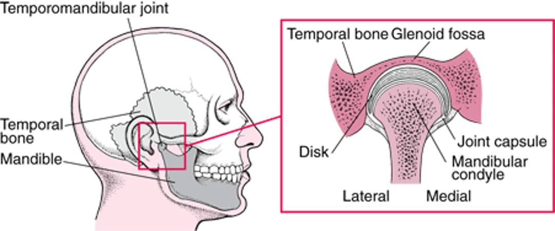
Internal derangements of the TMJ are a result of altered or disturbed movement of the mandibular condyle in the glenoid fossa or against the articular disk (see figure The Temporomandibular Joint). The disk, which is composed of dense fibrous connective tissue and shaped like a mature red blood cell, serves as a cushion between the bone surfaces. Unlike most joints, which are lined with hyaline cartilage, the TMJ bone surfaces are lined with fibrocartilage. Causes for the altered or disturbed movement include direct or indirect macrotrauma (eg, flexion-extension injury of the neck—also known as whiplash—which can cause referred pain), microtrauma (eg, due to parafunctional behaviors such as awake/sleep clenching and grinding of the teeth or persistent gum chewing), systemic disorders (eg, rheumatoid arthritis or other systemic rheumatic disorders), local or systemic infections, and acute malocclusion.
(See also Mandibular Dislocation, Temporal Bone Fractures and Jaw Tumors.)
The Temporomandibular Joint
The joint is formed by the mandibular condyle and the glenoid fossa of the temporal bone; a fibrocartilaginous articular disk functions as a cushion between the joint surfaces. |

Diagnosis of Temporomandibular Disorders
Clinical evaluation
Disorders of the temporomandibular joint (TMJ) must be distinguished from the many conditions that mimic them (see table Some Conditions That Mimic Temporomandibular Disorders). Pain exacerbated by finger pressure on the joint when the mouth is opened implicates the TMJ.
Patients are asked to describe the pain and designate painful areas. Typically, muscles of mastication (temporalis, masseter, and medial pterygoids) and the cervical and occipital muscles are palpated for general tenderness and trigger points (spots that radiate pain to another area). The lateral pterygoids cannot be directly palpated.
Patients are observed opening the mouth as wide as is comfortable. When patients open their mouth, the mandible typically deviates toward the painful side. Palpation and auscultation of the joint during opening and closing may reveal tenderness and catching and intracapsular sounds such as clicking/popping or crepitus.
Condylar motion can best be palpated by bilaterally placing the middle fingers on the preauricular areas (lateral poles of the condyle) and exerting gentle medial pressure or by placing the 5th fingers into the external ear canals and exerting very gentle forward pressure as patients move their jaw. The average-sized patient can open the mouth at least 40 mm (measured between the incisal edges of the upper and lower central incisors). To account for differences in patient size, a patient should typically be able to fit 3 fingers (index, middle, ring) into the mouth up to the distal interphalangeal joints.


