

Blepharitis is inflammation of the eyelid margins that may be acute or chronic. Symptoms and signs include itching and burning of the eyelid margins with redness and edema. Diagnosis is by history and physical examination. Treatment for all subtypes of blepharitis includes warm compresses and eyelid hygiene. Acute ulcerative blepharitis is usually also treated with topical antibiotics or systemic antivirals. Acute nonulcerative blepharitis is occasionally treated with topical glucocorticoids. Chronic disease is treated primarily with warm compresses and eyelid hygiene and occasionally oral antibiotics (eg, a tetracycline or azithromycin) for patients with refractory symptoms.Blepharitis is inflammation of the eyelid margins that may be acute or chronic. Symptoms and signs include itching and burning of the eyelid margins with redness and edema. Diagnosis is by history and physical examination. Treatment for all subtypes of blepharitis includes warm compresses and eyelid hygiene. Acute ulcerative blepharitis is usually also treated with topical antibiotics or systemic antivirals. Acute nonulcerative blepharitis is occasionally treated with topical glucocorticoids. Chronic disease is treated primarily with warm compresses and eyelid hygiene and occasionally oral antibiotics (eg, a tetracycline or azithromycin) for patients with refractory symptoms.
Etiology of Blepharitis

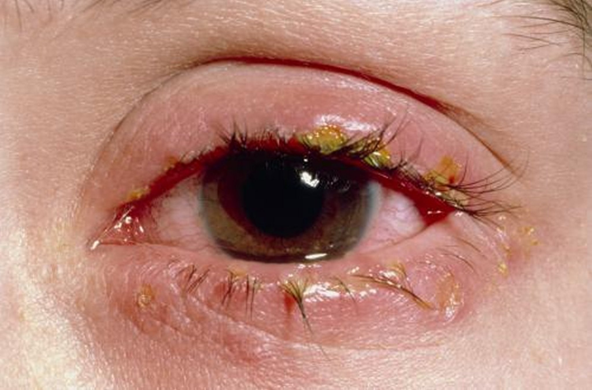
In this patient, eyelid margins have become edematous and erythematous, and eyelashes are crusted with dried serous fluid.
WESTERN OPHTHALMIC HOSPITAL/SCIENCE PHOTO LIBRARY
Blepharitis may be acute (ulcerative or nonulcerative) or chronic (caused by meibomian gland dysfunction, seborrheic blepharitis, Demodex infestation).
Acute blepharitis
Acute ulcerative blepharitis is usually caused by bacterial infection (usually staphylococcal) of the eyelid margin at the origins of the eyelashes; the lash follicles and the meibomian glands are also involved (1). It may also be due to a virus (eg, herpes simplex, varicella zoster). Bacterial infections typically have more crusting than the viral types, which usually have more of a clear serous discharge.
Acute nonulcerative blepharitis is usually caused by an allergic reaction involving the same area (eg, atopic blepharodermatitis and seasonal allergic blepharoconjunctivitis), and causes the following:
Intense itching and inflammation (usually along the edges of both eyelids)
Rubbing (a response to itching that can increase conjunctival itching and exacerbate atopic dermatitis [eczema] of the eyelid)
Contact sensitivity (dermatoblepharoconjunctivitis).
Chronic blepharitis
Chronic blepharitis is persistent inflammation of the eyelid margin that is typically multifactorial (1). Causes include persistent bacterial colonization (usually staphylococcal), meibomian gland dysfunction, underlying skin disease (eg, seborrheic dermatitis, rosacea), and Demodex infestation.
The most common cause of chronic blepharitis is meibomian gland dysfunction. Meibomian glands in the eyelid produce lipids (meibum) that reduce tear evaporation by forming a lipid layer on top of the aqueous tear layer. In meibomian gland dysfunction, the lipid composition is abnormal, and gland ducts and orifices become inspissated with hard, waxy plugs. Many patients have coexisting rosacea and recurrent hordeola or chalazia.
Many patients with seborrheic blepharitis have seborrheic dermatitis of the face and scalp or acne rosacea. Secondary bacterial colonization often occurs on the scales that develop on the eyelid margin. Meibomian glands can become obstructed.

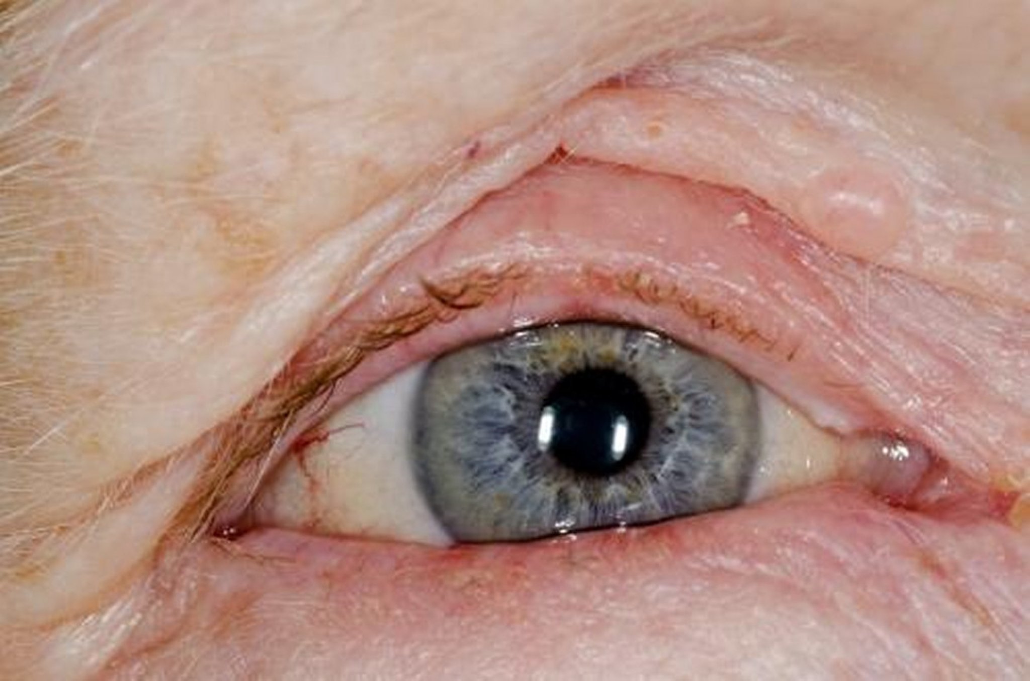
This patient has chronic blepharitis (inflammation with mild edema and erythema) of the eyelids, in this case due to atopic dermatitis (eczema). Note the inversion of the lower eyelid (entropion), which hides those lashes from view.
DR P. MARAZZI/SCIENCE PHOTO LIBRARY
Most patients with meibomian gland dysfunction or seborrheic blepharitis have increased tear evaporation and secondary keratoconjunctivitis sicca, also known as dry eye. Demodex mite (folliculorum and brevis species) infestation is also a common cause of chronic blepharitis.
Chronic blepharitis can also occur in patients with eyelid carcinoma (especially if blepharitis is unilateral and there is loss of eyelashes) or immune-mediated conditions such as ocular mucous membrane pemphigoid.
Etiology reference
1. Yeu E, Garg S, Ayres BD, Beckman K, et al. Current state and future perspectives in the diagnosis of eyelid margin disease: clinical review. J Cataract Refract Surg. 2024 Aug 1;50(8):868-875. doi: 10.1097/j.jcrs.0000000000001483.
Symptoms and Signs of Blepharitis
Symptoms common to all forms of blepharitis include itching and burning of the eyelid margins and conjunctival irritation with lacrimation, photosensitivity, and foreign body sensation. Itching is more common in allergic than infectious causes. Symptoms tend to be worse in the early morning compared with those of keratoconjunctivitis sicca, which tend to be worse toward the end of the day.
Acute blepharitis
In acute ulcerative blepharitis, small pustules may develop in eyelash follicles and eventually break down to form shallow marginal ulcers. Tenacious adherent crusts leave a bleeding surface when removed. During sleep, eyelids can become adhered together by dried secretions. Recurrent ulcerative blepharitis can cause eyelid scars and loss or misdirection (trichiasis) of eyelashes.
In acute nonulcerative blepharitis, eyelid margins become edematous and erythematous; eyelashes may become crusted with dried serous fluid.
Chronic blepharitis
In meibomian gland dysfunction, examination reveals dilated, inspissated gland orifices that, when pressed, exude a waxy, thick, yellowish secretion. In seborrheic blepharitis, greasy, easily removable scales develop on eyelid margins. Most patients with seborrheic blepharitis and meibomian gland dysfunction have symptoms of keratoconjunctivitis sicca, such as foreign body sensation, grittiness, eye strain and fatigue, and blurring with prolonged visual effort. The findings of eyelid erythema and debris on the eyelashes in blepharitis help distinguish it from keratoconjunctivitis sicca.
In Demodex infestation, patients may have chronic blepharitis that has not responded to standard treatments; symptoms include redness, inflammation, itching, and recurrent styes. Examination in Demodex infestation reveals cylindrical sleeves on the eyelashes, usually at the base.
Diagnosis of Blepharitis
Slit-lamp examination
Diagnosis is usually by slit-lamp examination. Chronic blepharitis that does not respond to treatment may require biopsy to exclude eyelid tumors or immune-mediated conditions that can simulate the condition.
Treatment of Blepharitis
Warm compresses and eyelid hygiene
For acute blepharitis, antimicrobials and sometimes topical glucocorticoids for acute nonulcerative blepharitis
For chronic blepharitis, sometimes topical or systemic antibiotics
An ectoparasiticide (eg, lotilaner) for suspected An ectoparasiticide (eg, lotilaner) for suspectedDemodex infestation
The mainstay of therapy for all subtypes of blepharitis is eyelid hygiene consisting of warm compresses and cleansing of the eyelid margins. This initial approach helps remove debris and excess oils from eyelid margins, which helps reduce inflammation and bacterial load. Additional therapy (eg, topical antibiotics) is guided by the underlying cause.
Acute blepharitis
Acute ulcerative blepharitis is treated with an antibiotic ointment (eg, bacitracin/polymyxin B or erythromycin) (Acute ulcerative blepharitis is treated with an antibiotic ointment (eg, bacitracin/polymyxin B or erythromycin) (1). Acute viral ulcerative blepharitis is treated with systemic antivirals (eg, for herpes simplex, acyclovir; for varicella zoster, famciclovir or valacyclovir).). Acute viral ulcerative blepharitis is treated with systemic antivirals (eg, for herpes simplex, acyclovir; for varicella zoster, famciclovir or valacyclovir).
Treatment of acute nonulcerative blepharitis begins with avoiding the offending action (eg, rubbing) or substance (eg, new eye drops). Warm compresses over the closed eyelid may relieve symptoms and speed resolution. If swelling persists > 24 hours, topical glucocorticoids (eg, fluorometholone ophthalmic ointment) can be used.24 hours, topical glucocorticoids (eg, fluorometholone ophthalmic ointment) can be used.
Chronic blepharitis
The initial treatment for both meibomian gland dysfunction and seborrheic blepharitis is directed toward eyelid hygiene and warm compresses, which melt the waxy plugs. Occasionally, eyelid massage is needed to extrude trapped secretions and coat the ocular surface.
If needed, additional treatment for seborrheic blepharitis includes gentle cleansing of the eyelid margin (lid scrubs) twice a day with a cotton swab dipped in a dilute solution of baby shampoo (2 to 3 drops in ½ cup of warm water) or with commercially available wipes and washes that contain ingredients such as a gentle eyelid cleanser, tea tree and coconut oils, or hypochlorous acid 0.01%. A topical antibiotic ointment (erythromycin, bacitracin/polymyxin B or drops such as azithromycin 1.0% or sulfacetamide 10%) may be added to reduce bacterial counts on the eyelid margin when cases are unresponsive to weeks of eyelid hygiene.If needed, additional treatment for seborrheic blepharitis includes gentle cleansing of the eyelid margin (lid scrubs) twice a day with a cotton swab dipped in a dilute solution of baby shampoo (2 to 3 drops in ½ cup of warm water) or with commercially available wipes and washes that contain ingredients such as a gentle eyelid cleanser, tea tree and coconut oils, or hypochlorous acid 0.01%. A topical antibiotic ointment (erythromycin, bacitracin/polymyxin B or drops such as azithromycin 1.0% or sulfacetamide 10%) may be added to reduce bacterial counts on the eyelid margin when cases are unresponsive to weeks of eyelid hygiene.
Blepharitis associated with Demodex infestation should be initially treated with topical low-concentration tea tree oil or hypochlorous acid (1). If the condition is unresponsive to these over-the-counter remedies, an ectoparasiticide (eg, lotilaner) can be prescribed; however, lotilaner is gaining favor as a first-line treatment (). If the condition is unresponsive to these over-the-counter remedies, an ectoparasiticide (eg, lotilaner) can be prescribed; however, lotilaner is gaining favor as a first-line treatment (2).
In patients with recurrent or refractory symptoms, oral antibiotics such as azithromycin or a tetracycline (eg, doxycycline) with alternative dosing regimens guided by an ophthalmologist may be required. Other treatments that may be effective for refractory meibomian gland dysfunction include intense pulsed light (IPL) therapy (In patients with recurrent or refractory symptoms, oral antibiotics such as azithromycin or a tetracycline (eg, doxycycline) with alternative dosing regimens guided by an ophthalmologist may be required. Other treatments that may be effective for refractory meibomian gland dysfunction include intense pulsed light (IPL) therapy (3) and thermal pulsation (4). In addition, oral omega-3 supplementation may improve dry eye symptoms of meibomian gland dysfunction, but the data are mixed (5).
Secondary keratoconjunctivitis sicca may also develop when dysfunctional meibomian glands produce a poor oil layer, resulting in increased tear evaporation. Tear supplements during the day, bland ointments (eg, petroleum jelly) at night and, if necessary, punctal plugs (inserts that obstruct the puncta and thus decrease tear drainage) are effective in most patients.
Treatment references
1. Amescua G, Akpek EK, Farid M, et al: Blepharitis Preferred Practice Pattern®. Ophthalmology 126(1):P56-P93. doi: 10.1016/j.ophtha.2018.10.019
2. Donnenfeld E, Nichols KK, Ayres BD, et al. The Demodex Expert Panel on Treatment and Eyelid Health (DEPTH) Consensus Regarding the Preferred Treatment for Demodex Blepharitis. Clin Ophthalmol. 2025;19:1893-1904. Published 2025 Jun 18. doi:10.2147/OPTH.S525681
3. Wladis EJ, Aakalu VK, Foster JA, et al. Intense Pulsed Light for Meibomian Gland Disease: A Report by the American Academy of Ophthalmology. Ophthalmology. 2020 Sep;127(9):1227-1233. doi: 10.1016/j.ophtha.2020.03.009.
4. Tao JP, Shen JF, Aakalu VK, et al. Thermal Pulsation in the Management of Meibomian Gland Dysfunction and Dry Eye: A Report by the American Academy of Ophthalmology. Ophthalmology. 2023 Dec;130(12):1336-1341. doi: 10.1016/j.ophtha.2023.07.009. Epub 2023 Aug 27.
5. Al-Namaeh M. A systematic review of the effect of omega-3 supplements on meibomian gland dysfunction. Ther Adv Ophthalmol. 2020;12:2515841420952188. Published 2020 Oct 16. doi:10.1177/2515841420952188
Prognosis for Blepharitis
Acute blepharitis most often responds to treatment but may recur, develop into chronic blepharitis, or both. Chronic blepharitis is indolent, recurrent, and resistant to treatment. Exacerbations are inconvenient, uncomfortable, and cosmetically unappealing but do not usually result in corneal scarring or vision loss. Long-term maintenance and preventive care with warm compresses and eyelid hygiene is critical.
Key Points
Common forms of blepharitis include acute ulcerative (often secondary to staphylococcal or herpes virus infection), acute nonulcerative (usually allergic), and chronic (often with meibomian gland dysfunction, seborrheic dermatitis, or Demodex infestation).
Secondary keratoconjunctivitis sicca usually accompanies chronic blepharitis.
Common symptoms include itching and burning of the eyelid margins and conjunctival irritation with lacrimation, photosensitivity, and foreign body sensation.
Diagnosis is usually by slit-lamp examination.
Consider alternate diagnoses, including eyelid carcinoma, if chronic blepharitis is unilateral.
Supportive treatments are indicated (eg, warm compresses, eyelid cleansing, and treatment of keratoconjunctivitis sicca as needed).
Specific medication treatments can include topical antimicrobials for acute ulcerative blepharitis, systemic antivirals if viral infection is suspected, and topical glucocorticoids for persistent acute nonulcerative blepharitis.
Treatment of chronic blepharitis includes treatment of keratoconjunctivitis sicca with tear supplements, use of warm compresses, and topical or systemic antibiotics as needed.
Drug Information for the Topic


