

The most common malignant tumor in the proximal two-thirds of the esophagus is squamous cell carcinoma; adenocarcinoma is the most common in the distal one-third. Symptoms are progressive dysphagia and weight loss. Diagnosis is by endoscopy, followed by PET-CT and endoscopic ultrasound for staging. Treatment varies with stage and generally includes surgery with or without chemotherapy and radiation. Long-term survival is poor except for patients with local disease.
Worldwide in 2022, esophageal cancer was the 11th most commonly diagnosed type of cancer and the 7th leading cause of cancer deaths with approximately 511,000 new cases and 445,000 deaths (1).
In the United States, esophageal cancer is much less commonly diagnosed. Esophageal cancer accounted for an estimated 22,370 new cancer diagnoses and 16,130 cancer deaths in the United States in 2024 (2).
The primary risk factors for esophageal cancer are
Alcohol ingestion
Tobacco use (in any form)
Gastroesophageal reflux disease (especially for adenocarcinoma)
Obesity (especially for adenocarcinoma)
Older age
Male sex
Barrett esophagus
Genetic syndromes (eg, tylosis, familial Barrett esophagus, Bloom syndrome, Fanconi anemia)
Other risk factors include achalasia, human papillomavirus infection, lye or other caustic substance ingestion (resulting in stricture), sclerotherapy, esophageal webs due to Plummer-Vinson syndrome, and irradiation of the esophagus.
References
1. Bray F, Laversanne M, Sung H, et al. Global cancer statistics 2022: GLOBOCAN estimates of incidence and mortality worldwide for 36 cancers in 185 countries. CA Cancer J Clin. 2024;74(3):229-263. doi:10.3322/caac.21834
2. Siegel RL, Giaquinto AN, Jemal A. Cancer statistics, 2024 [published correction appears in CA Cancer J Clin. 2024 Mar-Apr;74(2):203. doi: 10.3322/caac.21830]. CA Cancer J Clin. 2024;74(1):12-49. doi:10.3322/caac.21820
Types of Esophageal Cancer
Squamous cell carcinoma of the esophagus
Squamous cell carcinoma is the most common esophageal cancer worldwide, but, in the United States, adenocarcinoma is about twice as common (1). In the United States, it is 4 to 5 times more common among Black than White people, and 2 to 3 times more common among men than women (2).

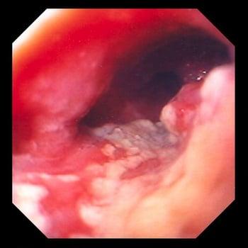
Squamous cell carcinoma typically manifests as an ulcerated, ragged mass compromising the lumen of the esophagus.
Adenocarcinoma of the esophagus
Adenocarcinoma occurs in the distal esophagus.
Its incidence is increasing; it accounts for approximately 80% of esophageal carcinoma in the United States (3). It is 4 times more common among White than Black people (4). Alcohol is not an important risk factor, but smoking is contributory.
Adenocarcinoma of the distal esophagus is difficult to distinguish from adenocarcinoma of the gastric cardia invading the distal esophagus.

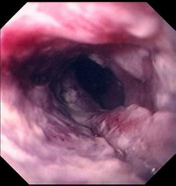
This image shows an ulcerated, constricting tumor located in the distal esophagus, which is highly suggestive of an adenocarcinoma arising from metaplastic columnar changes (Barrett esophagus).
Most adenocarcinomas arise in Barrett esophagus, which results from chronic gastroesophageal reflux disease and reflux esophagitis. In Barrett esophagus, a metaplastic, columnar, glandular, intestine-like mucosa with brush border and goblet cells replaces the normal stratified squamous epithelium of the distal esophagus during the healing phase of acute esophagitis when healing takes place in the continued presence of stomach acid. Obesity is associated with an increased risk of esophageal adenocarcinoma, probably because obesity is a contributing factor to reflux.
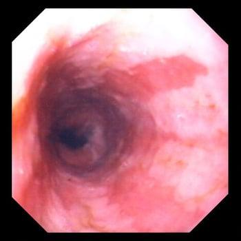
Most esophageal adenocarcinomas arise from Barrett esophagus. Barrett esophagus is replacement of normal squamous epithelium of the distal esophagus with metaplastic columnar epithelium during the healing phase of acute esophagitis. In this image, red-appearing bands of metaplastic epithelium can be seen extending proximally.

Other malignant tumors of the esophagus
Less common malignant tumors include spindle cell carcinoma (a poorly differentiated variant of squamous cell carcinoma), verrucous carcinoma (a well-differentiated variant of squamous cell carcinoma), pseudosarcoma, mucoepidermoid carcinoma, adenosquamous carcinoma, cylindroma (adenoid cystic carcinoma), primary oat cell carcinoma, choriocarcinoma, carcinoid tumor, sarcoma, and primary malignant melanoma.
Metastatic cancer to the esophagus is extremely rare and is largely relegated to case reports in the literature. A series of autopsy data from Japan cited an incidence of approximately 6.1% (5, 6). These tumors usually seed the loose connective tissue stroma around the esophagus, whereas primary esophageal cancers begin in the mucosa or submucosa.
Types of esophageal cancer references
1. Patel N, Benipal B. Incidence of esophageal cancer in the United States from 2001-2015: A United States cancer statistics analysis of 50 states. Cureus. 10(12):e3709, 2018. doi: 10.7759/cureus.3709
2. Morgan E, Soerjomataram I, Rumgay H, et al. The Global Landscape of Esophageal Squamous Cell Carcinoma and Esophageal Adenocarcinoma Incidence and Mortality in 2020 and Projections to 2040: New Estimates From GLOBOCAN 2020. Gastroenterology. 2022 Sep;163(3):649-658.e2. doi: 10.1053/j.gastro.2022.05.054
3. National Cancer Institute. Esophageal Cancer. Accessed February 4, 2025.
4. El-Serag HB, Mason AC, Petersen N, Key CR. Epidemiological differences between adenocarcinoma of the oesophagus and adenocarcinoma of the gastric cardia in the USA. Gut. 50(3):368–372, 2002. doi: 10.1136/gut.50.3.368
5. Mizobuchi S, Tachimori Y, Kato H, et al. Metastatic esophageal tumors from distant primary lesions: report of three esophagectomies and study of 1835 autopsy cases. Jpn J Clin Oncol. 1997;27(6):410-414. doi:10.1093/jjco/27.6.410
6. Shi H, Chen SY, Xie ZF, et al. Lung cancer metastasis-induced distal esophageal segmental spasm confirmed by individualized peroral endoscopic myotomy: A case report. World J Gastrointest Surg. 2024;16(10):3321-3327. doi:10.4240/wjgs.v16.i10.3321
Symptoms and Signs of Esophageal Cancer
Early-stage esophageal cancer tends to be asymptomatic. When the lumen of the esophagus becomes progressively constricted, dysphagia commonly occurs. The patient first has difficulty swallowing solid food, then semisolid food, and finally liquids and saliva; this steady progression suggests a growing malignant process rather than a spasm, benign ring, or peptic stricture. Chest pain may be present, usually radiating to the back.
Weight loss, even when the patient maintains a good appetite, is almost universal. Compression of the recurrent laryngeal nerve may lead to vocal cord paralysis and hoarseness. Nerve compression may cause spinal pain, hiccups, or paralysis of the diaphragm. Malignant pleural effusions or pulmonary metastasis may cause dyspnea. Intraluminal tumor involvement may cause odynophagia, vomiting, hematemesis, melena, iron deficiency anemia, aspiration, and cough. Fistulas between the esophagus and tracheobronchial tree may cause lung abscess and pneumonia. Other findings may include superior vena cava syndrome, malignant ascites, and bone pain.
Lymphatic spread to internal jugular, cervical, supraclavicular, mediastinal, and celiac nodes is common. The tumor usually metastasizes to lung and liver; less common sites include bone, heart, brain, adrenal glands, kidneys, and peritoneum.
Diagnosis of Esophageal Cancer
Endoscopy with biopsy
Abdominal and pelvic CT for staging
PET-CT and endoscopic ultrasound to complete staging
There are no screening tests. Patients suspected of having esophageal cancer should have endoscopy with cytology and biopsy. Although barium radiograph may show an obstructive lesion, endoscopy is required for biopsy and tissue diagnosis. Microsatellite instability (MSI) testing of the biopsy is now standard.
Patients in whom esophageal cancer is identified require CT of the chest, abdomen, and pelvis as well as whole-body positron emission tomography (PET)-CT to determine extent of tumor spread. If cross-sectional imaging results are negative for metastasis, endoscopic ultrasound should be done to determine the depth of the tumor in the esophageal wall and regional lymph node involvement. Findings guide therapy and help determine prognosis.
Basic blood tests, including complete blood count, electrolytes, and liver function, should be done.
Treatment of Esophageal Cancer
Surgical resection, often combined with chemotherapy and radiation
Endoscopic resection for certain small, early-stage tumors
Immunotherapy plus chemotherapy for certain advanced cancers
Esophageal cancer treatment decisions depend on tumor staging, size, location, and the patient’s wishes (many choose to forgo aggressive treatment).
General principles
Patients with stage 0 and early stage I (T1a) tumors are often amenable to endoscopic resection. Endoscopic resection avoids the high potential morbidity of esophageal resection. Tumors that are T1b are amenable to surgical resection alone without adjuvant therapies (chemotherapy or radiation). However, most esophageal cancers (T2 or higher, or any nodal involvement) are treated with combination chemotherapy and radiation (chemoradiation) before surgical resection (see table ). One study shows chemoradiation before surgery has a clear benefit to survival compared to surgery alone (1).
Another study showed that after surgery, immunotherapy (with nivolumab) added to neoadjuvant chemoradiation improved survival more than placebo (Another study showed that after surgery, immunotherapy (with nivolumab) added to neoadjuvant chemoradiation improved survival more than placebo (2).
Shallow (superficial) adenocarcinomas sometimes are cured by radiofrequency ablation.
Patients who are unable or unwilling to undergo surgery may receive some benefit from chemoradiation. Radiation or chemotherapy alone is of little benefit. Patients with stage IV disease require palliation and should not undergo surgery.
Staging Esophageal Adenocarcinoma*
Stage | Tumor (Maximum Penetration) | Regional Lymph Node Metastasis | Distant Metastasis |
|---|---|---|---|
0 | Tis | N0 | M0 |
I | T1 (T1a and T1b) | N0 | M0 |
IIA | T1 | N1 | M0 |
IIB | T2 | N0 | M0 |
III | T2 | N1 | M0 |
T3 or T4a | N0-N1 | M0 | |
IVA | T1-T4a | N2 | M0 |
T4b | N0-N2 | M0 | |
IVB | Any T | Any N | M1 |
* cTNM classification (clinical staging):
| |||
After treatment, patients typically are screened for recurrence by endoscopy and CT of the neck, chest, and abdomen at 6-month intervals for 2 to 3 years. Endoscopy is used after treatment with chemoradiation alone or endoscopic resection alone.
Patients with Barrett esophagus require intense long-term treatment for gastroesophageal reflux disease and endoscopic surveillance for malignant transformation at 3- to 12-month intervals depending on the degree of metaplasia.
Surgery
Superficial, early, noninvasive cancers (Tis, T1a, N0) may be treated with endoscopic mucosal resection or endoscopic submucosal dissection (usually by gastroenterologists at tertiary care centers) if the superficial nature of the lesion has been confirmed by endoscopic ultrasound. However, in the large majority of cases, en bloc resection for cure requires removal of the entire tumor, proximal and distal margins of normal tissue, all potentially malignant lymph nodes, and a portion of the proximal stomach sufficient to contain the distal draining lymphatics. The procedure requires gastric pull-up with esophagogastric anastomosis, small-bowel interposition, or colonic interposition. Pyloroplasty (surgical widening of the pylorus) is required to ensure proper gastric drainage because esophagectomy necessarily results in bilateral vagotomy. This extensive surgery may be poorly tolerated by patients > 75 years, particularly those with underlying cardiac or pulmonary disease (ejection fraction < 40%, or forced expiratory volume in 1 second [FEV1] < 1.5 L/minute). Overall, operative mortality is about 5%.
Complications of surgery include anastomotic leaks, fistulas, and strictures; bilious gastroesophageal reflux; and dumping syndrome. The burning chest pain of bile reflux after distal esophagectomy can be more annoying than the original symptom of dysphagia and may require subsequent Roux-en-Y jejunostomy for bile diversion. An interposed segment of small bowel or colon in the chest has a tenuous blood supply, and torsion, ischemia, or gangrene of the interposed bowel may result.
External beam radiation therapy
Radiation is usually used in combination with chemotherapy for patients who are poor candidates for curative surgery, including those with advanced disease. Radiation is contraindicated in patients with tracheoesophageal fistula because tumor shrinkage enlarges the fistula. Similarly, patients with vascular encasement by tumor may experience massive hemorrhage with tumor shrinkage.
During the early stages of radiation therapy, edema may worsen esophageal obstruction, dysphagia, and odynophagia. This problem may require preradiation dilation and/or placement of a stent. Some patients may require a temporary percutaneous gastrostomy feeding tube. Other adverse effects of radiation therapy include nausea, vomiting, anorexia, fatigue, esophagitis, excess esophageal mucus production, xerostomia, stricture, radiation pneumonitis, radiation pericarditis, myocarditis, and myelitis (spinal cord inflammation).
Chemotherapy
Tumors are poorly responsive to chemotherapy alone. Response rates (defined as ≥ 50% reduction in all measurable areas of tumor) vary from 10 to 40%, but responses generally are incomplete (minor shrinkage of tumor) and temporary. No medication is notably more effective than another.
Most commonly, cisplatin and 5-fluorouracil are used in combination. However, several other agents, including mitomycin, doxorubicin, vindesine, bleomycin, and methotrexate, also are active against squamous cell carcinoma.Most commonly, cisplatin and 5-fluorouracil are used in combination. However, several other agents, including mitomycin, doxorubicin, vindesine, bleomycin, and methotrexate, also are active against squamous cell carcinoma.
Immunotherapy
Immunotherapy has become increasingly used for esophageal cancer. Following neoadjuvant chemoradiation and then surgical resection, patients with residual disease receive postoperative immunotherapy (2). Immunotherapy plus chemotherapy is now recommended as first-line therapy for advanced esophageal squamous cell cancer and can be given regardless of programmed cell death ligand 1 (PD-L1) status. This treatment modality also is offered as first-line therapy for advanced esophageal adenocarcinoma, but patients with overexpression of PD-L1 had greater responses in randomized trials (combined positive score > 5) (3).
Palliation
Palliation is directed at reducing esophageal obstruction sufficiently to allow oral intake. Suffering caused by esophageal obstruction can be significant, with salivation and recurrent aspiration. Options include manual dilation procedures (bougienage), orally inserted stents, radiation therapy, laser photocoagulation, and photodynamic therapy. In some cases, cervical esophagostomy with feeding jejunostomy is required.
Relief provided by esophageal dilation rarely lasts more than a few days. Flexible metal mesh stents are more effective at maintaining esophageal patency. Some plastic-coated models can also be used to occlude malignant tracheoesophageal fistulas, and some are available with a valve that prevents reflux when the stent must be placed near the lower esophageal sphincter.
Endoscopic laser therapy can palliate dysphagia by burning a central channel through the tumor and can be repeated if needed. Photodynamic therapy uses an injection of porfimer sodium, a hematoporphyrin derivative that is taken up by tissues and acts as a photosensitizer. When activated by a laser beam directed on the tumor, this substance releases cytotoxic oxygen singlets that destroy tumor cells. Patients receiving this treatment must avoid sun exposure for 6 weeks after treatment because the skin is also sensitized to sunlight.Endoscopic laser therapy can palliate dysphagia by burning a central channel through the tumor and can be repeated if needed. Photodynamic therapy uses an injection of porfimer sodium, a hematoporphyrin derivative that is taken up by tissues and acts as a photosensitizer. When activated by a laser beam directed on the tumor, this substance releases cytotoxic oxygen singlets that destroy tumor cells. Patients receiving this treatment must avoid sun exposure for 6 weeks after treatment because the skin is also sensitized to sunlight.
Treatment references
1. Shapiro J, van Lanschot JJB, Hulshof MCCM, et al. Neoadjuvant chemoradiotherapy plus surgery versus surgery alone for oesophageal or junctional cancer (CROSS). Long-term results of a randomised controlled trial. Lancet Oncol. 16(9):1090–1098, 2015. doi: 10.1016/S1470-2045(15)00040-6
2. Kelly RJ, Ajani JA, Kuzdzal J, et al. Adjuvant nivolumab in resected esophageal or gastroesophageal junction cancer. N Engl J Med. 384(13):1191-1203, 2021. doi: 10.1056/NEJMoa2032125. Clarification and additional information. N Engl J Med. 388(7):672, 2023. doi:10.1056/NEJMoa2032125
3. Wang H, Xuan T, Chen Y, et al. Investigative therapy for advanced esophageal cancer using the option for combined immunotherapy and chemotherapy. Immunotherapy. 12(10):697–703, 2020. doi: 10.2217/imt-2020-0063
Prognosis for Esophageal Cancer
The prognosis for esophageal cancer has improved somewhat over time from advances in systemic therapies (including immunotherapy). However, this remains an aggressive disease with an overall 5-year survival rate of 22% (1). Five-year survival for cancer localized to the esophagus is 49%. Once it has spread to regional lymph nodes, this drops to 28%. Once there is distant metastasis, 5-year survival is 6%.
Prognosis reference
1. American Cancer Society. Cancer Facts & Figures 2024.
Key Points
Alcohol and tobacco are risk factors for squamous cell carcinoma; Barrett esophagus due to chronic reflux (often related to obesity) is a risk factor for adenocarcinoma.
Early-stage cancer is typically asymptomatic; initial symptoms are usually progressive dysphagia, which results from significant encroachment on the lumen, and sometimes chest discomfort.
Surgery for cure is extensive and often poorly tolerated by older patients and patients with comorbidities.
Palliation may involve stenting or endoscopic laser therapy to reduce obstruction and allow oral intake.
Overall, survival is poor (5-year survival: < 5%) because many patients present with advanced disease.
Drug Information for the Topic


