An evaluation of the knee includes a physical examination and sometimes arthrocentesis (see How To Do Knee Arthrocentesis).
(See also Evaluation of the Patient With Joint Symptoms.)
Physical Examination of the Knee
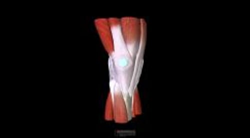
Inside the Knee

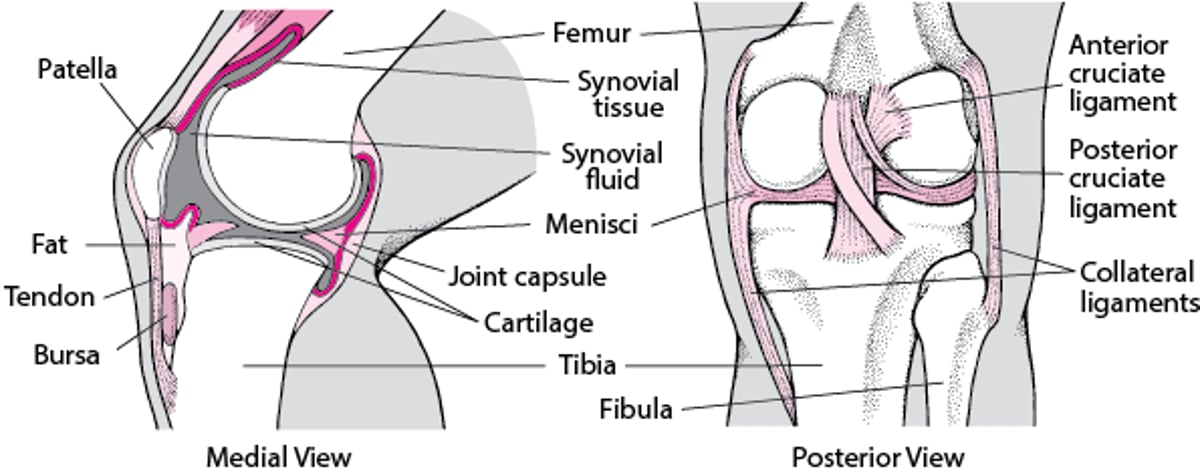
At the knee, gross deformities such as swelling (eg, joint effusion, popliteal cysts), quadriceps muscle atrophy, and joint instability may be obvious when the patient stands and walks. With the patient supine, the examiner should palpate the knee, identifying the patella, femoral condyles, tibial tuberosity, tibial plateau, fibular head, medial and lateral joint lines, popliteal fossa, and quadriceps and patellar tendons. The medial and lateral joint lines correspond to locations of the medial and lateral menisci and can be located by palpation while slowly flexing and extending the knee. Tender extra-articular bursae such as the anserine bursa below the medial joint line should be differentiated from true intra-articular disturbances.

Detection of small knee effusions is often difficult and is best accomplished using the bulge sign. The knee is fully extended and the leg slightly externally rotated while the patient is supine with muscles relaxed. The medial aspect of the knee is stroked to express any fluid away from this area. Placement of one hand on the suprapatellar pouch (within synovial tissue superior to the patella) and gentle stroking or pressing on the lateral aspect of the knee can create a fluid wave or bulge, visible medially when an effusion is present. Larger effusions can be identified visually or by balloting the patella. Comparison to the unaffected side is useful. Joint effusion can result from many joint diseases, including rheumatoid arthritis, osteoarthritis, gout, and trauma.
Full 180° extension of the knee is attempted to detect flexion contractures. The patella is tested for free, painless motion.