

Positron emission tomography (PET), a type of radionuclide scanning, uses compounds containing radionuclides that decay by releasing a positron (the positively charged antimatter equivalent of an electron). The released positron combines with an electron and produces 2 photons whose paths are 180° apart. Ring detector systems encircling the positron-emitting source simultaneously detect the 2 photons to localize the source and to produce color tomographic images of the area. Because PET incorporates positron-emitting radionuclides into metabolically active compounds, it can provide information about tissue function. Standard uptake value (SUV) indicates metabolic activity of a lesion; typically, the intensity of color is increased with higher SUVs.
The most commonly used compound in clinical PET is:
Fluorine-18 [18F]–labeled deoxyglucose (FDG)
FDG is an analog of glucose, and its uptake is proportional to glucose metabolic rates. A patient’s relative glucose metabolic rate (SUV) is calculated: The amount of FDG taken up from the injected dose is divided by the patient’s body weight.
Uses of PET
PET has several clinical indications, including:
Cancer (eg, staging and evaluating specific types of cancer and evaluating response to treatment), which accounts for the majority of PET usage
Cardiac function (eg, evaluating myocardial viability, detecting hibernating myocardium)
Neurologic function (eg, evaluation of dementia and seizures)
Variations of PET

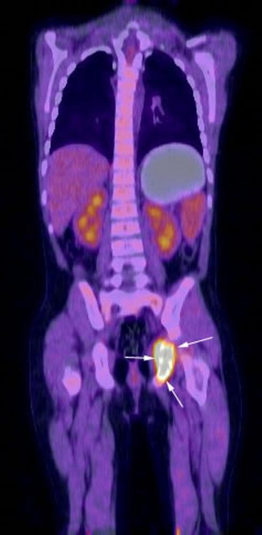
Coronal reconstruction image from FDG-PET-CT examination shows intense metabolic activity in the region of left ischium (arrows) corresponding to patient's known Langerhans cell histiocytosis.
Image courtesy of Hakan Ilaslan, MD.
PET-CT
Functional information provided by PET is superimposed on anatomic information provided by CT.
Disadvantages of PET
The typical effective radiation dose during PET is about 7 mSv. The effective radiation dose with PET-CT is 5 to 18 mSv.
Production of FDG requires a cyclotron. FDG has a short half-life (110 minutes); thus, shipment from the manufacturer and completion of the scan must occur very rapidly. The resulting expense, inconvenience, and impracticality greatly limit the availability of PET.


