


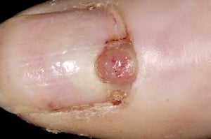
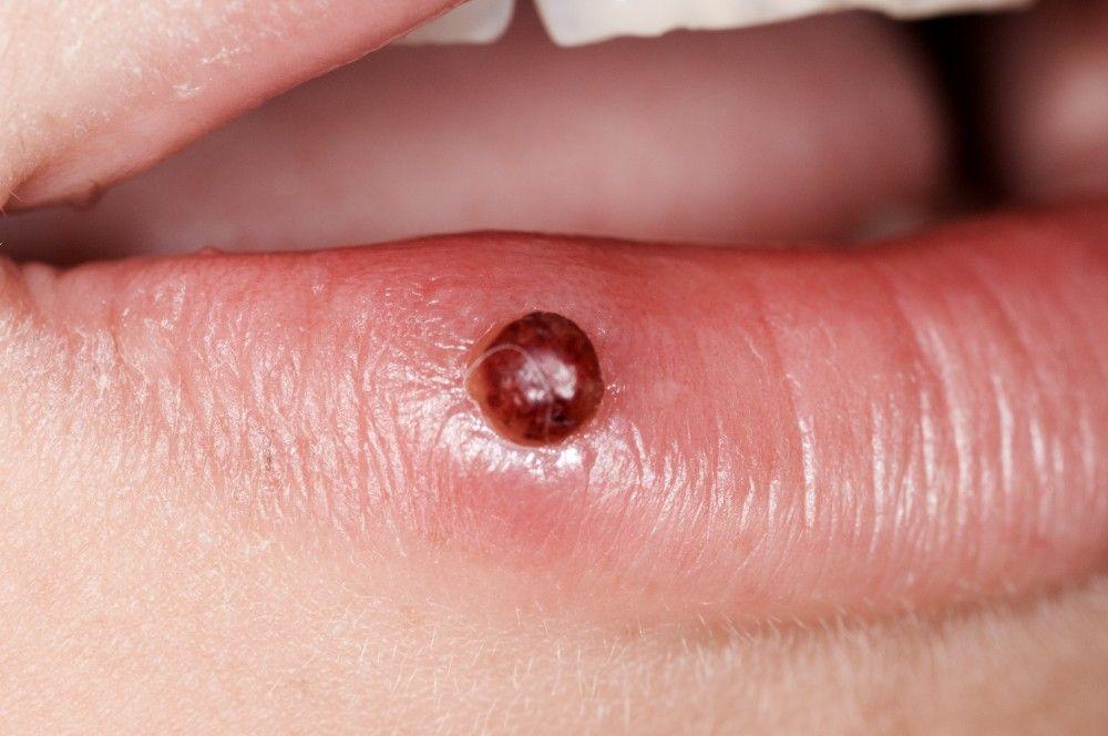
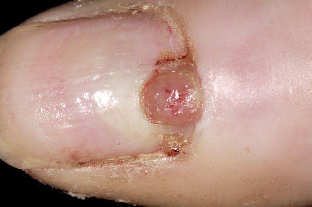
Pyogenic granulomas are fleshy, moist or crusty, usually scarlet vascular nodules composed of proliferating capillaries in an edematous stroma.
The lesion, composed of vascular tissue, is neither of bacterial origin nor a true granuloma. It develops rapidly, often at the site of recent injury (although injury may not be recalled), typically grows no larger than 2 cm in diameter, and probably represents a vascular and fibrous response to injury. There is no sex or age predilection.
The overlying epidermis is thin, and the lesion tends to be friable, bleeds easily, and does not blanch on pressure. The base may be pedunculated and surrounded by a collarette of epidermis.
During pregnancy, pyogenic granulomas may become large and exuberant (called gingival pregnancy tumors or telangiectatic epulis).
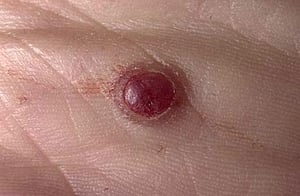
Image provided by Thomas Habif, MD.

DR P. MARAZZI/SCIENCE PHOTO LIBRARY
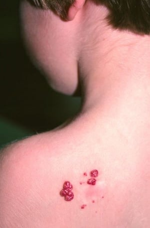
Image courtesy of Karen McKoy, MD.
DR P. MARAZZI/SCIENCE PHOTO LIBRARY

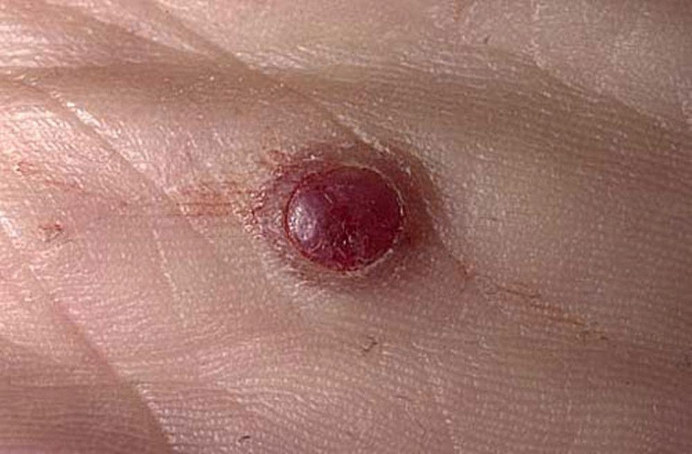
Image provided by Thomas Habif, MD.

DR P. MARAZZI/SCIENCE PHOTO LIBRARY


Image courtesy of Karen McKoy, MD.
DR P. MARAZZI/SCIENCE PHOTO LIBRARY
Diagnosis of Pyogenic Granulomas
Biopsy
Diagnosis of pyogenic granuloma involves biopsy and histologic examination. Histologic analysis is required for all removed tissue because these lesions occasionally resemble and must be differentiated from melanomas or other malignant tumors.
Treatment of Pyogenic Granulomas
Excision or curettage and electrodesiccation
Treatment of pyogenic granulomas consists of removal by excision or curettage and electrodesiccation, but the lesions may recur.


