


Tinea barbae is a dermatophyte infection of the beard area most often caused by Trichophyton mentagrophytes or T. verrucosum. Diagnosis is based on clinical appearance and examination of plucked hairs on potassium hydroxide wet mount. Treatment is with oral antifungals and sometimes prednisone.

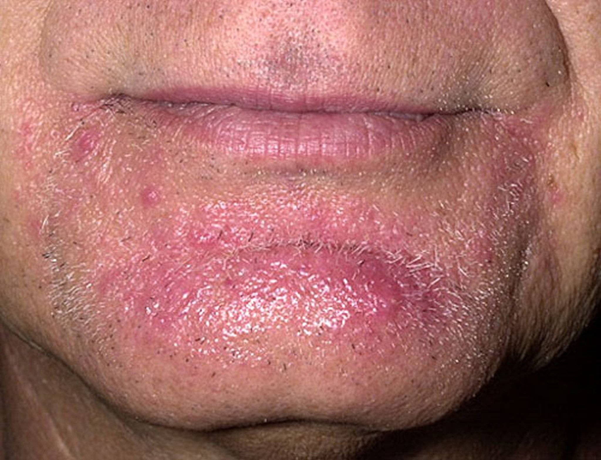
This photo shows follicular-based papules (some coalescent) and pustules in the beard area of the face caused by a dermatophyte infection.
Image provided by Thomas Habif, MD.
Tinea barbae is a dermatophytosis that manifests in the beard area as superficial annular lesions, but deeper skin infection similar to folliculitis may occur often due to Staphylococcus aureus (1). Risk factors for tinea barbae include recent shaving at barbershops, direct contact with infected animals (dairy cows or pets), and/or in the context of immunosuppression (2), or prior treatment of superficial fungal infections with topical glucocorticoids which can paradoxically promote the growth of the fungus. Other skin lesions include inflammatory papules, nodules, or plaques with or without crusting, and possible scarring. Tinea barbae may also manifest as a kerion (a large, boggy nodule caused by a severe inflammatory reaction to the dermatophyte) that can result in scarring hair loss.

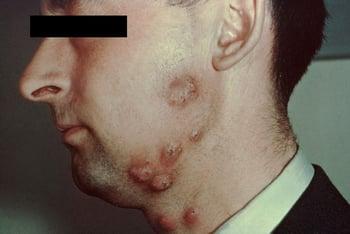
This photo shows large annular and erythematous lesions in the beard area of a patient with tinea barbae.
General references
1. Kaiser M, Abdin R, Yaghi M, et al. Beard Alopecia: An Updated and Comprehensive Review of Etiologies, Presentation and Treatment. J Clin Med. 2023;12(14):4793. doi:10.3390/jcm12144793
2. Bonifaz A, Ramírez-Tamayo T, Saúl A. Tinea barbae (tinea sycosis): experience with nine cases. J Dermatol. 2003;30(12):898-903. doi:10.1111/j.1346-8138.2003.tb00345.x
Diagnosis of Tinea Barbae
Primarily physical examination
Potassium hydroxide (KOH) wet mount
Rarely, fungal cultures or biopsy
Tinea barbae should be suspected based on characteristic clinical findings in patients with relevant risk factors (eg, animal contact, recent shaving at barbershops, immunosuppression).
The diagnosis of tinea barbae is established by identifying the fungal element (hyphae) on potassium hydroxide wet mount of plucked hairs, culture, or biopsy.
Differential diagnosis of follicular-based papules and pustules in the beard area includes bacterial folliculitis.
Treatment of Tinea Barbae
Oral antifungals
Sometimes oral glucocorticoids
Treatment of tinea barbae is with oral antifungals. Terbinafine and itraconazole are preferred agents that are often used for 4 to 6 weeks, depending on clinical response. Micronized griseofulvin is a reasonable alternative. (See table .)
If the lesions are severely inflamed, a short course of oral glucocorticoids such as prednisone should be added (to lessen symptoms and perhaps reduce the chance of scarring) and tapered over 2 weeks.
Drug Information for the Topic


