


Pyoderma gangrenosum is a chronic, neutrophilic, progressive skin necrosis of unknown etiology often associated with systemic illness and sometimes skin injury. Diagnosis is clinical. Treatment includes wound care and, based on severity, anti-inflammatory medications or immunosuppressants.
Pyoderma gangrenosum is a rare, noninfectious, neutrophilic dermatosis. It is often associated with systemic disease.
There are various subtypes, depending on the clinical progression of symptoms and site of occurrence:
Classic (ulcerative) subtype
Atypical (bullous) subtype
Pustular subtype
Vegetative subtype
Other subtypes (eg, peristomal, genital, extracutaneous)
Etiology of Pyoderma Gangrenosum
The etiology of pyoderma gangrenosum is incompletely understood, but it can be associated with various systemic illnesses in 50% of patients (1). These systemic diseases include inflammatory bowel disease, rheumatoid arthritis, cancers, and hematologic disorders (eg, monoclonal gammopathy of undetermined significance, myelodysplastic syndrome, polycythemia vera). It is thought to be mediated by abnormal responses in both the innate and adaptive immune systems (2).
A genetic predisposition may contribute to the development of pyoderma gangrenosum (2). Some patients with inherited autoinflammatory disorders are at increased risk. These disorders include PAPASH (pyogenic arthritis, pyoderma gangrenosum, acne, and suppurative hidradenitis) and PASH (pyoderma gangrenosum, acne, and suppurative hidradenitis without pyogenic arthritis). These syndromes are part of a spectrum that includes PAPA (pyogenic arthritis, pyoderma gangrenosum, and acne) syndrome (see also PAPA Syndrome).
Most patients are ≥ 55 years of age (3).
Pyoderma gangrenosum can manifest as various subtypes.
Etiology references
1. Ahronowitz I, Harp J, Shinkai K. Etiology and management of pyoderma gangrenosum: a comprehensive review. Am J Clin Dermatol. 2012;13(3):191-211. doi:10.2165/11595240-000000000-00000
2. Norouzi-Barough L, Biglari S, Sherkat R, et al. A Systematic Review of Mendelian Pyoderma Gangrenosum: Clinical and Genetic Characteristics in 120 Published Patients. Exp Dermatol. 2025;34(5):e70112. doi:10.1111/exd.70112
3. Xu A, Balgobind A, Strunk A, Garg A, Alloo A. Prevalence estimates for pyoderma gangrenosum in the United States: An age- and sex-adjusted population analysis. J Am Acad Dermatol. 2020;83(2):425-429. doi:10.1016/j.jaad.2019.08.001
Pathophysiology of Pyoderma Gangrenosum
The pathophysiology of pyoderma gangrenosum is poorly understood but may involve neutrophil chemotaxis. Interleukin-8 is overexpressed in lesions.
Ulcerations of pyoderma gangrenosum occur after trauma or injury to the skin in approximately 30% of patients; this process is termed pathergy (1).
Pathophysiology reference
1. Binus AM, Qureshi AA, Li VW, Winterfield LS. Pyoderma gangrenosum: a retrospective review of patient characteristics, comorbidities and therapy in 103 patients. Br J Dermatol. 2011;165(6):1244-1250. doi:10.1111/j.1365-2133.2011.10565.x
Symptoms and Signs of Pyoderma Gangrenosum
Pyoderma gangrenosum typically begins as an inflamed, erythematous papule, pustule, or nodule. The lesion, which may resemble a furuncle or an arthropod bite at this stage, ulcerates and expands rapidly, developing a swollen necrotic base and a raised dusky to violaceous border. An undermined border (ie, loss of underlying support tissue at the border) is common, if not pathognomonic. The ulcers can coalesce to form larger ulcers, often with cribriform or sieve-like scarring. Pathergy may be present.
Systemic symptoms such as fever and malaise are common.
Symptoms and signs can vary with the subtype.
Ulcerative (classic) subtype
In this most common subtype, ulcers form as described above, most commonly on the lower extremities or trunk, particularly the buttocks and perineum.

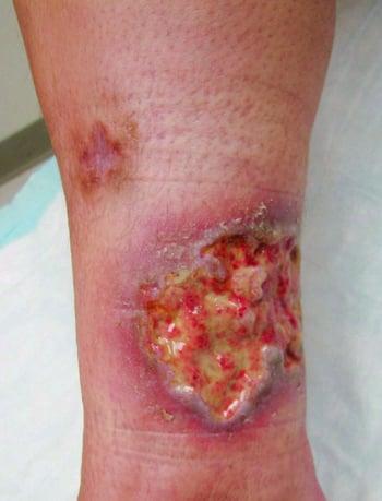
This photo shows an ulcerated lesion with a necrotic base and raised dusky to violaceous border on the leg.

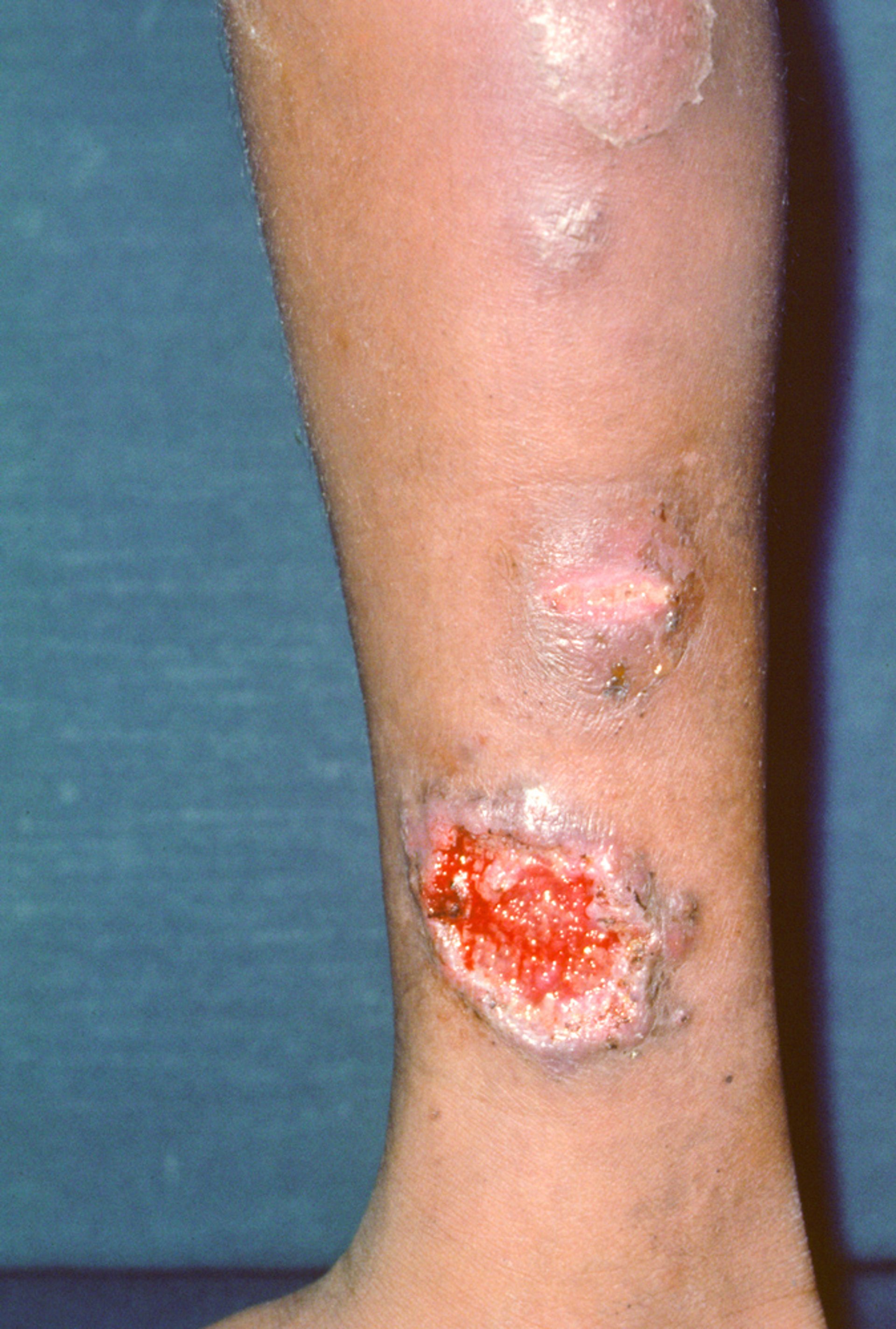
This photo shows an ulcerative type with a healing lesion at the top progressing to ulcerative lesions at the bottom.
Photo courtesy of Karen McKoy, MD.
Bullous (atypical) subtype
This less common subtype often develops in patients with hematologic disorders. Lesions usually begin as bullae that erode, becoming superficial ulcers. The arms and face are most often involved.

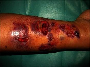
This photo shows multiple eroded bullae forming superficial lesions on a patient's arm.
Pustular subtype
This subtype tends to develop during exacerbations of inflammatory bowel disease. Painful pustules develop, surrounded by erythema. Arthralgias are common.
Vegetative (superficial granulomatous pyoderma) subtype
In this subtype, a single, indolent, mildly painful plaque or superficial ulcer develops, most often on the head or neck. The border is not undermined and the base is not necrotic.
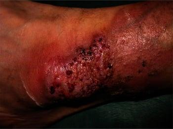
This photo shows a single superficial ulcer without raised borders or necrotic base.
Other subtypes
Pyoderma gangrenosum can also develop at other sites, such as around a stoma in patients who have inflammatory bowel disease (peristomal pyoderma gangrenosum), on the genitals (genital pyoderma gangrenosum), or in sites other than the skin, such as the bones, cornea, central nervous system, heart, intestine, liver, lungs, or muscle (extracutaneous pyoderma gangrenosum).
Diagnosis of Pyoderma Gangrenosum
Primarily history and physical examination
Sometimes, biopsy
The diagnosis of pyoderma gangrenosum is primarily clinical and is a diagnosis of exclusion (1). The PARACELSUS score is a validated diagnostic index that may aid in the diagnosis of pyoderma gangrenosum by incorporating clinical features, exclusion of alternative causes of ulceration, and associated systemic disease (2, 3). Expansion of ulceration after surgical debridement strongly suggests pyoderma gangrenosum.
Biopsies of lesions are not often diagnostic but may be supportive; many biopsies from the leading edge of a lesion show vasculitis with neutrophils and fibrin in superficial vessels.
Patients who have bullous (atypical) pyoderma gangrenosum should be monitored with periodic clinical assessment and complete blood count for development of a hematologic disorder.
Diagnosis references
1. Maverakis E, Ma C, Shinkai K, et al. Diagnostic Criteria of Ulcerative Pyoderma Gangrenosum: A Delphi Consensus of International Experts. JAMA Dermatol. 2018;154(4):461-466. doi:10.1001/jamadermatol.2017.5980
2. Jockenhöfer F, Wollina U, Salva KA, et al. The PARACELSUS score: a novel diagnostic tool for pyoderma gangrenosum. Br J Dermatol. 2019;180(3):615-620. doi:10.1111/bjd.16401
3. Moelleken M, Ortega-Loayza AG, Busch D, et al. Validation of PARACELSUS score performance for the diagnosis of pyoderma gangrenosum: An international multicenter study with 1403 cases. J Am Acad Dermatol. 2026;94(6):1738-1746. doi:10.1016/j.jaad.2026.02.101
Treatment of Pyoderma Gangrenosum
Wound care
Glucocorticoids
Tumor necrosis factor (TNF) inhibitors
Sometimes other anti-inflammatory medications or immunosuppressants
Avoidance of surgical debridement
The treatment of pyoderma gangrenosum requires a multimodal approach involving supportive wound care and topical and systemic immunomodulatory therapies.
Wound healing can be promoted with moisture-retaining occlusive dressings for less exudative plaques and absorptive dressings for highly exudative plaques. Biologic and other specialized dressings may be needed in refractory cases. Wet-to-dry dressings should be avoided.
Topical therapy with high-potency glucocorticoids or tacrolimus can aid in the resolution of superficial and early lesions. For more severe manifestations, prednisone 60 to 80 mg orally once a day is a common first-line therapy.
TNF inhibitors (eg, infliximab, adalimumab, etanercept) are effective, particularly in patients who have inflammatory bowel disease (1).
Other immunosuppressive therapies (ie, glucocorticoid-sparing agents) and other agents may also be used (2). Cyclosporine 3 mg/kg orally once a day is effective, particularly in rapidly progressive disease. Other glucocorticoid-sparing agents including azathioprine, methotrexate, and mycophenolate mofetil have been used.
Biologic therapies, particularly tumor necrosis factor (TNF) inhibitors such as infliximab, may be considered for refractory disease. Janus kinase (JAK) inhibitors such as tofacitinib and upadacitinib have also been used as treatment options for refractory pyoderma gangrenosum, although evidence remains limited. Therapies targeting the interleukin (IL)-1, IL-17 or IL-23 pathways have been reported in refractory cases but their efficacy is less well established.
Surgical debridement should be avoided because of the risk of pathergy and wound extension (3).
Treatment references
1. Zaman M, Martinez R, Mayur O, et al. Use of biologic therapies in the management of pyoderma gangrenosum: a systematic review. Arch Dermatol Res. 2024;316(8):539. Published 2024 Aug 19. doi:10.1007/s00403-024-03332-2
2. Partridge ACR, Bai JW, Rosen CF, et al. Effectiveness of systemic treatments for pyoderma gangrenosum: a systematic review of observational studies and clinical trials. Br J Dermatol. 2018;179(2):290-295. doi:10.1111/bjd.16485
3. Alavi A, French LE, Davis MD, et al. Pyoderma gangrenosum: An update on pathophysiology, diagnosis and treatment. Am J Clin Dermatol. 2017;18(3):355–372. doi: 10.1007/s40257-017-0251-7
Key Points
Pyoderma gangrenosum is often associated with a systemic disorder and is probably immune-mediated.
There are several subtypes; the ulcerative subtype (ie, necrotic base and raised violaceous border with undermined edge on a lower extremity, buttock, or perineum) is the most common.
The diagnosis of pyoderma gangrenosum is typically clinical.
Wound care should be optimized and surgical debridement avoided.
Medical management is with potent topical glucocorticoids or tacrolimus to treat early lesions and systemic glucocorticoids, tumor necrosis factor (TNF) inhibitors, or other anti-inflammatories or immunosuppressants to treat more severe manifestations.
Drug Information for the Topic







