


Onychomycosis refers to fungal infection of the nail plate, nail bed, or both. The nails typically are thickened and discolored yellow with subungual debris. The diagnosis is established by potassium hydroxide (KOH) wet mounts with microscopy, fungal culture, histopathology, or polymerase chain reaction. Treatment includes topical options such as ciclopirox lacquer, tavaborole solution, and efinaconazole solutions. Oral options include terbinafine, itraconazole, or fluconazole.
(See also Overview of Nail Disorders.)
Approximately 5.5% of the global population and 2 to 14% of people in the United States have onychomycosis (1, 2).
Risk factors for onychomycosis include:
Preexisting nail dystrophy (eg, in patients with mechanical nail trauma)
Older age
Male sex
Exposure to someone with tinea pedis or onychomycosis (eg, a family member or through public bathing)
Immunosuppression
Toenails are 10 times more commonly infected than fingernails. Approximately 65% of cases are caused by dermatophytes (eg, Trichophyton rubrum); dermatophyte infection of the nails is called tinea unguium (3). Many of the remaining cases are caused by nondermatophyte molds (eg, Aspergillus, Scopulariopsis, Fusarium). Immunocompromised patients and those with chronic mucocutaneous candidiasis may have candidal onychomycosis (which is more common on the fingers). Subclinical onychomycosis can also occur in patients with recurrent tinea pedis. Onychomycosis may predispose patients to lower extremity cellulitis.

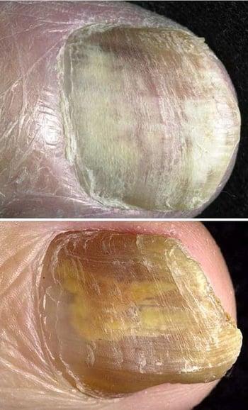
Onychomycosis is fungal infection of the nail plate, nail bed, or both. Infection may be distal subungual, with nail thickening and yellowing and accumulation of keratin and debris underneath the nail (top); proximal subungual (not pictured); or white superficial, with spreading of chalky white scale beneath the nail surface (bottom).
General references
1. Lipner SR, Scher RK. Onychomycosis: Clinical overview and diagnosis. J Am Acad Dermatol. 2019;80(4):835-851. doi:10.1016/j.jaad.2018.03.062
2. Lipner SR, Scher RK. Onychomycosis: Treatment and prevention of recurrence. J Am Acad Dermatol. 2019;80(4):853-867. doi:10.1016/j.jaad.2018.05.1260
3. Sigurgeirsson B, Baran R. The prevalence of onychomycosis in the global population: a literature study. J Eur Acad Dermatol Venereol. 2014;28(11):1480-1491. doi:10.1111/jdv.12323
Symptoms and Signs of Onychomycosis
Nails have asymptomatic patches of white or yellow discoloration and onycholysis with subungual debris. There are 3 common characteristic patterns:
Distal subungual: The nails thicken and yellow, keratin and debris accumulate distally and beneath the nails, and the nails separate from the nail bed (onycholysis).
Proximal subungual: A form that starts proximally and is a marker of immunosuppression.
White superficial: A chalky white scale slowly spreads beneath the nail surface.

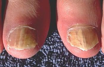
This photo shows onychomycosis of the great toenails. The right toenail shows classic distolateral (a distal subungual) onychomycosis (DLO), whereas the left toenail likely began as DLO and progressed to dermatophytoma (dense collection of hyphae that appears as a linear streak).
Diagnosis of Onychomycosis
Dermoscopic examination
Fungal culture or polymerase chain reaction (PCR)
Potassium hydroxide wet mount examination
Histopathologic examination of periodic acid-Schiff (PAS)– or Grocott-Gomori methenamine silver (GMS)-stained nail clippings and subungual debris
Onychomycosis is suspected by appearance in patients who also have tinea pedis; predictive clinical features include involvement of the 1st and 5th toenails on the same foot, and involvement of a single foot. Subclinical onychomycosis should be considered in patients with recurrent tinea pedis. Dermoscopy can be supportive, showing spikes or an aurora borealis pattern, but mycological confirmation is necessary.
Differentiation from psoriasis or lichen planus is important because the therapies differ. The prevalence of onychomycosis is significantly higher in patients with psoriasis compared to those without psoriasis (1). Confirmation to establish a fungal etiology is required prior to treatment.
The diagnosis is typically confirmed by microscopic examination and, unless microscopic findings are conclusive, culture of scrapings or PCR of clippings (2). Scrapings are taken from the most proximal position that can be accessed on the affected nail and are examined for hyphae on potassium hydroxide (KOH) wet mount and then cultured. PCR has become a more common technique to confirm the diagnosis of onychomycosis, especially if cultures are negative or a definitive diagnosis is required (3, 4). Histopathologic examination of PAS–stained or GMS nail clippings and subungual debris may also be helpful.
Obtaining an adequate sample of nail for culture can be difficult because the distal subungual debris, which is easy to sample, often does not contain living fungus. Therefore, removing the distal portion of the nail with clippers before sampling or using a small curette to reach more proximally beneath the nail increases the yield.
Diagnosis references
1. Liu M, Kang Y, Zhang R. The Prevalence of Onchomycosis in Psoriasis Patients: A Systematic Review and Meta-Analysis. Mycoses. 2025;68(2):e70035. doi:10.1111/myc.70035
2. Gupta AK, Mays RR, Versteeg SG, et al: Update on current approaches to diagnosis and treatment of onychomycosis. Expert Rev Anti Infect Ther 16(12):929–938, 2018. doi: 10.1080/14787210.2018.1544891
3. Joyce A, Gupta AK, Koenig L, et al: Fungal Diversity and Onychomycosis: An analysis of 8,816 toenail samples using quantitative PCR and next-generation sequencing. J Am Podiatr Med Assoc 109(1):57–63, 2019. doi: 10.7547/17-070
4. Haghani I, Shams-Ghahfarokhi M, Dalimi Asl A, et al: Molecular identification and antifungal susceptibility of clinical fungal isolates from onychomycosis (uncommon and emerging species). Mycoses 62(2):128–143, 2019. doi: 10.1111/myc.12854
Treatment of Onychomycosis
Oral terbinafine or itraconazole
Topical treatments (eg, efinaconazole, tavaborole, ciclopirox 8%, amorolfine)
Onychomycosis should generally be treated. Treatment can be deferred in patients who are very old or debilitated or those with contraindications. Some proposed indications for treatment include the following:
Previous ipsilateral cellulitis
Diabetes or other risk factors for cellulitis
Presence of bothersome symptoms (eg, pain, social embarrassment, difficulty with daily activities)
Treatment of onychomycosis is dependent upon the number of nails involved, severity of disease (surface area, thickness), the presence of comorbidities, and patient preference. Oral treatment options are oral terbinafine or itraconazole. Both treatments are effective. Oral fluconazole may also be an option, with fewer potential side effects compared to itraconazole. Azoles are associated with lower clinical as well as mycological cure rates compared to terbinafine (1). It may not be necessary to treat until complete clinical resolution because these medications remain bound to the nail plate and will continue to be effective after oral administration is stopped. Recurrence rates are high without post-treatment prophylaxis, which is typically achieved by the application of antifungal cream to the feet. The affected nail will not revert to normal, but newly growing nail will appear normal; this process can take 12 to 18 months.
Topical agents efinaconazole and tavaborole can penetrate the nail plate and are more effective than older topical agents.
Topical antifungal nail lacquer containing efinaconazole 10%, ciclopirox 8%, or amorolfine 5% (not available in the United States) may be effective as primary treatment when disease is mild and there are few nails affected. It can be used for more severe disease with adjuncts such as softening agents (urea) or debridement. Topical treatment is particularly effective in children due to their thinner and faster growing nails compared to adults. Investigative treatments include new delivery systems for terbinafine.
To limit relapse, the patient should trim nails short, dry feet after bathing, wear absorbent socks, and use an antifungal cream. Old shoes may harbor a high density of spores and, if feasible, should be discarded. Ultraviolet sanitizers could be used to treat old shoes.
Treatment reference
1. Kreijkamp-Kaspers S, Hawke KL, van Driel ML. Oral Medications to Treat Toenail Fungal Infection. JAMA. 2018;319(4):397-398. doi:10.1001/jama.2017.20160
Key Points
Onychomycosis is highly prevalent, particularly among older men and patients with compromised distal circulation, nail dystrophies, and/or tinea pedis.
Suspect the diagnosis based on appearance and the pattern of nail involvement and confirm it by microscopy and culture or PCR.
Treatment is warranted if onychomycosis causes complications or troublesome symptoms.
If treatment is warranted, consider terbinafine (azoles are an alternative) and measures to prevent recurrence (eg, limiting moisture, discarding old shoes, trimming nails short).
Drug Information for the Topic


