

Lichen sclerosus is an inflammatory dermatosis of unknown cause, possibly autoimmune, that usually affects the anogenital area. Diagnosis is usually clinical and supported by skin biopsy. Treatment is with potent topical glucocorticoids.
Lichen sclerosus (LS) is a chronic, inflammatory dermatosis. The disease is characterized by a bimodal age distribution, being most common in postmenopausal women followed by men, prepubertal children, and adolescents.
The etiology remains incompletely understood. Evidence supports a multifactorial pathogenesis involving genetic predisposition, hormonal factors (women of childbearing age are largely spared), autoimmune mechanisms (notably Th1-mediated immune responses with heightened production of IFN-gamma), and possibly trauma or local irritation (1). Infectious etiologies are unlikely to contribute.
General reference
1. De Luca DA, Papara C, Vorobyev A, et al. Lichen sclerosus: The 2023 update. Front Med (Lausanne). 2023;10:1106318. Published 2023 Feb 16. doi:10.3389/fmed.2023.1106318
Signs and Symptoms of Lichen Sclerosus
Lichen sclerosus (LS) primarily affects the anogenital region but can also be generalized in 6 to 20% of patients (1). The earliest signs are skin fragility, bruising, and sometimes blistering. Lesions typically cause mild to severe itching. When LS manifests in children, the appearance may be mistaken for injuries resulting from sexual abuse. With time, the involved tissue becomes atrophic, thinned, hypopigmented (there may be flecks of postinflammatory hyperpigmentation), fissured, and scaly. Hyperkeratotic and fibrotic forms exist.
Severe and longstanding cases cause scarring and distortion or absorption of normal anogenital architecture. In women, this distortion can even lead to total destruction of the labia minora and clitoris. In men, phimosis or fusion of the foreskin to the coronal sulcus can occur.

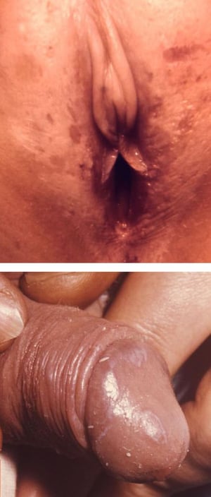
This image shows atrophy, hypopigmentation, and hyperpigmentation of the labia (top) and atrophy and hypopigmentation of the glans of the penis (bottom) resulting from lichen sclerosus.
This image shows atrophy, hypopigmentation, and hyperpigmentation of the labia (top) and atrophy and hypopigmentation o
Images courtesy of Joe Miller (top) and Brian Hill (bottom) via the Public Health Image Library of the Centers for Disease Control and Prevention.


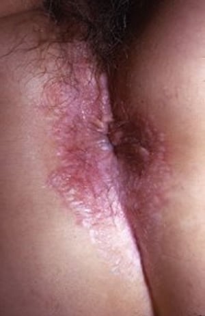
This photo shows atrophy and hypopigmentation in the vulvar and perianal areas.
This photo shows atrophy and hypopigmentation in the vulvar and perianal areas.
Image provided by E. Laurie Tolman, MD.

This photo shows scarring, atrophy, and distorted anatomy in the perianal area.
This photo shows scarring, atrophy, and distorted anatomy in the perianal area.
Image provided by E. Laurie Tolman, MD.

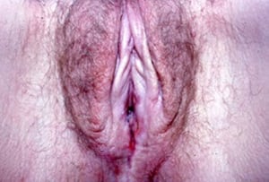
This photo shows porcelain white patches and areas of purpura in the labia minora, as well as early labial fusion.
This photo shows porcelain white patches and areas of purpura in the labia minora, as well as early labial fusion.
Image provided by E. Laurie Tolman, MD.

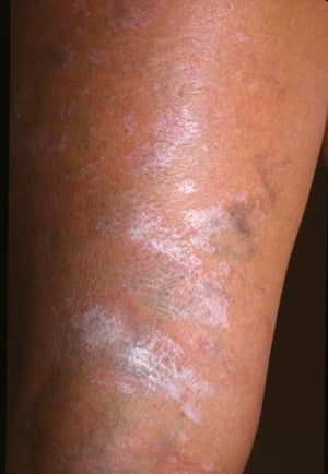
This image shows hypopigmented, porcelain white patches of lichen sclerosus.
This image shows hypopigmented, porcelain white patches of lichen sclerosus.
Image courtesy of Karen McKoy, MD.
This image shows atrophy, hypopigmentation, and hyperpigmentation of the labia (top) and atrophy and hypopigmentation of the glans of the penis (bottom) resulting from lichen sclerosus.
This image shows atrophy, hypopigmentation, and hyperpigmentation of the labia (top) and atrophy and hypopigmentation o
Images courtesy of Joe Miller (top) and Brian Hill (bottom) via the Public Health Image Library of the Centers for Disease Control and Prevention.
This photo shows atrophy and hypopigmentation in the vulvar and perianal areas.
This photo shows atrophy and hypopigmentation in the vulvar and perianal areas.
Image provided by E. Laurie Tolman, MD.
This photo shows scarring, atrophy, and distorted anatomy in the perianal area.
This photo shows scarring, atrophy, and distorted anatomy in the perianal area.
Image provided by E. Laurie Tolman, MD.
This photo shows porcelain white patches and areas of purpura in the labia minora, as well as early labial fusion.
This photo shows porcelain white patches and areas of purpura in the labia minora, as well as early labial fusion.
Image provided by E. Laurie Tolman, MD.
This image shows hypopigmented, porcelain white patches of lichen sclerosus.
This image shows hypopigmented, porcelain white patches of lichen sclerosus.
Image courtesy of Karen McKoy, MD.
Signs and symptoms reference
1. Kirtschig G. Lichen Sclerosus-Presentation, Diagnosis and Management. Dtsch Arztebl Int. 2016;113(19):337-343. doi:10.3238/arztebl.2016.0337
Diagnosis of Lichen Sclerosus
Primarily physical examination
Sometimes biopsy
Diagnosis of lichen sclerosus (LS) can usually be based on appearance, especially in advanced cases; however, biopsy should be performed on any anogenital dermatosis that does not resolve with mild conventional therapy. In addition to treatment refractory cases, it is especially important to biopsy any area that becomes thickened or ulcerated, because LS is associated with an increased frequency of squamous cell carcinoma (1).
Diagnosis reference
1. Leis M, Singh A, Li C, et al: Risk of vulvar squamous cell carcinoma in lichen sclerosus and lichen planus: A systematic review. J Obstet Gynaecol Can 44(2):182–192, 2022. doi: 10.1016/j.jogc.2021.09.023
Treatment of Lichen Sclerosus
Topical glucocorticoids
Topical glucocorticoid-sparing agents
For recalcitrant lichen sclerosus, oral retinoids, methotrexate, and hydroxychloroquineFor recalcitrant lichen sclerosus, oral retinoids, methotrexate, and hydroxychloroquine
First-line treatment of lichen sclerosus consists of potent topical glucocorticoids (medications that otherwise should be used with extreme caution in this area). High-potency steroids may be necessary to induce remission (eg, clobetasol) (First-line treatment of lichen sclerosus consists of potent topical glucocorticoids (medications that otherwise should be used with extreme caution in this area). High-potency steroids may be necessary to induce remission (eg, clobetasol) (1). The disease course is generally intractable, so long-term treatment and follow-up are necessary. Topical glucocorticoid-sparing agents such as topical calcineurin inhibitors are alternative options for patients who are intolerant of or refractory to glucocorticoids. Emerging strategies may include the use of oral JAK inhibitors (eg, ruxolitinib). ). The disease course is generally intractable, so long-term treatment and follow-up are necessary. Topical glucocorticoid-sparing agents such as topical calcineurin inhibitors are alternative options for patients who are intolerant of or refractory to glucocorticoids. Emerging strategies may include the use of oral JAK inhibitors (eg, ruxolitinib).
For recalcitrant lichen sclerosus, systemic options may include oral retinoids, methotrexate, and hydroxychloroquine. For recalcitrant lichen sclerosus, systemic options may include oral retinoids, methotrexate, and hydroxychloroquine.
Monitoring for progression to squamous cell carcinoma and sexual dysfunction and providing psychological support are indicated.
Treatment reference
1. De Luca DA, Papara C, Vorobyev A, et al. Lichen sclerosus: The 2023 update. Front Med (Lausanne). 2023;10:1106318. Published 2023 Feb 16. doi:10.3389/fmed.2023.1106318
Key Points
Lichen sclerosus can cause anogenital bruising, itching, or blistering early, and atrophy and scarring later.
The diagnosis should be considered in patients with persistent anogenital dermatosis.
Treatment is with long-term, high-potency topical glucocorticoids, close monitoring, and sexual and psychological support.
The risk of subsequent squamous cell carcinoma is increased.
Drug Information for the Topic


