


Pityriasis rubra pilaris is a rare chronic disorder that causes hyperkeratotic yellowing of the skin, including the trunk, extremities, and, particularly, the palms and soles. Red follicular papules typically merge to form red-orange scaling plaques and confluent areas of erythema with islands of normal skin between lesions. Diagnosis is clinical. Treatment is aimed at relieving symptoms.
Pityriasis rubra pilaris is a rare, chronic, inflammatory papulosquamous dermatosis characterized by the presence of hyperkeratotic follicular papules, palmoplantar keratoderma, and orange-red scaly plaques with well-demarcated "islands of sparing". There is a slight male predominance (1).
The cause of pityriasis rubra pilaris is largely idiopathic (2). Genetic mutations are thought to contribute, particularly gain-of-function mutations in the CARD14 gene and more rarely, loss-of-function mutations in KRT32. However, it has been associated with infections, autoimmune diseases, and malignancies.
Pathogenesis involves dysregulation of the Th17 axis causing aberrant NF-κB signaling and increased levels of IL-23, overlapping with mechanisms seen in psoriasis.
The 2 most common forms of the disorder are:
Juvenile classic (characterized by autosomal dominant inheritance and childhood onset)
Adult classic (characterized by no apparent inheritance and adult onset)
Atypical (nonclassic) forms exist in both age groups. Atypical forms are characterized by unusual morphology, chronicity, and recalcitrance to therapy compared to classic pityriasis rubra pilaris. Sunlight, HIV or another infection, minor trauma, or an autoimmune disorder may trigger a flare-up.

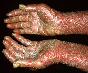
Pityriasis rubra pilaris can manifest as hyperkeratotic yellowing of the palms and soles.
Pityriasis rubra pilaris can manifest as hyperkeratotic yellowing of the palms and soles.
Image provided by Thomas Habif, MD.

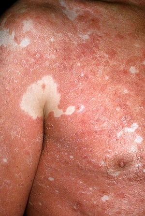
Pityriasis rubra pilaris can manifest as red follicular papules that coalesce to form red-orange scaling plaques and confluent areas of erythema with islands of normal skin between lesions.
Pityriasis rubra pilaris can manifest as red follicular papules that coalesce to form red-orange scaling plaques and co
Image provided by Thomas Habif, MD.

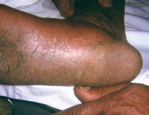
This image shows hyperkeratosis of the sole characteristic of pityriasis rubra pilaris.
This image shows hyperkeratosis of the sole characteristic of pityriasis rubra pilaris.
Image courtesy of Karen McKoy, MD.
Pityriasis rubra pilaris can manifest as hyperkeratotic yellowing of the palms and soles.
Pityriasis rubra pilaris can manifest as hyperkeratotic yellowing of the palms and soles.
Image provided by Thomas Habif, MD.
Pityriasis rubra pilaris can manifest as red follicular papules that coalesce to form red-orange scaling plaques and confluent areas of erythema with islands of normal skin between lesions.
Pityriasis rubra pilaris can manifest as red follicular papules that coalesce to form red-orange scaling plaques and co
Image provided by Thomas Habif, MD.
This image shows hyperkeratosis of the sole characteristic of pityriasis rubra pilaris.
This image shows hyperkeratosis of the sole characteristic of pityriasis rubra pilaris.
Image courtesy of Karen McKoy, MD.
General references
1. Ross NA, Chung HJ, Li Q, et al. Epidemiologic, Clinicopathologic, Diagnostic, and Management Challenges of Pityriasis Rubra Pilaris: A Case Series of 100 Patients. JAMA Dermatol. 2016;152(6):670-675. doi:10.1001/jamadermatol.2016.0091
2. Joshi TP, Duvic M. Pityriasis Rubra Pilaris: An Updated Review of Clinical Presentation, Etiopathogenesis, and Treatment Options. Am J Clin Dermatol. 2024;25(2):243-259. doi:10.1007/s40257-023-00836-x
Diagnosis of Pityriasis Rubra Pilaris
Primarily history and physical examination
Sometimes biopsy
The diagnosis of pityriasis rubra pilaris is by clinical appearance and may be supported by biopsy and histopathological examination showing alternating orthokeratosis and parakeratosis in a checkerboard pattern, with focal acantholytic dyskeratosis (1).
Biopsy is performed when the clinical diagnosis is unclear (eg, when the patient is erythrodermic at presentation). Pityriasis rubra pilaris is often misdiagnosed and multiple biopsies may be performed before the diagnosis is established.
Differential diagnose of pityriasis rubra pilaris include the following:
Seborrheic dermatitis (in children)
Psoriasis (when disease occurs on the scalp, elbows, and knees)
Diagnosis reference
1. Klein A, Landthaler M, Karrer S. Pityriasis rubra pilaris: a review of diagnosis and treatment. Am J Clin Dermatol. 2010;11(3):157-170. doi:10.2165/11530070-000000000-00000
Treatment of Pityriasis Rubra Pilaris
Symptom relief (eg, with emollients, topical lactic acid, and topical glucocorticoids)
Sometimes systemic therapy (eg, oral retinoids, methotrexate, cyclosporine, mycophenolate mofetil [MMF], azathioprine)
Sometimes biologics (eg, guselkumab, ixekizumab, secukinumab)
Treatment of pityriasis rubra pilaris is exceedingly challenging and often empiric (1). The disorder may be ameliorated but almost never cured; classic forms of the disorder resolve slowly over 3 years, whereas nonclassic forms persist.
Scaling may be reduced with emollients or 12% lactic acid under occlusive dressing, followed by topical glucocorticoids.
Oral retinoids (eg, acitretin, isotretinoin, etretinate) are considered first-line, with systemic glucocorticoids sometimes used for short-term control. Biologics targeting IL-23 (eg, guselkumab, risankizumab, ustekinumab) and IL-17 (ixekizumab, secukinumab, or both) have shown promise in refractory cases (1, 2). Tumor necrosis factor-alpha inhibitors may also be helpful (3).
Phototherapy has a variable response rate and may be used with caution due to risk of photoexacerbation (an isomorphic response also known as "Koebnerization"). Oral vitamin A, cyclosporine, MMF, and azathioprine have also been used (4).
Spontaneous remission occurs in up to 80% of classic adult cases within 1 to 3 years (5).
Treatment references
1. Tran TS, Nguyen TM, Lehman JS, et al. Treatment of pityriasis rubra pilaris with systemic and biological agents: A single-center retrospective cohort of 76 patients. J Am Acad Dermatol. 2025;92(6):1362-1365. doi:10.1016/j.jaad.2025.01.017
2. Napolitano M, Abeni D, Didona B. Biologics for pityriasis rubra pilaris treatment: A review of the literature. J Am Acad Dermatol 79(2):353–359.e11, 2018. doi: 10.1016/j.jaad.2018.03.036
3. Kromer C, Sabat R, Celis D, Mössner R. Systemic therapies of pityriasis rubra pilaris: a systematic review. J Dtsch Dermatol Ges. 2019;17(3):243-259. doi:10.1111/ddg.13718
4. Engelmann C, Elsner P, Miguel D. Treatment of pityriasis rubra pilaris type I: A systematic review. Eur J Dermatol 29(5):524-537, 2019. doi: 10.1684/ejd.2019.3641
5. Klein A, Landthaler M, Karrer S. Pityriasis rubra pilaris: a review of diagnosis and treatment. Am J Clin Dermatol. 2010;11(3):157-170. doi:10.2165/11530070-000000000-00000
Drug Information for the Topic


