Manifestations of diabetic retinopathy include microaneurysms, intraretinal hemorrhage, exudates, macular edema, macular ischemia, neovascularization, vitreous hemorrhage, and traction retinal detachment. Symptoms may not develop until late in the disease. Diagnosis is by funduscopy; further details are elucidated by color fundus photography, fluorescein angiography, and optical coherence tomography. Treatment includes control of blood glucose and blood pressure. Ocular treatments include retinal laser photocoagulation, intravitreal injection of antivascular endothelial growth factor medications (eg, aflibercept, ranibizumab, bevacizumab), intraocular glucocorticoids, vitrectomy, or a combination.
Pathophysiology of Diabetic Retinopathy
Diabetic retinopathy is a major cause of blindness, particularly among working-age adults. The degree of retinopathy is highly correlated with:
Duration of diabetes
Blood glucose levels
Blood pressure levels
Pregnancy can worsen retinopathy, in part due to impaired blood glucose control.
Clinically significant macular edema can occur with nonproliferative or proliferative retinopathy and is the most common cause of vision loss due to diabetic retinopathy (1).
Nonproliferative retinopathy
Nonproliferative retinopathy (also called background retinopathy) develops first and causes increased capillary permeability, microaneurysms, hemorrhages, exudates, macular ischemia, and macular edema (thickening of the retina caused by fluid leakage from capillaries).
Proliferative retinopathy
Proliferative retinopathy develops after nonproliferative retinopathy and is more severe; it may lead to vitreous hemorrhage and traction retinal detachment. Proliferative retinopathy is characterized by abnormal new vessel formation (neovascularization), which occurs on the inner (vitreous) surface of the retina and may extend into the vitreous cavity and cause vitreous hemorrhage. Neovascularization is often accompanied by preretinal fibrous tissue, which, along with the vitreous, can contract, resulting in traction retinal detachment. Neovascularization may also occur in the anterior segment of the eye on the iris; neovascular membrane growth in the anterior chamber angle of the eye at the peripheral margin of the iris can occur, and this growth leads to neovascular glaucoma. Vision loss with proliferative retinopathy may be severe.
Pathophysiology reference
1. Singh R, Walia A, Kaur J, Kumar P, Verma I, Rani N. Diabetic Retinopathy - Pathophysiology to Treatment: A Review. Curr Diabetes Rev. 2025;21(3):58-67. doi:10.2174/0115733998259940231105200251
Symptoms and Signs of Diabetic Retinopathy
Nonproliferative retinopathy
Vision symptoms are caused by macular edema or macular ischemia. However, patients may not have vision loss even with advanced retinopathy. The first signs of nonproliferative retinopathy are:
Capillary microaneurysms
Dot and blot retinal hemorrhages
Hard exudates
Cotton-wool spots (soft exudates)

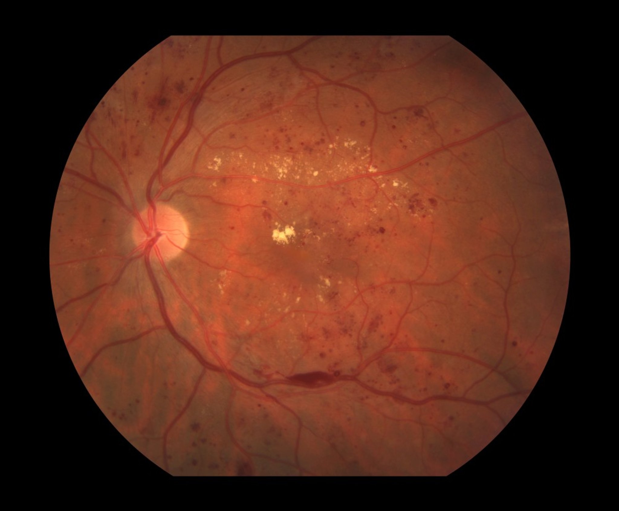
Funduscopic features of nonproliferative diabetic retinopathy include retinal hemorrhages and hard exudates (yellow patches).
Paul Whitten/SCIENCE PHOTO LIBRARY
Hard exudates are discrete, yellow particles within the retina. When present, they suggest chronic edema. Cotton-wool spots are areas of microinfarction of the retinal nerve fiber layer that lead to retinal opacification; they are fuzzy-edged and white and obscure underlying vessels.
Signs in later stages are:
Macular edema (seen on slit-lamp biomicroscopy as elevation and blurring of retinal layers)
Venous dilation and intraretinal microvascular abnormalities
Proliferative retinopathy
Symptoms may include blurred vision, floaters (black spots) or flashing lights (photopsias) in the field of vision, and sudden, severe, painless vision loss. These symptoms are typically caused by vitreous hemorrhage or traction retinal detachment.
Proliferative retinopathy, unlike nonproliferative retinopathy, causes formation of fine preretinal vessel neovascularization visible on the optic nerve or retinal surface. Macular edema or retinal hemorrhage may be visible on funduscopy.

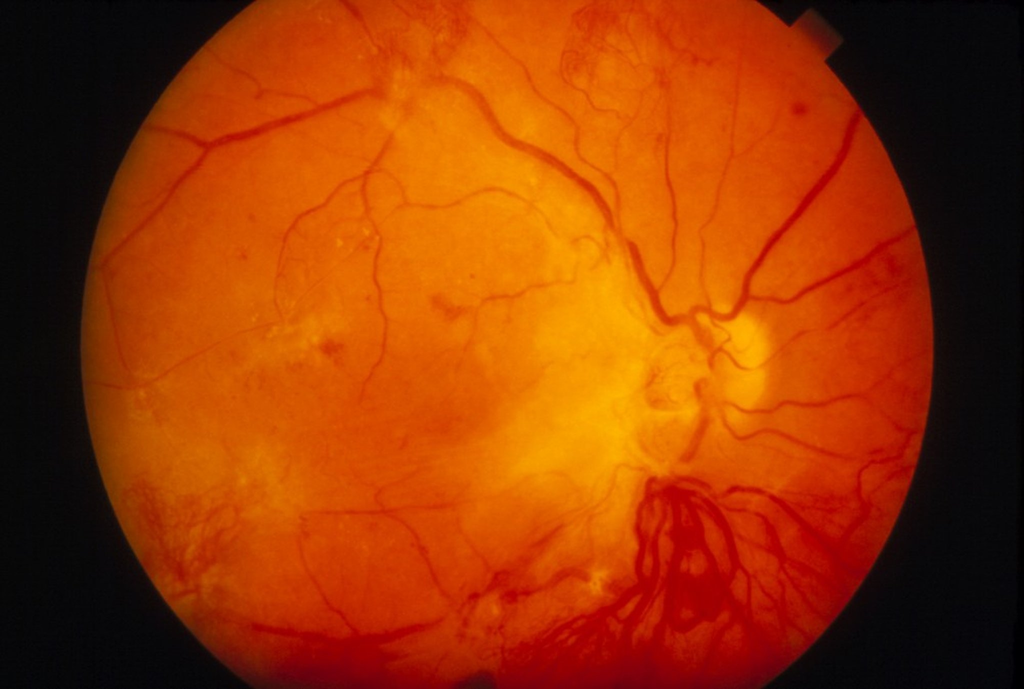
The key funduscopic feature of proliferative diabetic retinopathy is neovascularization, seen around the optic disc.
WESTERN OPHTHALMIC HOSPITAL/SCIENCE PHOTO LIBRARY
Diagnosis of Diabetic Retinopathy
Funduscopy
Color fundus photography
Fluorescein angiography
Optical coherence tomography
Diagnosis is by funduscopy. Color fundus photography helps grade the level of retinopathy. Fluorescein angiography is used to determine the extent of retinopathy, to develop a treatment plan, and to monitor the results of treatment. Optical coherence tomography is also useful to assess severity of macular edema and treatment response.
Screening
Diabetic retinopathy occurs over time in nearly all patients with diabetes (1). Because early detection is important, all patients with type 2 diabetes mellitus and patients who have had type 1 diabetes for more than 3 to 5 years should have an annual dilated ophthalmologic examination. Patients with diabetes who are pregnant should be examined every trimester. Vision symptoms (eg, blurred vision) are indications for ophthalmologic referral.
Diagnosis reference
1. Vujosevic S, Aldington SJ, Silva P, et al. Screening for diabetic retinopathy: new perspectives and challenges. Lancet Diabetes Endocrinol. 2020;8(4):337-347. doi:10.1016/S2213-8587(19)30411-5
Treatment of Diabetic Retinopathy
Control of blood glucose and blood pressure
For macular edema, intraocular injection of antivascular endothelial growth factor (anti-VEGF) medications, intraocular glucocorticoid implants, focal laser, and/or vitrectomy
For high-risk or complicated proliferative retinopathy, anti-VEGF medications, panretinal laser photocoagulation, and sometimes vitrectomy
Control of blood glucose and blood pressure are critical (1, 2); intensive control of blood glucose slows progression of retinopathy (3). Clinically significant diabetic macular edema is treated with intraocular injection of anti-VEGF medications (eg, ranibizumab, bevacizumab, aflibercept, high-dose aflibercept), and/or with focal laser photocoagulation (4). Intravitreal faricimab, which is a dual inhibitor of VEGF-A and angiopoietin-2, is also available for the treatment of diabetic macular edema and appears similar to aflibercept (5). An intraocular dexamethasone implant and intravitreal triamcinolone can treat eyes with persistent macular edema; an intraocular fluocinolone implant is also available in some countries. Vitrectomy can help in recalcitrant diabetic macular edema (6).
Proliferative diabetic retinopathy with high-risk characteristics of vitreous hemorrhage, extensive preretinal neovascularization, or anterior segment neovascularization/neovascular glaucoma should be treated with panretinal laser photocoagulation (7). Studies also support the use of intravitreal anti-VEGF medications in the treatment of proliferative diabetic retinopathy (8). These treatments significantly reduce the risk of severe vision loss. In a study comparing anti-VEGF treatment with panretinal photocoagulation, visual acuity and visual field preservation at year 2 was better in the anti-VEGF group, but there was no significant difference at year 5 (8). Additionally, the effect of panretinal laser photocoagulation persists and the retina remains protected even if there is inadvertent loss to followup.
Panretinal laser photocoagulation may also be used in select cases of severe nonproliferative retinopathy (9); however, usually panretinal laser photocoagulation can be delayed until proliferative retinopathy develops.
Vitrectomy can help preserve and often restore lost vision in patients with any of the following (3):
Persistent vitreous hemorrhage
Extensive preretinal membrane formation
Traction retinal detachment
Recalcitrant diabetic macular edema
Treatment references
1. American Diabetes Association Professional Practice Committee for Diabetes. 12. Retinopathy, Neuropathy, and Foot Care: Standards of Care in Diabetes-2026. Diabetes Care. 2026;49(Supplement_1):S261-S276. doi:10.2337/dc26-S012
2. Mohamed Q, Gillies MC, Wong TY. Management of diabetic retinopathy: A systematic review. JAMA. 2007;98(8):902-916. doi: 10.1001/jama.298.8.902
3. Flaxel CJ, Adelman RA, Bailey ST, et al. Diabetic Retinopathy Preferred Practice Pattern®. Ophthalmology. 2020;127(1):P66-P145. doi:10.1016/j.ophtha.2019.09.025
4. The Diabetic Retinopathy Clinical Research Network. Aflibercept, bevacizumab, or ranibizumab for diabetic macular edema. N Engl J Med. 2015;372(13):1193-1203. doi:10.1056/NEJMoa1414264
5. Wykoff CC, Abreu F, Adamis AP, et al. Efficacy, durability, and safety of intravitreal faricimab with extended dosing up to every 16 weeks in patients with diabetic macular oedema (YOSEMITE and RHINE): Two randomised, double-masked, phase 3 trials. Lancet. 2022;399(10326):741-755. doi: 10.1016/S0140-6736(22)00018-6
6. Kralinger MT, Pedri M, Kralinger F, Tet al. Long-term outcome after vitrectomy for diabetic macular edema. Ophthalmologica. 2006;220(3):147-152. doi: 10.1159/000091756
7. The Diabetic Retinopathy Study Research Group. Photocoagulation treatment of proliferative diabetic retinopathy. Clinical application of Diabetic Retinopathy Study (DRS) findings, DRS Report Number 8. Ophthalmology. 1981;88(7):583-600.
8. Gross JG, Glassman AR, Liu D, et al. Five-Year Outcomes of Panretinal Photocoagulation vs Intravitreous Ranibizumab for Proliferative Diabetic Retinopathy: A Randomized Clinical Trial. JAMA Ophthalmol. 2018;136(10):1138-1148. doi:10.1001/jamaophthalmol.2018.3255
9. Japanese Society of Ophthalmic Diabetology, Subcommittee on the Study of Diabetic Retinopathy Treatment. Multicenter randomized clinical trial of retinal photocoagulation for preproliferative diabetic retinopathy. Jpn J Ophthalmol. 2012;56(1):52-59. doi: 10.1007/s10384-011-0095-2
Prevention of Diabetic Retinopathy
Control of blood glucose and blood pressure is critical; intensive control of blood glucose delays onset of retinopathy (1).
Prevention reference
1. Mohamed Q, Gillies MC, Wong TY. Management of diabetic retinopathy: A systematic review. JAMA. 2007;298(8):902-916. doi: 10.1001/jama.298.8.902
Key Points
Features of diabetic retinopathy include microaneurysms, intraretinal hemorrhage, exudates, cotton-wool spots, macular edema, macular ischemia, neovascularization, vitreous hemorrhage, and traction retinal detachment.
Symptoms may not develop until damage is advanced.
Test patients who have diabetic retinopathy with color fundus photography, fluorescein angiography, and optical coherence tomography.
Screen all patients with type 2 diabetes with an annual dilated ophthalmologic examination; initiate annual ophthalmologic screening for patients with type 1 diabetes 3 to 5 years after disease onset.
Treat patients with macular edema with intraocular anti-VEGF medications (eg, ranibizumab, aflibercept, bevacizumab), intraocular corticosteroid implants, focal laser photocoagulation, and/or vitrectomy.
Treat patients with high-risk or complicated proliferative retinopathy with panretinal laser photocoagulation, anti-VEGF medications, and/or sometimes vitrectomy.
Drug Information for the Topic