


In patients with diabetes mellitus, poorly controlled hyperglycemia over years leads to multiple, primarily vascular, complications that affect small vessels (microvascular), large vessels (macrovascular), or both.
The mechanisms by which vascular disease develops include (1):
Glycosylation of serum and tissue proteins with formation of advanced glycation end products
Superoxide production
Activation of protein kinase C, a signaling molecule that increases vascular permeability and causes endothelial dysfunction
Accelerated hexosamine biosynthetic and polyol pathways leading to sorbitol accumulation within tissues
Hypertension and dyslipidemias that commonly accompany diabetes mellitus
Arterial microthromboses
Proinflammatory and prothrombotic effects of hyperglycemia and hyperinsulinemia that impair vascular autoregulation
Microvascular disease underlies 3 common and devastating manifestations of diabetes mellitus:
Microvascular disease may also impair skin healing, so that even minor breaks in skin integrity can develop into deeper ulcers and easily become infected, particularly in the lower extremities. Intensive control of plasma glucose can prevent or delay many of these complications but may not reverse them once established.
Macrovascular disease involves atherosclerosis of large vessels, which can lead to:
Immune dysfunction is another major complication and develops from the direct effects of hyperglycemia on cellular immunity. Patients with diabetes mellitus are particularly susceptible to bacterial and fungal infections.

General reference
1. Paul S, Ali A, Katare R. Molecular complexities underlying the vascular complications of diabetes mellitus - A comprehensive review. J Diabetes Complications. 2020;34(8):107613. doi:10.1016/j.jdiacomp.2020.107613
Monitoring for Long-Term Complications
The timing of initial screening for microvascular complications varies depending on the type of diabetes and the patient's age, whereas screening for most macrovascular complications begins at diagnosis. (See Type 1 Diabetes - Complications and Type 2 Diabetes - Complications.) Once screening has been initiated, typical monitoring for complications includes (1, 2, 3):
Retinopathy: Dilated and comprehensive eye examination every 1 to 2 years
Nephropathy: Spot urine albumin-to-creatinine ratio and estimated glomerular filtration rate (eGFR) annually
Hypertension: Blood pressure measurement at every visit
Neuropathy and foot ulcers: Foot examination with pulses, reflexes, temperature or pinprick sensation, vibration, and monofilament testing annually
Atherosclerotic cardiovascular disease: Lipid profile annually; additional screening depending on signs, symptoms, and additional risk factors
All men should be asked about the presence of erectile dysfunction, which is caused by microvascular, macrovascular, and/or neurogenic causes in diabetes.
Initial evaluation for patients with type 2 diabetes should also include screening for peripheral artery disease, metabolic dysfunction-associated steatotic liver disease and steatohepatitis, and heart failure, as appropriate. Liver disease screening is performed every 1 to 2 years with the blood test-based FIB-4 fibrosis risk estimation tool (4).
Many physicians consider baseline ECG important given the risk of heart disease.
Monitoring complications references
1. American Diabetes Association Professional Practice Committee. 10. Cardiovascular Disease and Risk Management: Standards of Care in Diabetes-2025. Diabetes Care. 2025;48(1 Suppl 1):S207-S238. doi:10.2337/dc25-S010
2. American Diabetes Association Professional Practice Committee. 11. Chronic Kidney Disease and Risk Management: Standards of Care in Diabetes-2025. Diabetes Care. 2025;48(1 Suppl 1):S239-S251. doi:10.2337/dc25-S011
3. American Diabetes Association Professional Practice Committee. 12. Retinopathy, Neuropathy, and Foot Care: Standards of Care in Diabetes-2025. Diabetes Care. 2025;48(1 Suppl 1):S252-S265. doi:10.2337/dc25-S012
4. American Diabetes Association Professional Practice Committee. 4. Comprehensive Medical Evaluation and Assessment of Comorbidities: Standards of Care in Diabetes-2025. Diabetes Care. 2025;48(1 Suppl 1):S59-S85. doi:10.2337/dc25-S004
Diabetic Retinopathy
Diabetic retinopathy is a common cause of adult blindness in the United States (1). It is characterized initially by retinal capillary microaneurysms (background retinopathy) and later by neovascularization (proliferative retinopathy) and macular edema. There are no early symptoms, but focal blurring, vitreous or retinal detachment, and partial or total vision loss can eventually develop; rate of progression is highly variable.
Screening and diagnosis are by retinal examination performed by an ophthalmologist, which should be performed regularly (usually annually) in both type 1 and type 2 diabetes (1). Early detection and treatment are critical to preventing vision loss.
Treatment for all patients includes intensive glycemic and blood pressure control. Vascular endothelial growth factor (VEGF) inhibitors (eg, aflibercept, bevacizumab, ranibizumab, brolucizumab, faricimab), and sometimes focal laser photocoagulation, are used for macular edema and can also be used for proliferative retinopathy. Panretinal laser photocoagulation is used for proliferative diabetic retinopathy and sometimes severe nonproliferative diabetic retinopathy.
Diabetic retinopathy reference
1. American Diabetes Association Professional Practice Committee. 12. Retinopathy, Neuropathy, and Foot Care: Standards of Care in Diabetes-2025. Diabetes Care. 2025;48(1 Suppl 1):S252-S265. doi:10.2337/dc25-S012
Diabetic Nephropathy
Diabetic nephropathy, a form of chronic kidney disease, occurs in 20 to 40% of adults with diabetes (1). It is characterized by thickening of the glomerular basement membrane, mesangial expansion, and glomerular sclerosis. These changes cause glomerular hypertension and progressive decline in glomerular filtration rate (GFR). Systemic hypertension may accelerate progression. The disease is usually asymptomatic until nephrotic syndrome or renal failure develops.
Diagnosis is by detection of urinary albumin or decline in GFR. Once screening has started, urinary albumin level should be monitored annually so that nephropathy can be detected early (1). Monitoring can be performed by measuring the albumin:creatinine ratio on a spot urine specimen or total urinary albumin in a 24-hour collection. A ratio ≥ 30 mg/g ( ≥ 3.4 mg/mmol) or an albumin excretion of 30 to 299 mg/day signifies moderately increased albuminuria (previously called microalbuminuria) and early diabetic nephropathy. An albumin excretion ≥ 300 mg/day is considered severely increased albuminuria (previously called macroalbuminuria), or overt proteinuria, and signifies more advanced diabetic nephropathy. Typically a urine dipstick is positive only if the protein excretion exceeds 300 to 500 mg/day. GFR is considered abnormal when < 60 mL/min/1.73 m2.
Treatment is rigorous glycemic control combined with blood pressure control.
An angiotensin-converting enzyme (ACE) inhibitor or an angiotensin II receptor blocker (ARB) should be used at the earliest sign of albuminuria (albumin:creatinine ratio ≥ 30 mg/g [ ≥ 3.4 mg/mmol]) and in patients with hypertension, to prevent progression of kidney disease because these medications lower intraglomerular blood pressure and thus have renoprotective effects (1). However, these medications have not been shown to be beneficial for primary prevention (ie, in patients who do not have albuminuria or hypertension) (2, 3, 4).
Sodium glucose cotransporter- 2 (SGLT-2) inhibitors also delay progression of kidney disease and reduce cardiovascular risk, and should be prescribed in patients with diabetic nephropathy who have an estimated glomerular filtration rate (eGFR) ≥ 20 mL/minute and a urine albumin:creatinine ratio ≥ 200 mg/g (1). Glucagon-like peptide-1 (GLP-1) receptor agonists, which have also been shown to have renoprotective effects, can be considered if SGLT-2 inhibitors are contraindicated, or for additional glycemic and cardiovascular benefit in patients with diabetic kidney disease (5).
Finerenone, a nonsteroidal mineralocorticoid receptor antagonist, has also been shown to decrease the risk of progression of diabetic kidney disease and cardiovascular events and can be used in addition to, or instead of, an ACE inhibitor or ARB, particularly in patients at higher risk of cardiovascular disease (5, 6).
Diabetic nephropathy references
1. American Diabetes Association Professional Practice Committee. 11. Chronic Kidney Disease and Risk Management: Standards of Care in Diabetes-2025. Diabetes Care. 2025;48(1 Suppl 1):S239-S251. doi:10.2337/dc25-S011
2. Bilous R, Chaturvedi N, Sjølie AK, et al. Effect of candesartan on microalbuminuria and albumin excretion rate in diabetes: three randomized trials. Ann Intern Med. 2009;151(1):11-W4. doi:10.7326/0003-4819-151-1-200907070-00120
3. Randomised placebo-controlled trial of lisinopril in normotensive patients with insulin-dependent diabetes and normoalbuminuria or microalbuminuria. The EUCLID Study Group. Lancet. 1997;349(9068):1787-1792.
4. Mauer M, Zinman B, Gardiner R, et al. Renal and retinal effects of enalapril and losartan in type 1 diabetes. N Engl J Med. 2009;361(1):40-51. doi:10.1056/NEJMoa0808400
5. Agarwal A, Mustafa R, Manja V, et al. Cardiovascular, kidney related, and weight loss effects of therapeutics for type 2 diabetes: a living clinical practice guideline. BMJ. 2025;390:e082071. doi:10.1136/bmj-2024-082071
6. Veneti S, Tziomalos K. The Role of Finerenone in the Management of Diabetic Nephropathy. Diabetes Ther. 2021;12(7):1791-1797. doi:10.1007/s13300-021-01085-z
Diabetic Neuropathy
Diabetic neuropathy is the result of nerve ischemia due to microvascular disease, direct effects of hyperglycemia on neurons, and intracellular metabolic changes that impair nerve function. There are multiple types, including:
Symmetric polyneuropathy (with small- and large-fiber variants)
Cranial neuropathy
Symmetric polyneuropathy is most common and affects the distal feet and hands (stocking-glove distribution); it manifests as paresthesias, dysesthesias, or a painless loss of sense of touch, vibration, proprioception, or temperature. In the lower extremities, these symptoms can lead to blunted perception of foot trauma due to ill-fitting shoes and abnormal weight bearing, which can in turn lead to foot ulceration and infection or to fractures, subluxation, and dislocation or destruction of normal foot architecture (Charcot arthropathy). Small-fiber neuropathy is characterized by pain, numbness, and loss of temperature sensation with preserved vibration and position sense. Patients are prone to foot ulceration and neuropathic joint degeneration and have a high incidence of autonomic neuropathy. Predominant large-fiber neuropathy is characterized by muscle weakness, loss of vibration and position sense, and lack of deep tendon reflexes. Atrophy of intrinsic muscles of the feet and foot drop can occur.
Autonomic neuropathy can cause orthostatic hypotension, exercise intolerance, resting tachycardia, dysphagia, nausea and vomiting (due to gastroparesis), constipation and/or diarrhea (including dumping syndrome), fecal incontinence, urinary retention and/or incontinence, erectile dysfunction and retrograde ejaculation, and decreased vaginal lubrication.
Radiculopathies most often affect the proximal lumbar (L2 through L4) nerve roots, causing pain, weakness, and atrophy of the lower extremities (diabetic amyotrophy), or the proximal thoracic (T4 through T12) nerve roots, causing abdominal pain (thoracic polyradiculopathy).
Cranial neuropathies cause diplopia, ptosis, and anisocoria when they affect the third cranial nerve or motor palsies when they affect the fourth or sixth cranial nerve.
Mononeuropathies cause finger weakness and numbness (median nerve) or foot drop (peroneal nerve). Patients with diabetes are also prone to nerve compression disorders, such as carpal tunnel syndrome. Mononeuropathies can occur in several places simultaneously (mononeuritis multiplex). All tend to affect older patients predominantly and usually abate spontaneously over months; however, nerve compression disorders do not.
Diagnosis of symmetric polyneuropathy is by detection of sensory deficits and diminished ankle reflexes. Loss of ability to detect the light touch of a nylon monofilament identifies patients at highest risk of foot ulceration (see figure ). Alternatively, a 128-Hz tuning fork can be used to assess vibratory sense on the dorsum of the first toe.
Electromyography and nerve conduction studies may be needed to evaluate all forms of neuropathy and are sometimes used to exclude other causes of neuropathic symptoms, such as radiculopathy not caused by diabetes and carpal tunnel syndrome.
Management of neuropathy involves a multidimensional approach including glycemic control, regular foot care, and management of pain. Strict glycemic control may lessen neuropathy. Treatments to relieve symptoms include topical capsaicin cream, tricyclic antidepressants (eg, amitriptyline), serotonin-norepinephrine reuptake inhibitors (eg, duloxetine), and antiseizure medications (eg, pregabalin, gabapentin). Patients with sensory loss should examine their feet daily to detect minor foot trauma and prevent it from progressing to limb-threatening infection (1).
Diabetic foot screening
Diabetic foot complications (skin changes, ulceration, infection, gangrene) are common, and are attributable not only to microvascular neuropathy but also to macrovascular peripheral artery disease and relative immunosuppression. These complications can ultimately lead to lower extremity amputations.


A patient with diabetes often develops microvascular disease, which may impair skin healing, so that even minor breaks in skin integrity can develop into deeper ulcers and easily become infected, particularly in the lower extremities.
© Springer Science+Business Media
Foot examination should be performed at least annually for impaired sense of pressure, vibration, pain, or temperature, which is characteristic of peripheral neuropathy. Pressure sense is best tested with a monofilament esthesiometer (see figure ) (1). The entire foot, and especially skin beneath the metatarsal heads, should be examined for skin cracking and signs of ischemia or infection, such as ulcerations, gangrene, fungal nail infections, deceased pulses, and hair loss.
Diabetic Foot Screening
A 10-g monofilament esthesiometer is touched to specific sites on each foot and is pushed until it bends. This test provides a constant, reproducible pressure stimulus (usually a 10-g force), which can be used to monitor change in sensation over time. Both feet are tested, and presence (+) or absence (−) of sensation at each site is recorded. |


Diabetic neuropathy reference
1. American Diabetes Association Professional Practice Committee. 12. Retinopathy, Neuropathy, and Foot Care: Standards of Care in Diabetes-2025. Diabetes Care. 2025;48(1 Suppl 1):S252-S265. doi:10.2337/dc25-S012
Macrovascular Disease
Large-vessel atherosclerosis is a result of the hyperinsulinemia, dyslipidemia, and hyperglycemia characteristic of diabetes mellitus. Manifestations are:
Diagnosis is made by history and physical examination, or by screening. Asymptomatic adults with diabetes and risk factors (age ≥ 65 years, smoking, diabetes duration of ≥ 10 years, any microvascular disease, foot complications, or other end-organ damage) should be screened for peripheral arterial disease using ankle-brachial index (ABI) testing (1).
Treatment is rigorous control of atherosclerotic risk factors, including rigorous management of plasma glucose, lipids, and blood pressure, combined with smoking cessation, and treatment with an SGLT2 inhibitor or GLP-1 receptor agonist, aspirin and possibly rivaroxaban if indicated, and statins (1, 2, 3). A multifactorial approach that includes management of glycemic control, hypertension, and dyslipidemia may be effective in reducing the rate of cardiovascular events. In contrast with microvascular disease, intensive control of plasma glucose alone has been shown to reduce risk in type 1 diabetes but not in type 2 (4, 5, 6). SGLT2 inhibitors and GLP-1 receptor agonists decrease the risk of major adverse cardiovascular events.
Macrovascular disease references
1. Das SR, Bonaca MP, Creager MA, et al. Management of Peripheral Artery Disease in Adults With Diabetes: 2025 ACC Scientific Statement: A Report of the American College of Cardiology. J Am Coll Cardiol. Published online December 17, 2025. doi:10.1016/j.jacc.2025.11.027
2. American Diabetes Association Professional Practice Committee. 10. Cardiovascular Disease and Risk Management: Standards of Care in Diabetes-2025. Diabetes Care. 2025;48(1 Suppl 1):S207-S238. doi:10.2337/dc25-S010
3. Agarwal A, Mustafa R, Manja V, et al. Cardiovascular, kidney related, and weight loss effects of therapeutics for type 2 diabetes: a living clinical practice guideline. BMJ. 2025;390:e082071. doi:10.1136/bmj-2024-082071
4. ACCORD Study Group; ACCORD Eye Study Group, Chew EY, et al. Effects of medical therapies on retinopathy progression in type 2 diabetes [published correction appears in N Engl J Med. 2011 Jan 13;364(2):190] [published correction appears in N Engl J Med. 2012 Dec 20;367(25):2458]. N Engl J Med. 2010;363(3):233-244. doi:10.1056/NEJMoa1001288
5. Nathan DM, Cleary PA, Backlund JY, et al. Intensive diabetes treatment and cardiovascular disease in patients with type 1 diabetes. N Engl J Med. 2005;353(25):2643-2653. doi:10.1056/NEJMoa052187
6. Zoungas S, Chalmers J, Neal B, et al. Follow-up of blood-pressure lowering and glucose control in type 2 diabetes. N Engl J Med. 2014;371(15):1392-1406. doi:10.1056/NEJMoa1407963
Heart failure and cardiomyopathy
Diabetic cardiomyopathy is thought to result from many factors, including epicardial atherosclerosis, hypertension and left ventricular hypertrophy, microvascular disease, endothelial and autonomic dysfunction, obesity, and metabolic disturbances. Patients develop heart failure due to impairment in left ventricular systolic and diastolic function and are more likely to develop heart failure after myocardial infarction.
Patients with type 2 diabetes and heart failure, either with reduced or preserved ejection fraction should be treated with an SGLT2 inhibitor. Those with symptomatic heart failure with preserved ejection fraction and obesity may also receive a GLP-1 receptor agonist (1).
Heart failure and cardiomyopathy reference
1. American Diabetes Association Professional Practice Committee. 9. Pharmacologic Approaches to Glycemic Treatment: Standards of Care in Diabetes-2025. Diabetes Care. 2025;48(1 Suppl 1):S181-S206. doi:10.2337/dc25-S009
Infection
Patients with poorly controlled diabetes mellitus are prone to bacterial and fungal infections because of adverse effects of hyperglycemia on the function of granulocytes and T cells. In addition to an overall increase in risk for infectious diseases, individuals with diabetes have an increased susceptibility to mucocutaneous fungal infections (eg, oral and vaginal candidiasis) and bacterial foot infections (including osteomyelitis), which are typically exacerbated by lower extremity vascular insufficiency and diabetic neuropathy. Hyperglycemia is a well-established risk factor for surgical site infections.
People with diabetes have a higher risk for severe illness or death from SARS-CoV-2 (COVID-19) infection; either type 1 diabetes or type 2 diabetes is an independent risk factor for COVID-related death (1). In people with diabetes infected with COVID-19, higher blood glucose levels have been associated with poorer outcomes, including higher mortality. Several studies have reported an increased risk of new-onset type 1 diabetes associated with COVID-19 (2); however, other studies have suggested this effect in studies was due to a delay in diagnosis due to pandemic lockdowns.
Infection references
1. Hartmann-Boyce J, Rees K, Perring JC, et al. Risks of and From SARS-CoV-2 Infection and COVID-19 in People With Diabetes: A Systematic Review of Reviews [published correction appears in Diabetes Care. 2022 Jun 2;45(6):1489]. Diabetes Care. 2021;44(12):2790-2811. doi:10.2337/dc21-0930
2. D'Souza D, Empringham J, Pechlivanoglou P, Uleryk EM, Cohen E, Shulman R. Incidence of Diabetes in Children and Adolescents During the COVID-19 Pandemic: A Systematic Review and Meta-Analysis. JAMA Netw Open. 2023;6(6):e2321281. doi:10.1001/jamanetworkopen.2023.21281
Metabolic Dysfunction-Associated Steatotic Liver Disease (MASLD)
Metabolic dysfunction-associated steatotic liver disease (MASLD, formerly nonalcoholic fatty liver disease [NAFLD]), is an important comorbidity of type 2 diabetes. Some studies have found that over half of patients with type 2 diabetes have MASLD (1). MASLD can also occur in patients with metabolic syndrome, obesity, and dyslipidemia in the absence of diabetes mellitus.
Diagnosis of MASLD requires evidence of hepatic steatosis by imaging or histology, and a lack of other causes of fat accumulation (such as alcohol consumption or medications that cause fat accumulation) (2). MASLD occurs when there is ≥ 5% hepatic steatosis but no evidence of hepatocellular injury. In contrast, metabolic dysfunction-associated steatohepatitis (MASH), formerly nonalcoholic steatohepatitis (NASH), requires both hepatic steatosis (≥ 5%) and inflammation with hepatocyte injury. Fibrosis may also occur in MASH and can lead to cirrhosis. The pathogenesis of MASLD is not well understood but is clearly related to insulin resistance, leading to accumulation of triglycerides in the liver.
Individuals with type 2 diabetes can be screened for fibrosis by calculating the fibrosis 4 index for liver fibrosis (FIB-4 index) from age, aminotransferase levels, and platelet count. Further testing to look for fibrosis, with transient elastography or fibrosis blood markers can be performed if the FIB-4 score shows indeterminate or high risk of fibrosis.

The mainstays of treatment are diet, exercise, and weight loss. If there is biopsy-proven MASH or a high risk for liver fibrosis (based on noninvasive tests), there is evidence of benefit of pioglitazone and GLP-1 receptor agonists, or a dual GIP /GLP-1 receptor agonist for MASH, so these agents are preferred for glycemic management, and combination therapy can be considered (3, 4). Additionally, adults with type 2 diabetes or prediabetes with moderate (F2) or advanced (F3) liver fibrosis on liver histology, or by a validated imaging-based or blood-based test, should be considered for treatment with a thyroid hormone receptor-beta agonist. MASH/ MASLD with fibrosis F2 or greater should also be referred to a gastroenterologist or hepatologist with MASLD experience. In patients with diabetes and evidence of MASH, use of pioglitazone and/or a GLP-1 receptor agonist or dual GIP/GLP-1 receptor agonist has been shown to slow progression of fibrosis (5, 6, 7, 8). Other medications known to induce weight loss in diabetes (eg, SGLT-2 inhibitors) can be used.
MASLD references
1. Lee YH, Cho Y, Lee BW, et al. Nonalcoholic Fatty Liver Disease in Diabetes. Part I: Epidemiology and Diagnosis [published correction appears in Diabetes Metab J 2019 Oct;43(5):731]. Diabetes Metab J. 2019;43(1):31-45. doi:10.4093/dmj.2019.0011
2. Cusi K, Isaacs S, Barb D, et al. American Association of Clinical Endocrinology Clinical Practice Guideline for the Diagnosis and Management of Nonalcoholic Fatty Liver Disease in Primary Care and Endocrinology Clinical Settings: Co-Sponsored by the American Association for the Study of Liver Diseases (AASLD). Endocr Pract. 2022;28(5):528-562. doi:10.1016/j.eprac.2022.03.010
3. American Diabetes Association Professional Practice Committee. 9. Pharmacologic Approaches to Glycemic Treatment: Standards of Care in Diabetes-2025. Diabetes Care. 2025;48(1 Suppl 1):S181-S206. doi:10.2337/dc25-S009
4. Patel Chavez C, Cusi K, Kadiyala S. The Emerging Role of Glucagon-like Peptide-1 Receptor Agonists for the Management of NAFLD. J Clin Endocrinol Metab. 2022;107(1):29-38. doi:10.1210/clinem/dgab578
5. Armstrong MJ, Gaunt P, Aithal GP, et al. Liraglutide safety and efficacy in patients with non-alcoholic steatohepatitis (LEAN): a multicentre, double-blind, randomised, placebo-controlled phase 2 study. Lancet. 2016;387(10019):679-690. doi:10.1016/S0140-6736(15)00803-X
6. Belfort R, Harrison SA, Brown K, et al. A placebo-controlled trial of pioglitazone in subjects with nonalcoholic steatohepatitis. N Engl J Med. 2006;355(22):2297-2307. doi:10.1056/NEJMoa060326
7. Cusi K, Orsak B, Bril F, et al. Long-Term Pioglitazone Treatment for Patients With Nonalcoholic Steatohepatitis and Prediabetes or Type 2 Diabetes Mellitus: A Randomized Trial. Ann Intern Med. 2016;165(5):305-315. doi:10.7326/M15-1774
8. Newsome PN, Buchholtz K, Cusi K, et al. A Placebo-Controlled Trial of Subcutaneous Semaglutide in Nonalcoholic Steatohepatitis. N Engl J Med. 2021;384(12):1113-1124. doi:10.1056/NEJMoa2028395
Other Complications of Diabetes Mellitus
Certain musculoskeletal disorders are more common in patients with diabetes, including muscle infarction, carpal tunnel syndrome, Dupuytren contracture, adhesive capsulitis, and sclerodactyly.
Patients with diabetes may also develop:
Ophthalmologic disease unrelated to diabetic retinopathy (eg, cataracts, glaucoma, corneal abrasions, optic neuropathy)
Hepatobiliary diseases (eg, cirrhosis, gallstones)
Dermatologic disease (eg, tinea infections, lower-extremity ulcers, diabetic dermopathy, necrobiosis lipoidica diabeticorum, diabetic systemic sclerosis, vitiligo, granuloma annulare, acanthosis nigricans and skin tags [a sign of insulin resistance])
Periodontal disease
Sexual dysfunction, including erectile dysfunction


Necrobiosis lipoidica is associated with diabetes. Lesions most often appear on the legs and begin as erythematous papules that develop into atrophic, waxy, and yellow or brown lesions.
Necrobiosis lipoidica is associated with diabetes. Lesions most often appear on the legs and begin as erythematous papu
Photo provided by Thomas Habif, MD.


Acanthosis nigricans is skin thickening and pigmentation most typically developing in the axillae and nape of the neck (top); in people with dark skin, the skin may have a leathery appearance (bottom). It is most often a skin manifestation of impaired glucose tolerance, but it may reflect internal cancer, especially if onset is rapid and distribution is widespread.
Acanthosis nigricans is skin thickening and pigmentation most typically developing in the axillae and nape of the neck
Photos provided by Thomas Habif, MD.
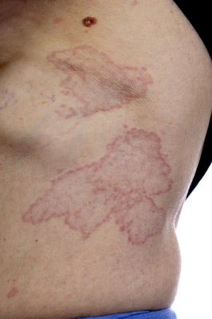
This image shows erythematous bumps arranged in a ring or circular pattern on the torso of a patient with diabetes.
This image shows erythematous bumps arranged in a ring or circular pattern on the torso of a patient with diabetes.
SCIENCE PHOTO LIBRARY
Necrobiosis lipoidica is associated with diabetes. Lesions most often appear on the legs and begin as erythematous papules that develop into atrophic, waxy, and yellow or brown lesions.
Necrobiosis lipoidica is associated with diabetes. Lesions most often appear on the legs and begin as erythematous papu
Photo provided by Thomas Habif, MD.
Acanthosis nigricans is skin thickening and pigmentation most typically developing in the axillae and nape of the neck (top); in people with dark skin, the skin may have a leathery appearance (bottom). It is most often a skin manifestation of impaired glucose tolerance, but it may reflect internal cancer, especially if onset is rapid and distribution is widespread.
Acanthosis nigricans is skin thickening and pigmentation most typically developing in the axillae and nape of the neck
Photos provided by Thomas Habif, MD.
This image shows erythematous bumps arranged in a ring or circular pattern on the torso of a patient with diabetes.
This image shows erythematous bumps arranged in a ring or circular pattern on the torso of a patient with diabetes.
SCIENCE PHOTO LIBRARY
Key Points
Microvascular complications of diabetes include kidney disease (nephropathy), retinopathy, and neuropathy. Neuropathy can lead to foot ulcers.
Macrovascular complications of diabetes involve atherosclerosis of the coronary, cerebral, and peripheral arteries.
Other important complications include heart failure and metabolic dysfunction-associated steatotic liver disease (primarily in type 2 diabetes).
Guideline-based screening for and treatment of complications is a critical part of diabetes care.
More Information
The following English-language resources may be useful. Please note that The Manual is not responsible for the content of these resources.
American Diabetes Association: Standards of Medical Care in Diabetes Diabetes Care. 48 (Supplement 1): S1-S336, 2025.
Drug Information for the Topic


