

Retinal detachment is separation of the neurosensory retina from the underlying retinal pigment epithelium. The most common cause is a retinal break (a tear or, less commonly, a hole—rhegmatogenous detachment). Symptoms are decreased peripheral or central vision, often described as a curtain or dark cloud coming across the field of vision. Associated symptoms can include painless vision disturbances, including flashing lights and increased floaters. Traction and serous retinal detachments (not involving retinal breaks) cause central or peripheral vision loss. Diagnosis is by funduscopy; ultrasonography may help determine the presence and type of retinal detachment if it cannot be seen with funduscopy. Immediate treatment is imperative if rhegmatogenous retinal detachment is acute and threatens central vision. Treatment of rhegmatogenous detachment may include sealing retinal breaks (by laser or cryotherapy), supporting the breaks with scleral buckling, pneumatic retinopexy, and/or vitrectomy.
Etiology of Retinal Detachment
There are 3 types of detachment: rhegmatogenous (which involves a retinal break), traction, and serous (exudative) detachment. Traction and serous retinal detachments do not involve a break and are called nonrhegmatogenous.
Rhegmatogenous detachment is the most common. Risk factors include the following (1):
Myopia
Previous cataract surgery
Ocular trauma
Lattice retinal degeneration
A family history of retinal detachment
Traction retinal detachment can be caused by vitreoretinal traction due to preretinal fibrous membranes as may occur in proliferative diabetic or sickle cell retinopathy.
Serous detachment results from transudation of fluid into the subretinal space. Causes include severe uveitis, especially in Vogt-Koyanagi-Harada disease, choroidal hemangiomas, and primary or metastatic choroidal cancers (see Cancers Affecting the Retina).
Etiology reference
1. Flaxel CJ, Adelman RA, Bailey ST, et al: Posterior vitreous detachment, retinal breaks, and lattice degeneration Preferred Practice Pattern®. Ophthalmology 127(1):P145-P181, 2020.
Symptoms and Signs of Retinal Detachment
Retinal detachment is painless. Early symptoms of rhegmatogenous detachment may include dark or irregular vitreous floaters (particularly a sudden increase), flashes of light (photopsias), and blurred vision. As detachment progresses, the patient often notices a curtain, veil, or grayness in the field of vision. If the macula is involved, central vision becomes poor. Patients may have simultaneous vitreous hemorrhage. Traction and serous (exudative) retinal detachments can cause blurriness of vision, but they may not cause any symptoms in the early stages.

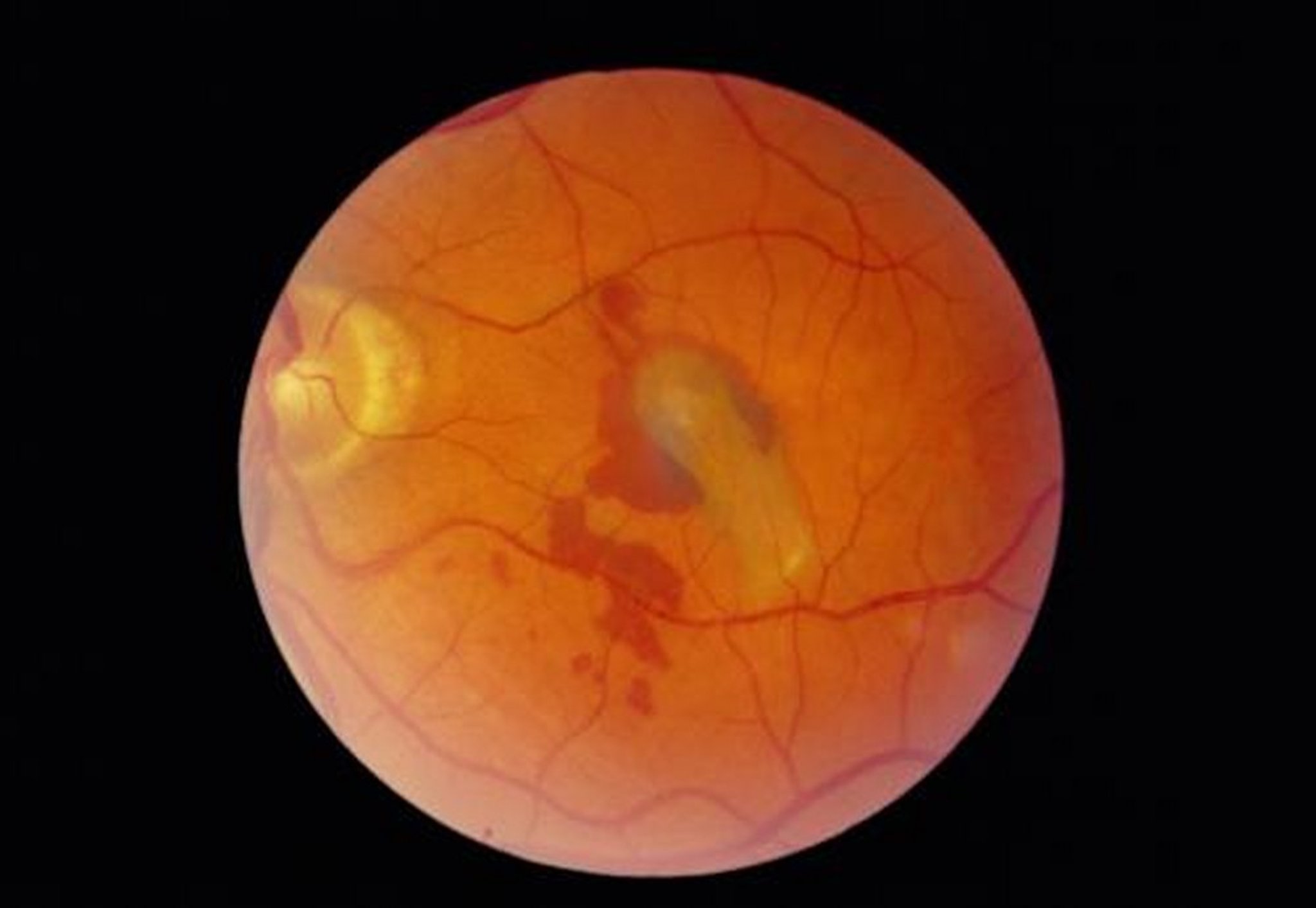
The area of retinal detachment in this photo is seen at the center. Symptoms include blurry vision, light flashes (photopsias), and floaters.
PAUL PARKER/SCIENCE PHOTO LIBRARY
Diagnosis of Retinal Detachment
Indirect ophthalmoscopy with pupillary dilation
Retinal detachment should be suspected in patients, particularly those at risk, who have any of the following:
Sudden increase or change in floaters
Photopsias
Curtain or veil across the visual field
Any sudden, unexplained loss of vision
Vitreous hemorrhage that obscures the retina
Indirect ophthalmoscopy shows the retinal detachment and can differentiate the subtypes of retinal detachment in nearly all cases. Direct funduscopy using a handheld ophthalmoscope can miss some retinal detachments, which may be peripheral. Peripheral fundus examination, using either indirect ophthalmoscopy with scleral depression, the slit lamp with the eye in extreme positions of gaze, or using a 3-mirror lens, should be done.
If vitreous hemorrhage (which may be due to a retinal tear), cataract, corneal opacification, or traumatic injury obscures the retina, retinal detachment should be suspected and B-scan ultrasonography should be done.
Treatment of Retinal Detachment
Sealing retinal breaks
Scleral buckling
Pneumatic retinopexy
Vitrectomy
Although often localized, retinal detachments due to retinal breaks can expand to involve the entire retina if they are not treated promptly (1). Any patient with a suspected or established retinal detachment should be examined urgently by an ophthalmologist.
Pearls & Pitfalls
|
Rhegmatogenous detachment is treated with one or more methods, depending on the cause and location of the lesion. These methods involve sealing the retinal breaks by laser or cryotherapy. In scleral buckling, a piece of silicone is placed on the sclera, which indents the sclera and pushes the retina inward, thereby relieving vitreous traction on the retina. During this procedure, fluid may be drained from the subretinal space. Pneumatic retinopexy (intravitreal injection of gas) and vitrectomy are other treatments. Retinal breaks without detachment can be sealed by laser photocoagulation or transconjunctival cryopexy. Nearly all rhegmatogenous detachments can be reattached surgically.
Nonrhegmatogenous detachments due to vitreoretinal traction may be treated by vitrectomy; exudative detachments due to uveitis may respond to systemic corticosteroids or systemic immunosuppression (eg, methotrexate, azathioprine, anti-tumor necrosis factor [TNF] medications). Alternatively, transudative detachments due to uveitis can be treated locally with a periocular corticosteroid injection, intravitreal corticosteroid injection, or an intravitreal dexamethasone implant. Primary and metastatic due to vitreoretinal traction may be treated by vitrectomy; exudative detachments due to uveitis may respond to systemic corticosteroids or systemic immunosuppression (eg, methotrexate, azathioprine, anti-tumor necrosis factor [TNF] medications). Alternatively, transudative detachments due to uveitis can be treated locally with a periocular corticosteroid injection, intravitreal corticosteroid injection, or an intravitreal dexamethasone implant. Primary and metastaticchoroidal cancers also require treatment. Choroidal hemangiomas may respond to localized photocoagulation or photodynamic therapy.
Treatment reference
1. Flaxel CJ, Adelman RA, Bailey ST, et al: Posterior vitreous detachment, retinal breaks, and lattice degeneration Preferred Practice Pattern®. Ophthalmology 127(1):P145-P181, 2020.
Key Points
Risk factors for rhegmatogenous retinal detachments include myopia, previous cataract surgery, ocular trauma, and lattice retinal degeneration.
All forms of retinal detachment eventually blur vision; early symptoms of rhegmatogenous detachment can include irregular vitreous floaters (particularly with a sudden increase) and flashes of light (photopsias).
Arrange for urgent indirect ophthalmoscopy by an ophthalmologist to diagnose retinal detachment if patients have a sudden increase or change in floaters; photopsias; a curtain or veil across the visual field; any sudden, unexplained loss of vision; or if vitreous hemorrhage obscures the retina.
Treat rhegmatogenous detachment by sealing retinal breaks (with laser or cryotherapy), by sealing the breaks with scleral buckling, with pneumatic retinopexy, and/or with vitrectomy.
Nonrhegmatogenous detachment due to vitreoretinal traction can be treated with vitrectomy and transudative detachments due to uveitis can be treated with local or systemic corticosteroids or systemic immunosuppressants.
Drug Information for the Topic


