Uterine prolapse is descent of the uterus toward or past the introitus. Apical prolapse is descent of the vaginal vault (vaginal cuff) after hysterectomy. Symptoms include vaginal pressure and fullness. Diagnosis is clinical. Treatment includes reduction, pessaries, and surgery.
In uterine prolapse, the cervix descends and can be visualized on pelvic examination or, with severe prolapse, it can be visualized beyond the introitus. In patients who have had a total hysterectomy (cervix and uterus removed), the vaginal vault may prolapse. With severe vaginal vault prolapse, the vagina can completely evert.
Symptoms and Signs of Uterine and Apical Prolapse
Symptoms tend to be minimal with mild uterine or apical prolapse. In more severe uterine or apical prolapse, vaginal or pelvic fullness, pressure, sexual dysfunction, and a sensation of organs falling out are common. The most common presenting symptom is a vaginal bulge, although this may be intermittent because spontaneous reduction can occur. Lower back pain may develop. Incomplete emptying of the bladder and constipation are possible.
If vaginal or cervical mucosa protrudes beyond the vagina, it may become dried, thickened, chronically inflamed, edematous, and ulcerated. Ulcers may be painful or bleed and need to be differentiated from vulvovaginal infection or dermatosis.

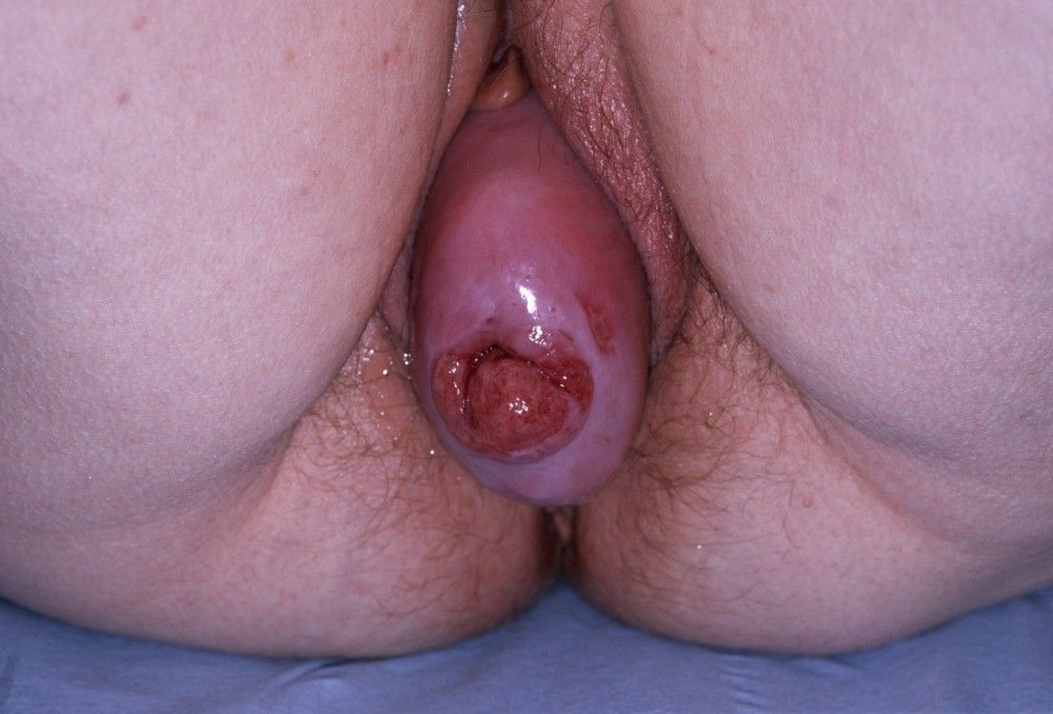
This photo shows stage III prolapse of the uterus.
DR P. MARAZZI/SCIENCE PHOTO LIBRARY
Cystocele or rectocele is usually also present.

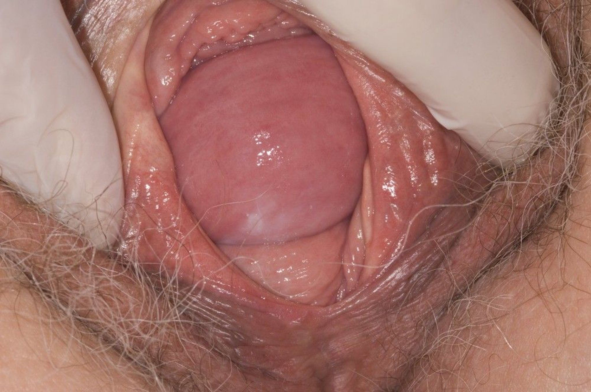
This photo shows prolapse of anterior and posterior vaginal walls.
JIM VARNEY/SCIENCE PHOTO LIBRARY
Urinary incontinence is also commonly present. Alternatively, the descending pelvic organs may intermittently obstruct urine flow, causing urinary retention and overflow incontinence and masking stress incontinence. Urinary frequency and urge incontinence may accompany uterine or vaginal prolapse.
Diagnosis of Uterine and Apical Prolapse
Pelvic examination at rest and while a patient strains
Diagnosis of uterine or vaginal apical prolapse is made with pelvic examination speculum and bimanual pelvic examination with the patient at rest and then with the patient straining. The Pelvic Organ Prolapse-Quantification (POP-Q) system is usually used to document severity.
Concomitant urinary incontinence or urinary retention requires evaluation.
Treatment of Uterine and Apical Prolapse
For mild symptomatic prolapse, pessaries
Surgical repair of supporting structures if necessary, usually combined with hysterectomy
Uterine prolapse
Asymptomatic prolapse does not require treatment. Symptomatic uterine prolapse past the introitus will not respond to pelvic floor exercises. A pessary is a good first-line treatment option. Surgery can be offered to patients who do not wish to or are unable to use a pessary (1).
Surgery for uterovaginal prolapse can be performed transvaginally or transabdominally using various techniques. Factors determining choice of technique include surgeon experience and patient preference. Techniques may include 1 or a combination of the following:
Hysterectomy
Surgical repair of the pelvic support structures (colporrhaphy)
Suspension of the top of the vagina (suturing of the upper vagina to a stable structure nearby)
Colpocleisis (closure of the vagina after removal of the uterus or with the uterus in place [Le Fort procedure])
Regardless of the surgical route, symptoms often recur, especially along the anterior vaginal wall.
Surgery is delayed until all ulcers, if present, have healed.
Vaginal apical prolapse
Vaginal apical prolapse is treated similarly to uterine prolapse.
If women are not good candidates for prolonged surgery (eg, if they have serious comorbidities) and are not planning future vaginal intercourse, they may be offered colpocleisis (suturing the vagina closed). Advantages of vaginal closure include short duration of surgery, low risk of perioperative morbidity, and very low risk of prolapse recurrence (2).
Urinary incontinence requires concurrent treatment.
Pessaries
Pessaries are devices that are inserted into the vagina to maintain normal anatomy and reduction of the prolapsed structures, resulting in improved objective and subjective benefit (3). Pessaries are typically made of silicone and vary in shape and size; some are inflatable.


To fit a pessary, a clinician should perform a POP-Q examination to guide pessary selection. The pessary is then inserted and the patient is asked to walk around to assess comfort, and empty her bladder to ensure the pessary does not obstruct the urethra. The clinician should provide instructions for removing, cleaning, and reinserting the pessary, and arrange a follow-up visit in 1 month. Proper size, fit, and position are important, because a pessary can cause vaginal ulceration with bleeding if it is does not fit correctly and vaginal discharge if it is not cleaned regularly (at least monthly if not more frequently).
The frequency of follow-up visits for a patient with a pessary is based on the patient's ability to independently manage the pessary. Those patients who are able to remove, clean, and replace the pessary themselves at least weekly may be seen annually, while those who are unable to provide self-care will need more frequent visits (4).
Treatment references
1. Pelvic Organ Prolapse. ACOG Practice Bulletin, Number 214. Obstet Gynecol. 2019 (reaffirmed 2024);134(5):e126-e142. doi:10.1097/AOG.0000000000003519
2. Berger AA, Bretschneider CE, Gregory WT, Sung V. Longitudinal Reoperation Risk After Apical Prolapse Procedures in Women Aged 65 Years and Older. Obstet Gynecol. 2024;143(3):411-418. doi:10.1097/AOG.0000000000005511
3. Sansone S, Sze C, Eidelberg A, et al. Role of Pessaries in the Treatment of Pelvic Organ Prolapse: A Systematic Review and Meta-analysis. Obstet Gynecol. 2022;140(4):613-622. doi:10.1097/AOG.0000000000004931
4. Vaginal Pessary Use and Management for Pelvic Organ Prolapse: Developed by the joint writing group of the American Urogynecologic Society and the Society of Urologic Nurses and Associates. Individual writing group members are noted in the Acknowledgments section. Urogynecology (Phila). 2023;29(1):5-20. doi:10.1097/SPV.0000000000001293
Key Points
Uterine prolapse is descent of the uterus toward or past the introitus. Vaginal apical prolapse is descent of the vaginal vault (vaginal cuff) after hysterectomy.
Symptoms include pelvic or vaginal fullness, pressure, and a sensation of organs falling out. Organs may bulge into the vaginal canal or through the vaginal opening (introitus), particularly during straining or coughing.
The descending pelvic organs may intermittently obstruct urine flow, causing urinary retention and overflow incontinence and masking stress incontinence.
Diagnose uterine or vaginal apical prolapse with speculum and bimanual pelvic examination with the patient at rest and then with the patient straining.
First-line conservative treatment is with pessaries; treat surgically if women prefer surgery to a pessary.