


Mycosis fungoides and Sézary syndrome are uncommon chronic T-cell non-Hodgkin lymphomas primarily affecting the skin and occasionally the lymph nodes.
(See also Overview of Lymphoma and Non-Hodgkin Lymphomas.)
The 2 main types of cutaneous T-cell lymphomas are
Mycosis fungoides
Sézary syndrome
They comprise less than 5% of all lymphoma cases.
Cutaneous T-cell lymphomas are insidious in onset. Patients may initially present with a chronic, pruritic rash that is difficult to diagnose even with biopsies. This prodrome may exist for several years until the diagnosis of cutaneous T-cell lymphoma is finally made.
The lesions of mycosis fungoides are characterized as patches, plaques, or tumor nodules; the nodules often ulcerate and become infected.
In Sézary syndrome, the skin is typically diffusely erythematous with cracking of the palms and soles. In patients with dark skin, erythema may be subtle.
Lymphadenopathy is usually mild to moderate. Symptoms are related mostly to the skin, with fevers, night sweats, and unintentional weight loss coming later in the disease course.

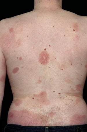
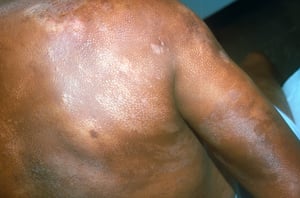
This photo shows erythematous patches on the back of a patient with mycosis fungoides.
This photo shows erythematous patches on the back of a patient with mycosis fungoides.
DR P. MARAZZI/SCIENCE PHOTO LIBRARY

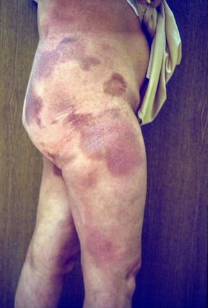
Photo courtesy of Karen McKoy, MD.
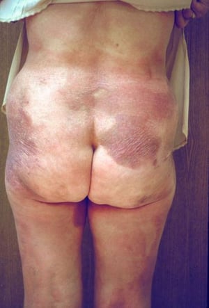
Photo courtesy of Karen McKoy, MD.

Mycosis fungoides (cutaneous T-cell lymphoma) can be difficult to distinguish from nonmalignant chronic dermatoses. Accurate diagnosis can be made only by a detailed clinical history (no progression of nonmalignant lesions over time) and biopsy with microscopic evaluation.
Mycosis fungoides (cutaneous T-cell lymphoma) can be difficult to distinguish from nonmalignant chronic dermatoses. Acc
(Courtesy of Libby Edwards, MD, Charlotte, NC.) By permission of the publisher. From Banks P, et al. In Atlas of Clinical Hematology. Edited by JO Armitage. Philadelphia, Current Medicine, 2004. Available at www.images.md.

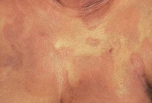
This photo shows hypopigmented and hyperpigmented patches.
This photo shows hypopigmented and hyperpigmented patches.
Photo courtesy of Karen McKoy, MD.

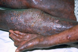
This photo shows patches and cutaneous tumors with ulceration.
This photo shows patches and cutaneous tumors with ulceration.
Photo courtesy of Karen McKoy, MD.

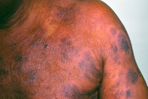
This photo shows erythematous and hyperpigmented plaques.
This photo shows erythematous and hyperpigmented plaques.
Photo courtesy of Karen McKoy, MD.

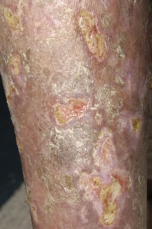
Ulcerations can sometimes develop in patients with mycosis fungoides.
Ulcerations can sometimes develop in patients with mycosis fungoides.
DR P. MARAZZI/SCIENCE PHOTO LIBRARY
This photo shows erythematous patches on the back of a patient with mycosis fungoides.
This photo shows erythematous patches on the back of a patient with mycosis fungoides.
DR P. MARAZZI/SCIENCE PHOTO LIBRARY
Photo courtesy of Karen McKoy, MD.
Photo courtesy of Karen McKoy, MD.
Mycosis fungoides (cutaneous T-cell lymphoma) can be difficult to distinguish from nonmalignant chronic dermatoses. Accurate diagnosis can be made only by a detailed clinical history (no progression of nonmalignant lesions over time) and biopsy with microscopic evaluation.
Mycosis fungoides (cutaneous T-cell lymphoma) can be difficult to distinguish from nonmalignant chronic dermatoses. Acc
(Courtesy of Libby Edwards, MD, Charlotte, NC.) By permission of the publisher. From Banks P, et al. In Atlas of Clinical Hematology. Edited by JO Armitage. Philadelphia, Current Medicine, 2004. Available at www.images.md.
This photo shows hypopigmented and hyperpigmented patches.
This photo shows hypopigmented and hyperpigmented patches.
Photo courtesy of Karen McKoy, MD.
This photo shows patches and cutaneous tumors with ulceration.
This photo shows patches and cutaneous tumors with ulceration.
Photo courtesy of Karen McKoy, MD.
This photo shows erythematous and hyperpigmented plaques.
This photo shows erythematous and hyperpigmented plaques.
Photo courtesy of Karen McKoy, MD.
Ulcerations can sometimes develop in patients with mycosis fungoides.
Ulcerations can sometimes develop in patients with mycosis fungoides.
DR P. MARAZZI/SCIENCE PHOTO LIBRARY
Diagnosis of Cutaneous T-cell Lymphomas
Skin biopsy
Peripheral blood smear and flow cytometry for circulating malignant T-cells (Sézary cells)
For staging, lymph node biopsy and CT of chest, abdomen, and pelvis or FDG-PET (fluorodeoxyglucose-positron emission tomography)
Diagnosis is based on skin biopsy, but histology may be equivocal early in the course because of insufficient quantities of lymphoma cells. The malignant cells are mature CD4+ T cells that may have lost common T-cell markers such as CD7.
Characteristic Pautrier microabscesses may be present in the epidermis on skin punch biopsies. In some cases, a leukemic phase is characterized by the appearance of malignant T cells with serpentine nuclei in the peripheral blood (Sézary cells). These malignant T cells in the blood can be detected on a routine Wright-stained smear or by flow cytometry.
After diagnosis, stage is determined to guide therapy. The commonly used ISCL/EORTC (International Society of Cutaneous Lymphomas/European Organization of Research and Treatment of Cancer) staging system incorporates physical examination findings, histopathology results, and results of imaging tests (1, 2).
Diagnosis references
1. Olsen EA: Evaluation, diagnosis, and staging of cutaneous lymphoma. Dermatol Clin 33(4):643–654, 2015. doi: 10.1016/j.det.2015.06.001
2. Willemze R, Cerroni L, Kempf W, et al: The 2018 update of the WHO-EORTC classification for primary cutaneous lymphomas. Blood133 (16):1703–1714, 2019. doi: 10.1182/blood-2018-11-881268
Treatment of Cutaneous T-cell Lymphomas
Radiation therapy, topical chemotherapy, phototherapy, or topical corticosteroids
Sometimes systemic chemotherapy
Treatment of Sézary syndrome and mycosis fungoides is similar. Treatments can be divided into
Skin-directed (topical chemotherapy, phototherapy, retinoids, radiation therapy)
Systemic therapies—traditional chemotherapy and other targeted therapies (eg, histone deacetylase [HDAC] inhibitors, brentuximab vedotin)
Patients are managed by a team of dermatologists, radiation oncologists, and hematology/oncology specialists.
Skin-directed therapies are used first and are often effective for years. As lesions become more resistant, or in patients with Sézary syndrome, systemic therapies are used. Lesions may become infected, and the clinician must always consider an infectious cause for any skin flare.
Electron beam radiation therapy, in which most of the energy is absorbed in the first 5 to 10 mm of tissue, and topical nitrogen mustard have proved highly effective. Plaques may also be treated with sunlight and topical corticosteroids.
Systemic treatment with alkylating agents and folic acid antagonists produces transient tumor regression, but systemic treatment is primarily used when other therapies have failed, after relapse, or in patients with documented extranodal or extracutaneous disease. HDAC inhibitors (vorinostat, romidepsin, panobinostat, belinostat), which induce cancer cell cycle arrest, differentiation, and cell death can be given IV or orally. Extracorporeal photophoresis with a chemosensitive drug has shown modest success. Brentuximab vedotin is an antibody-drug conjugate medication with activity against Hodgkin lymphoma and cutaneous T cell lymphoma (1).
Treatment reference
1. Prince HM, Kim YH, Horwitz SM, et al: Brentuximab vedotin or physician's choice in CD30-positive cutaneous T-cell lymphoma (ALCANZA): an international, open-label, randomised, phase 3, multicentre trial. Lancet 2017 Aug 5;390(10094):555-566. doi: 10.1016/S0140-6736(17)31266-7
Prognosis for Cutaneous T-cell Lymphomas
Most patients are > 50 years at diagnosis. Survival rates vary markedly depending on stage at diagnosis. Patients who receive treatment for stage IA disease have a life expectancy analogous to that of similar people without mycosis fungoides. In one study, patients with stage II disease (10% or more skin involved) had a relative survival of 67% at 10 years, compared with 41% for those with stage IV disease (generalized erythroderma) (1).
Prognosis reference
1. Zackheim HS, Amin S, Kashani-Sabet M, McMillan A: Prognosis in cutaneous T-cell lymphoma by skin stage: long-term survival in 489 patients. J Am Acad Dermatol 1999 Mar;40(3):418-25. doi: 10.1016/s0190-9622(99)70491-3
More Information
The following English language resource provides information for clinicians and support and information for patients. THE MANUAL is not responsible for the content of this resource.
Leukemia & Lymphoma Society: Resources for Healthcare Professionals: provides educational resources for health care practitioners as well as information for patient referrals
Drug Information for the Topic


