


Impetigo is a superficial skin infection with crusting or bullae caused by streptococci, staphylococci, or both. Ecthyma is an ulcerative form of impetigo. Diagnosis is clinical. Treatment is with topical and sometimes oral antibiotics.
(See also Overview of Bacterial Skin Infections.)
Impetigo is a common, superficial bacterial infection of the epidermis usually caused by Staphylococcus aureus and/or Streptococcus pyogenes. It is highly contagious. No predisposing lesion is identified in most patients, but impetigo may occur after any type of break in the skin.
Impetigo may be nonbullous (70% of cases) or bullous (30% of cases) (1). S. aureus is the predominant cause of nonbullous impetigo and the cause of all bullous impetigo. Bullae are caused by exfoliative toxins produced by staphylococci. Methicillin-resistant S. aureus (MRSA) has been isolated in many cases of impetigo.
Ecthyma is an ulcerative, deep form of impetigo that penetrates through the epidermis into the dermis (see figure ). It commonly develops on the lower extremities and is associated with scarring.
General risk factors for impetigo and ecthyma are a moist environment, poor hygiene, and/or chronic nasopharyngeal carriage of staphylococci or streptococci. Immunocompromise and a disrupted cutaneous barrier, such as in patients with underlying atopic dermatitis or extensive xerosis, may contribute to the development of impetigo.
General reference
1. Hartman-Adams H, Banvard C, Juckett G. Impetigo: diagnosis and treatment. Am Fam Physician. 2014;90(4):229-235.
Symptoms and Signs of Impetigo and Ecthyma
Nonbullous impetigo typically manifests as clusters of vesicles or pustules that rupture and develop a honey-colored crust (exudate from the lesion base) over the lesions. Smaller lesions may coalesce into larger crusted plaques.
Bullous impetigo is similar except that vesicles typically enlarge rapidly to form bullae. The bullae burst and expose larger bases, which become covered with honey-colored varnish or crust.

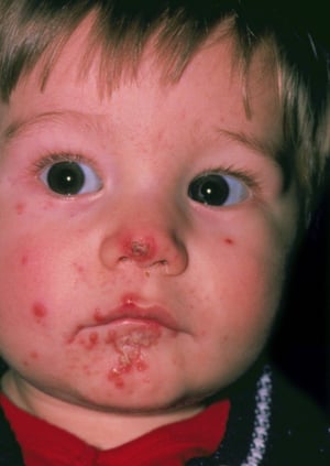
This photo shows clusters of vesicles and pustules with developing honey-colored crust on the nose.
This photo shows clusters of vesicles and pustules with developing honey-colored crust on the nose.
DR P. MARAZZI/SCIENCE PHOTO LIBRARY

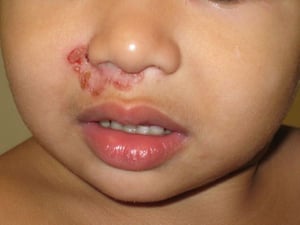
This image shows crusted perinasal plaques of impetigo on the face of a child.
This image shows crusted perinasal plaques of impetigo on the face of a child.
Image courtesy of Wingfield Rehmus, MD, MPH.

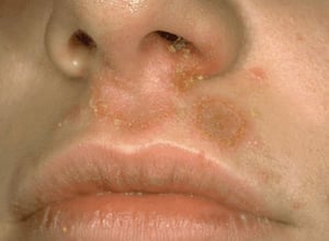
This photo shows clusters of vesicles or pustules that have ruptured and developed a honey-colored crust.
This photo shows clusters of vesicles or pustules that have ruptured and developed a honey-colored crust.
Image provided by Thomas Habif, MD.

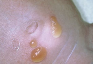
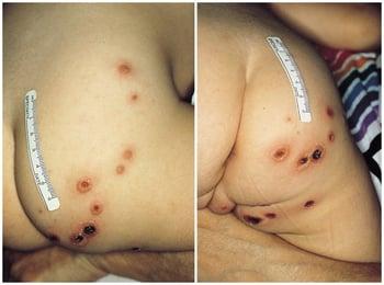
This photo shows bullous impetigo on an infant's abdomen, which has manifested as small, coalesced pustules that eventually formed into yellow bullae.
This photo shows bullous impetigo on an infant's abdomen, which has manifested as small, coalesced pustules that eventu
SCIENCE PHOTO LIBRARY
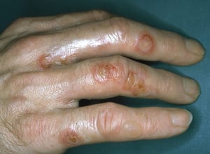
This photo shows bullous impetigo on a person's fingers. The bullae burst and expose larger bases, which become covered with honey-colored varnish or crust.
This photo shows bullous impetigo on a person's fingers. The bullae burst and expose larger bases, which become covered
Image provided by Thomas Habif, MD.
This photo shows clusters of vesicles and pustules with developing honey-colored crust on the nose.
This photo shows clusters of vesicles and pustules with developing honey-colored crust on the nose.
DR P. MARAZZI/SCIENCE PHOTO LIBRARY
This image shows crusted perinasal plaques of impetigo on the face of a child.
This image shows crusted perinasal plaques of impetigo on the face of a child.
Image courtesy of Wingfield Rehmus, MD, MPH.
This photo shows clusters of vesicles or pustules that have ruptured and developed a honey-colored crust.
This photo shows clusters of vesicles or pustules that have ruptured and developed a honey-colored crust.
Image provided by Thomas Habif, MD.
This photo shows bullous impetigo on an infant's abdomen, which has manifested as small, coalesced pustules that eventually formed into yellow bullae.
This photo shows bullous impetigo on an infant's abdomen, which has manifested as small, coalesced pustules that eventu
SCIENCE PHOTO LIBRARY
This photo shows bullous impetigo on a person's fingers. The bullae burst and expose larger bases, which become covered with honey-colored varnish or crust.
This photo shows bullous impetigo on a person's fingers. The bullae burst and expose larger bases, which become covered
Image provided by Thomas Habif, MD.
Ecthyma is a form of impetigo characterized by small, purulent, shallow, punched-out ulcers with thick, brown-black crusts and surrounding erythema.

This photo shows small, pustular, shallow, punched-out ulcers with thick, brown-black crusts and surrounding erythema.
Impetigo and ecthyma can cause mild pain or discomfort. Pruritus is common; scratching may spread infection, inoculating adjacent and nonadjacent skin.
Diagnosis of Impetigo and Ecthyma
Clinical evaluation
Gram stain, culture, and antibiotic sensitivity testing of purulent material
The diagnosis of impetigo and ecthyma is typically based on their characteristic appearance.
Gram stain and culture of lesion exudate are recommended to identify S. aureus (including MRSA) or beta-hemolytic streptococcal species in impetigo and ecthyma (1). However, initiating empiric treatment is reasonable for typical cases.
Diagnosis reference
1. Stevens DL, Bisno AL, Chambers HF, et al. Practice guidelines for the diagnosis and management of skin and soft tissue infections: 2014 update by the Infectious Diseases Society of America. Clin Infect Dis. 2014;59(2):e10-e52. doi:10.1093/cid/ciu444
Treatment of Impetigo and Ecthyma
Topical mupirocin, retapamulin, fusidic acid, or ozenoxacin
Sometimes oral antibiotics (eg, ecthyma; extensive, recurrent impetigo; immunocompromise)
Bullous and nonbullous impetigo can be treated with either topical or oral antibiotics (1). However, oral antibiotics are preferred for patients with numerous lesions and for immunocompromised patients at risk of complications, when topical therapy is impractical (eg, extensive disease), and during outbreaks affecting several people to help reduce transmission. Ecthyma requires treatment with oral antibiotics. Any affected areas should be washed gently with soap and water several times a day to remove any crusts.
The treatment for localized impetigo is a topical antistaphylococcal antibiotic for 5 to 7 days. Mupirocin is most commonly used in the United States. Alternatives include ozenoxacin, retapamulin, and fusidic acid; the latter two are not available in the United States. Topical treatment (eg, mupirocin, fusidic acid) is as effective or more effective than oral treatment for localized impetigo (2).
Oral antibiotics include dicloxacillin or cephalexin for 7 days. In penicillin-allergic patients, clindamycin 300 mg 4 times a day or erythromycin 250 mg 4 times a day may be used, but resistance to both antibiotics is an increasing problem.
The use of initial empiric therapy against MRSA is not typically advised unless there is compelling clinical evidence (eg, contact with a person who has a documented case, exposure to a documented outbreak, severe and/or extensive disease). Treatment of MRSA should be directed by culture and sensitivity test results; clindamycin, sulfamethoxazole/trimethoprim, and doxycycline are reasonable empiric oral antibiotic options.
Other therapy includes restoring a normal cutaneous barrier in patients with underlying atopic dermatitis or extensive xerosis using topical emollients and glucocorticoids if warranted. Decolonization in chronic staphylococcal nasal carriers may be effectively achieved by administering topical antibiotics (mupirocin), which causes a significant reduction in the rate of S. aureus infection (3); however, whether such decolonization lowers rates of recurrent impetigo specifically is less clear.
Treatment references
1. Stevens DL, Bisno AL, Chambers HF, et al. Practice guidelines for the diagnosis and management of skin and soft tissue infections: 2014 update by the Infectious Diseases Society of America. Clin Infect Dis. 2014;59(2):e10-e52. doi:10.1093/cid/ciu444
2. Koning S, van der Sande R, Verhagen AP, et al. Interventions for impetigo. Cochrane Database Syst Rev. 2012;1(1):CD003261. Published 2012 Jan 18. doi:10.1002/14651858.CD003261.pub3
3. van Rijen M, Bonten M, Wenzel R, Kluytmans J. Mupirocin ointment for preventing Staphylococcus aureus infections in nasal carriers. Cochrane Database Syst Rev. 2008;2008(4):CD006216. Published 2008 Oct 8. doi:10.1002/14651858.CD006216.pub2
Prognosis for Impetigo and Ecthyma
Prompt recovery usually follows timely treatment. Treatment delays can cause cellulitis, lymphangitis, furunculosis, and hyperpigmentation or hypopigmentation with or without scarring.
Children aged 2 to 4 years are at risk of acute postinfectious glomerulonephritis if nephritogenic strains of group A streptococci are involved (types 49, 42, 2, 55, 57, and 60); postinfectious glomerulonephritis seems to be more common among people in resource-limited regions and in the southern United States compared to other regions (1, 2). It is unlikely that treatment with antibiotics prevents postinfectious glomerulonephritis.
Prognosis references
1. Rodriguez-Iturbe B, Haas M. Post-Streptococcus pyogenes Glomerulonephritis. In: Ferretti JJ, Stevens DL, Fischetti VA, eds. Streptococcus pyogenes: Basic Biology to Clinical Manifestations. 2nd ed. Oklahoma City (OK): University of Oklahoma Health Sciences Center; September 9, 2022.
2. Satoskar AA, Parikh SV, Nadasdy T. Epidemiology, pathogenesis, treatment and outcomes of infection-associated glomerulonephritis. Nat Rev Nephrol. 2020;16(1):32-50. doi:10.1038/s41581-019-0178-8
Key Points
Staphylococcus aureus causes most nonbullous impetigo and all bullous impetigo.
Honey-colored crusted lesions are characteristic of bullous and nonbullous impetigo.
Culturing lesions (to identify methicillin-resistant S. aureus [MRSA]) and the nose (to identify a potential nasal reservoir) is generally recommended.
Treat most cases with topical antibiotics; systemic antibiotic therapy is required for severe, persistent, or recurrent impetigo; outbreaks; immunocompromised patients; and ecthyma.
Drug Information for the Topic


