


Tularemia is a febrile disease caused by the gram-negative bacterium Francisella tularensis; it may resemble typhoid fever. Symptoms are a primary local ulcerative lesion, regional lymphadenopathy, profound systemic symptoms, and, occasionally, atypical pneumonia. Diagnosis is primarily epidemiologic and clinical and supported by serologic tests. Treatment is with streptomycin, gentamicin, chloramphenicol, ciprofloxacin, or doxycycline.
There are 7 clinical syndromes associated with tularemia (see table ); clinical manifestations vary by the type of exposure to the organism.
The causative organism, F. tularensis, is a small, pleomorphic, nonmotile, nonsporulating, aerobic, gram-negative bacillus. It enters the body by
Ingestion of contaminated food or water
Bite of an infected arthropod vector (ticks, deer flies, fleas)
Inhalation
Direct contact with infected tissues or material
Tularemia does not spread from person to person.
Types of Tularemia
Type | Frequency | Comment |
|---|---|---|
Ulceroglandular | Most common | Primary lesions on the hands or fingers with regional lymphadenitis |
Typhoidal* | Common | Systemic illness without indication of the site of inoculation or localized infection |
Oculoglandular | Uncommon | Conjunctivitis with inflammation of ipsilateral preauricular, submandibular, or cervical lymph nodes, probably caused by inoculation of an eye from an infected finger or hand |
Glandular | Rare | Regional lymphadenitis but no primary lesion and often cervical adenopathy, suggesting oral ingestion of bacteria |
Pneumonic† | Uncommon | Infiltrates with asymmetric hilar adenopathy, with or without bloody pleural effusion |
Oropharyngeal | Rare | Sore throat and cervical adenopathy due to ingestion of contaminated food or water |
Septicemic* | Rare | Severe systemic illness with hypotension, acute respiratory distress syndrome (ARDS), disseminated intravascular coagulation, and multiorgan dysfunction |
* Hematogenous spread to various organs (eg, lungs, bone, pericardium, peritoneum, heart valves, meninges) may also occur. | ||
† Tularemic pneumonia may be primary or may complicate any form of tularemia. | ||
The organism can penetrate apparently unbroken skin but may actually enter through microlesions.
Two types of F. tularensis cause most cases of tularemia:
Type A: This type is a more virulent serotype for humans; it usually occurs in rabbits, hares, and rodents in the United States and Canada.
Type B: This type usually causes a mild ulceroglandular infection and occurs in rodents and in aquatic environments throughout the Northern Hemisphere, including North America, Europe, and Asia.
Hunters, butchers, farmers, and fur handlers are most commonly infected. In winter months, most cases result from contact (especially during skinning) with infected wild rabbits and hares. In summer months, infection usually follows handling of other infected animals or birds or bites of infected ticks or other arthropods. Rarely, cases result from eating undercooked infected meat, drinking contaminated water, or mowing fields in endemic areas. In the Western United States, ticks, deer flies, horse flies, and direct contact with infected animals are other sources of infection. Human-to-human transmission has not been reported. Laboratory workers are at particular risk because infection is readily acquired during normal handling of infected specimens.
Tularemia is considered a possible agent of bioterrorism because inhalation of as few as 10 organisms in the form of an aerosol can cause severe pneumonia.
In disseminated cases, characteristic focal necrotic lesions in various stages of evolution are scattered throughout the body. They are 1 mm to 8 cm in diameter and whitish yellow; they are seen externally as the primary lesions on the fingers, eyes, or mouth and commonly occur in lymph nodes, spleen, liver, kidneys, and lungs. In pneumonia, necrotic foci occur in the lungs. Although severe systemic toxicity may occur, no toxins have been demonstrated.
Symptoms and Signs of Tularemia
Onset of tularemia is sudden, occurring 1 to 10 (usually 2 to 4) days after exposure, with headache, chills, nausea, vomiting, fever of 39.5 to 40° C, and severe prostration. Extreme weakness, recurring chills, and drenching sweats develop. Clinical manifestations depend to some extent on the type of exposure (see table ).
Within 24 to 48 hours, an inflamed papule appears at the site of exposure (finger, arm, eye, roof of the mouth), except in glandular or typhoidal tularemia. The papule rapidly becomes pustular and ulcerates, producing a clean ulcer crater with a scanty, thin, colorless exudate. Ulcers are usually single on the extremities but multiple in the mouth or eyes. Usually, only 1 eye is affected.
Regional lymph nodes enlarge and may suppurate and drain profusely.
A typhoid-like state frequently develops by the fifth day, and the patient may develop atypical pneumonia, sometimes accompanied by delirium.

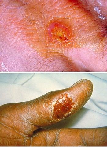
In ulceroglandular tularemia, an inflamed papule appears on the finger or hand (top); it then rapidly becomes pustular and ulcerates, producing a clean ulcer crater with scanty, thin, colorless exudates (bottom).
Pneumonic tularemia
Pneumonic tularemia can occur after inhalation or by hematogenous spread from another type of tularemia; it develops in 10 to 15% of ulceroglandular tularemia cases and in about 50% of typhoidal tularemia cases.
Although signs of consolidation are frequently present, reduced breath sounds and occasional rales may be the only physical findings in tularemic pneumonia. A dry, nonproductive cough is associated with a retrosternal burning sensation. A nonspecific roseola-like rash may appear at any stage of the disease. Splenomegaly and perisplenitis may occur.
In untreated cases, temperature remains elevated for 3 to 4 weeks and resolves gradually.
Mediastinitis, lung abscess, and meningitis are rare complications.
Diagnosis of Tularemia
Cultures
Acute and convalescent serologic and polymerase chain reaction (PCR) testing
Diagnosis of tularemia is suspected based on a history of contact with rabbits, hares, or rodents or exposure to arthropod vectors, the sudden onset of symptoms, and the characteristic primary lesion and regional lymphadenopathy.
Patients should have cultures of blood and relevant clinical material (eg, sputum, lesions); routine cultures may be negative, and the laboratory should be notified that tularemia is suspected so that appropriate media can be used and appropriate safety precautions are ensured.
Acute and convalescent antibody titers should be done 2 weeks apart. A 4-fold rise or a single acute tube agglutination titer > 1:160 is diagnostic. The serum of patients with brucellosis may cross-react to F. tularensis antigens but usually in much lower titers. Fluorescent antibody or immunohistochemical staining is used by some laboratories. PCR testing can provide a rapid diagnosis.
Leukocytosis is common, but the white blood cell count may be normal with an increase only in the proportion of polymorphonuclear leukocytes.
Because this organism is highly infectious, samples and culture media from patients suspected of having tularemia should be handled with extreme caution and, if possible, processed by a high-level biosafety containment-equipped laboratory with a level 3 rating.
Treatment of Tularemia
Streptomycin or gentamicin (plus ciprofloxacin, doxycycline, or chloramphenicol for meningitis)
The preferred antibiotic for moderate to severe disease is
Streptomycin 10 mg/kg IM every 12 hours for 7 to 10 days for adults (not to exceed 2 g/day) and 15 to 20 mg/kg IM every 12 hours for 7 to 10 days for children (not to exceed 2 g/day)
Gentamicin 1.7 mg/kg IM or IV every 8 hours for 7 to 10 days for adults and 1.7 mg/kg every 8 hours or 2.5 mg/kg every 12 hours for 7 to 10 days for children is often used as first-line therapy (instead of streptomycin) for moderate to severe disease because of its safety (less ototoxicity) and availability.
Alternatives to streptomycin include the following:
Ciprofloxacin 500 mg orally every 12 hours for 10 to 14 days (for mild to moderate disease)
Doxycycline 100 mg orally every 12 hours for 14 to 21 days (for mild to moderate disease)
Chloramphenicol 15 to 25 mg/kg IV every 6 hours for 14 to 21 days (used only for meningitis because there are more effective and safer alternatives)
Adults who have evidence of meningitis are given streptomycin or gentamicin in the dosage above for moderate to severe disease plus ciprofloxacin 400 mg IV every 8 to 12 hours, doxycycline 100 mg IV every 12 hours, or chloramphenicol 15 to 25 mg/kg IV every 6 hours (not to exceed 4 g/day) for 14 to 21 days. Children who have evidence of meningitis are given gentamicin in the dosage above for moderate to severe disease plus ciprofloxacin 6.7 to 10 to 30 mg/kg every 8 hours or 10 to 15 mg/kg every 12 hours (not to exceed 1.2 g/day) or doxycycline 1.1 to 2.2 mg/kg every 12 hours (not to exceed 200 mg/day) for 14 to 21 days.
In a mass casualty setting if parenteral treatment is not feasible, oral doxycycline or ciprofloxacin may be used for adults and children. However, relapses occasionally occur with all of these antibiotics, and they may not prevent node suppuration.
Continuous wet saline dressings are beneficial for primary skin lesions and may diminish the severity of lymphangitis and lymphadenitis. Surgical drainage of large abscesses is rarely necessary unless therapy is delayed.
In ocular tularemia, applying warm saline compresses and using dark glasses give some relief. In severe cases, 2% homatropine 1 to 2 drops every 4 hours may relieve symptoms.
Intense headache usually responds to oral analgesics.
Prognosis for Tularemia
Case fatality is almost nil in treated cases and approximately 6% in untreated cases of ulceroglandular tularemia. Case fatality rates are higher for type A infection and for typhoidal, septicemic, and pneumonic tularemia; they are as high as 50% for untreated cases (1). Death usually results from overwhelming infection, pneumonia, meningitis, or peritonitis.
Relapses can occur in inadequately treated cases.
One attack confers immunity.
Prognosis reference
1. Nigrovic LE, Wingerter SL. Tularemia. Infect Dis Clin North Am. 2008;22(3):489-ix. doi:10.1016/j.idc.2008.03.004
Prevention of Tularemia
When entering endemic areas, people should use tick-proof clothing and repellents. A thorough search for ticks should be done after leaving tick-infested areas. Ticks should be removed at once (see sidebar ).
When handling rabbits, hares, and rodents, especially in endemic areas, people should wear protective clothing, including rubber gloves and face masks, because organisms may be present in the animal and in tick feces on the animal’s fur. Wild birds and game must be thoroughly cooked before eating.
Water that may be contaminated must be disinfected before use.
No vaccine is currently available, although one is currently under review by the U.S. Food and Drug Administration (FDA) (1).
Antibiotic prophylaxis with 14 days of oral doxycycline or ciprofloxacin is recommended after high-risk exposure (eg, a laboratory accident, terrorism event).
Prevention reference
1. Centers for Disease Control and Prevention (CDC): Tularemia: Prevention. Accessed March 25, 2024.
Key Points
F. tularensis is a highly infectious organism; in the United States and Canada, the main reservoirs are wild rabbits, hares, and rodents.
Tularemia can be acquired in many ways, including direct contact with infected animals (particularly rabbits and hares) or birds, bites of infected arthropods, inadvertent contact with laboratory specimens, or, rarely, inhalation of an infectious aerosol or ingestion of contaminated meat or water.
Patients have a fever of 39.5 to 40° C and other constitutional symptoms (eg, headache, chills, nausea, vomiting, severe prostration) along with specific manifestations related to the organ affected; skin lesions and/or lymphadenitis are most common, and pneumonia may occur.
Diagnose using cultures of blood and relevant clinical material; acute and convalescent antibody titers, polymerase chain reaction (PCR) testing, and certain staining techniques may also be helpful.
Treat with streptomycin or gentamicin (plus ciprofloxacin, doxycycline, or chloramphenicol for meningitis).
Take appropriate precautions in endemic areas, including tick avoidance strategies, use of protective gear while handling rabbits, hares, and rodents, and thorough cooking of wild birds and game.
Drug Information for the Topic


