


Nocardiosis is an acute or chronic, often disseminated, suppurative or granulomatous infection caused by various aerobic soil saprophytes of the gram-positive bacilli genus Nocardia. Pneumonia is typical, but skin and central nervous system infections are common. Diagnosis is by culture and special stains. Treatment is usually with sulfonamides.
Nocardia are obligate aerobic, actinomycetous, partially acid-fast, beaded, branching, gram-positive bacilli. The genus Nocardia has many species.
N. asteroides usually causes pulmonary and disseminated infection.
N. brasiliensis most commonly causes skin infection, particularly in tropical climates.
Infection occurs via inhalation or by direct inoculation of the skin.
Other Nocardia species sometimes cause localized or, occasionally, systemic infections.
Nocardiosis occurs worldwide in all age groups, but its incidence is higher in older adults and immunocompromised patients. Person-to-person and animal-to-human transmission are not known to occur. In the United States, there are approximately 500 to 1,000 new cases of nocardiosis infection annually (1).
Nocardia is found in standing water, decaying plants, and soil. Infection is via inhalation of dust that contains the bacteria or by direct inoculation of the skin when contaminated soil or water enters through a cut or scrape.
Risk factors
Predisposing risk factors for nocardiosis include:
Lymphoreticular cancers
Organ transplantation
High-dose glucocorticoid or other immunosuppressive therapy
Underlying pulmonary disease
Diabetes mellitus
Alcohol use disorder
However, up to 40% of patients with nocardiosis have no identified immunocompromise (1).
Nocardiosis is also an opportunistic infection in patients with advanced HIV infection.
General reference
1. Centers for Disease Control and Prevention: Nocardiosis: Clinical Overview of Nocardiosis. January 31, 2025. Accessed July 17, 2025.
Symptoms and Signs of Nocardiosis
Nocardiosis usually begins as a subacute pulmonary infection that resembles actinomycosis, but Nocardia are more likely to disseminate locally or hematogenously than Actinomyces. Dissemination with abscess formation may involve any organ but most commonly affects the brain, skin, kidneys, bone, or muscle.

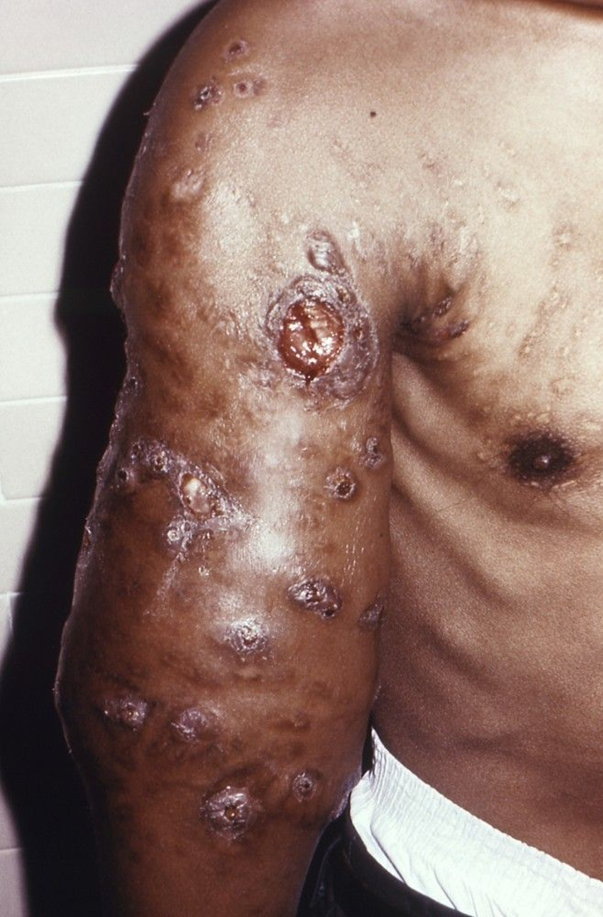
This photo shows nocardiosis infection of the upper extremity with swelling and multiple subcutaneous nodules.
Image courtesy of Dr. Libero Ajello via the Public Health Image Library of the Centers for Disease Control and Prevention.
The most common symptoms of pulmonary involvement—cough, fever, chills, chest pain, weakness, anorexia, and weight loss—are nonspecific and may resemble those of tuberculosis or suppurative pneumonia. Pleural effusion may also occur.
Metastatic brain abscesses, occurring in approximately 37% of cases, usually cause severe headaches and focal neurologic abnormalities (1). Infection may be acute, subacute, or chronic.
Primary cutaneous nocardiosis usually occurs in immunocompetent patients 1 to 3 weeks after infection, sometimes as a primary local inoculation. Skin or subcutaneous abscesses occur frequently. They may appear as:
Firm cellulitis
Lymphocutaneous syndrome
An actinomycetoma
Cutaneous nocardiosis can present as an erythematous, tender, warm, swollen area similar to cellulitis, but it may also involve firm nodules or abscesses, particularly around a site of injury where the bacteria entered the skin (2).
The lymphocutaneous syndrome consists of a primary pyoderma lesion and lymphatic nodules resembling sporotrichosis.
An actinomycetoma due to nocardiosis begins as a nodule, suppurates, spreads along fascial planes, and drains through chronic fistulas.

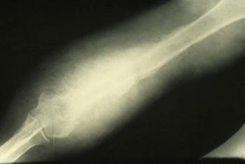
Actinomycetoma due to nocardiosis is a slowly progressive, destructive infection of the cutaneous and subcutaneous tissues, fascia, and, as seen here, bone. Actinomycetoma starts as a nodule, suppurates, spreads along fascial plains, and drains through chronic fistulas.
Symptoms and signs references
1. Averbuch D, De Greef J, Duréault A, et al. Nocardia Infections in Hematopoietic Cell Transplant Recipients: A Multicenter International Retrospective Study of the Infectious Diseases Working Party of the European Society for Blood and Marrow Transplantation [published correction appears in Clin Infect Dis. 2025 Feb 5;80(1):239. doi: 10.1093/cid/ciae606.]. Clin Infect Dis. 2022;75(1):88-97. doi:10.1093/cid/ciab866
2. Tariq EF, Anwar MM, Khan UA. Primary Cutaneous Nocardiosis: A Rare Presentation of Nocardiosis. Cureus. 2019;11(10):e5860. doi:10.7759/cureus.5860
Diagnosis of Nocardiosis
Microscopic examination or culture
Acid-fast staining
Sometimes molecular nucleic acid tests (multilocus sequence analysis)
Diagnosis of nocardiosis is based on identification of Nocardia species in tissue or in culture of samples from localized lesions identified by physical examination, radiographs, or other imaging studies. Clumps of beaded, right-angled, branching filaments of gram-positive bacteria (which may be weakly acid-fast using the modified Kinyoun technique) are often seen. Both blood and tissue cultures may take up to 2 weeks to grow. Molecular nucleic acid tests such as multilocus sequence analysis may be used (1). Rarely,matrix-assisted laser desorption/ionization time-of-flight mass spectrometry (MALDI-TOF MS) can be useful in accurately identifying Nocardia species (2).
Diagnosis references
1. Xiao M, Pang L, Chen SC, et al. Accurate Identification of Common Pathogenic Nocardia Species: Evaluation of a Multilocus Sequence Analysis Platform and Matrix-Assisted Laser Desorption Ionization-Time of Flight Mass Spectrometry. PLoS One. 2016;11(1):e0147487. Published 2016 Jan 25. doi:10.1371/journal.pone.0147487
2. Liu Y, Wu SY, Deng J, et al. Application of MALDI-TOF mass spectrometry for identification of Nocardia species. BMC Microbiol. 2024;24(1):358. Published 2024 Sep 20. doi:10.1186/s12866-024-03483-2
Treatment of Nocardiosis
Trimethoprim/sulfamethoxazole
Sometimes amikacin, imipenem, or meropenem
Sulfamethoxazole/trimethoprim orally every 6 to 12 hours (dependent on the extent of the disease) is the treatment of choice (1). The duration of treatment depends on the type of infection and predisposing risk factors of the infected patient. In immunocompromised patients and patients with disseminated disease, trimethoprim/sulfamethoxazole should be used with amikacin, imipenem, or meropenem pending species identification and susceptibility testing results.
When sulfonamide hypersensitivity, sulfamethoxazole/trimethoprim resistance, or refractory infection is present, amikacin, a tetracycline (particularly minocycline), imipenem/cilastatin, meropenem, ceftriaxone, cefotaxime, extended-spectrum fluoroquinolones (eg, moxifloxacin), or dapsone can be used. Tigecycline may be an effective alternative. Linezolid in combination with another agent may be appropriate in certain cases (eg, if using amikacin and there is concern for nephrotoxicity). In vitro susceptibility data should guide the choice of alternative antibiotics.
Cutaneous infections should also be treated initially with trimethoprim/sulfamethoxazole (2). Combination therapy with other agents as for refractory infection should be considered in patients with severe cutaneous lesions or immunocompromise. Localized abscesses typically require incision, drainage, and surgical debridement.
Treatment references
1. Yang J, Ren HT, Wang J, et al. Clinical characteristics, susceptibility profiles, and treatment of nocardiosis: a multicenter retrospective study in 2015-2021. Int J Infect Dis. 2023;130:136-143. doi:10.1016/j.ijid.2023.02.023
2. Stevens DL, Bisno AL, Chambers HF, et al. Practice guidelines for the diagnosis and management of skin and soft tissue infections: 2014 update by the Infectious Diseases Society of America. Clin Infect Dis. 2014;59(2):147-159. doi:10.1093/cid/ciu296
Prognosis for Nocardiosis
Without treatment, pulmonary nocardiosis and disseminated nocardiosis can be fatal.
Among patients who are treated with appropriate antibiotics, the case fatality rate is highest in immunocompromised patients with disseminated infections and is lower in immunocompetent patients with lesions restricted to the lungs (1).
Nocardial cutaneous infections typically require prolonged therapy, often ranging from 6 to 24 months, depending on the severity and extent of the infection (2).
Prognosis references
1. Steinbrink J, Leavens J, Kauffman CA, Miceli MH. Manifestations and outcomes of nocardia infections: Comparison of immunocompromised and nonimmunocompromised adult patients. Medicine (Baltimore). 2018;97(40):e12436. doi:10.1097/MD.0000000000012436
2. Stevens DL, Bisno AL, Chambers HF, et al. Practice guidelines for the diagnosis and management of skin and soft tissue infections: 2014 update by the Infectious Diseases Society of America. Clin Infect Dis. 2014;59(2):147-159. doi:10.1093/cid/ciu296
Key Points
Immunosuppression and chronic pulmonary disease are predisposing factors, but up to 40% of patients have no preexisting disease.
Pneumonia is typical, but skin and central nervous system infections are also common; hematogenous spread can involve almost any organ.
Treat with trimethoprim/sulfamethoxazole (or one of the numerous alternatives) for a prolonged duration.
Drug Information for the Topic


