


Blunt facial trauma can fracture the mandible or other bones of the midface. Symptoms depend on the location of the fracture. Radiographs or CT are diagnostic. Treatment may include surgery and/or external fixation.
Fractures of the mandible are suspected in patients with post-traumatic malocclusion or focal swelling and tenderness over a segment of the mandible. Other clues include defects (stepoff) of the dental occlusal surface, alveolar ridge disruptions, and anesthesia in the distribution of the inferior alveolar or mental nerve. Some fractures result in palpable instability. Fractures of the mandibular condyle usually cause preauricular pain, swelling, and limited opening of the mouth (trismus). With a unilateral condylar fracture, the jaw deviates to the affected side when the mouth is opened.
Fractures of the midface, which includes the maxilla, zygoma, orbit, and nasal bones, can cause irregularity in the smooth contour of the cheeks, malar eminences, zygomatic arch, or orbital rims. Traumatic malocclusion and upper alveolar ridge fractures may suggest a maxillary fracture that involves the occlusal surface.
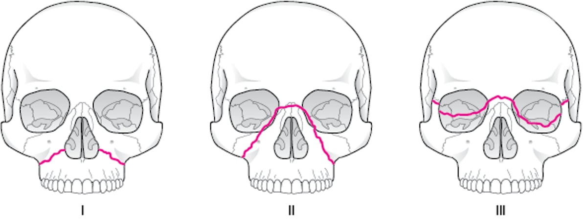
The Le Fort classification (see figure ) can be used to describe midface fractures (1). In combination or as an alternative, classification systems may be used that focus on specific midface components, such as the naso-orbito-ethmoidal complex or zygomaticomaxillary complex.
Orbital floor fracture is suggested by infraorbital nerve anesthesia, enophthalmos, or diplopia. An injury near the orbit requires an eye examination that includes at minimum an assessment of visual acuity, pupils, extraocular movements, and intraocular pressure (see also Blowout Fracture).
Zygomatic arch fracture is suggested by trismus and a defect on palpation of the zygomatic arch. A depression on the ipsilateral cheek may or may not be visible initially due to swelling.
Brain injury and fractured cervical vertebrae are possible when trauma has been severe enough to fracture facial bones. In major impact injuries, hemorrhage and edema due to a facial fracture may compromise the airway.
Le Fort Classification of Midface Fractures
I: Only the lower maxilla; II: The infraorbital rim; III: Complete detachment of the midface from the skull (craniofacial dissociation). |

Diagnosis of Mandibular and Midface Fractures
Radiograph and/or CT

This panoramic radiograph shows a lucency (arrow) at the angle of the mandible, representing a fracture.
SCIENCE PHOTO LIBRARY

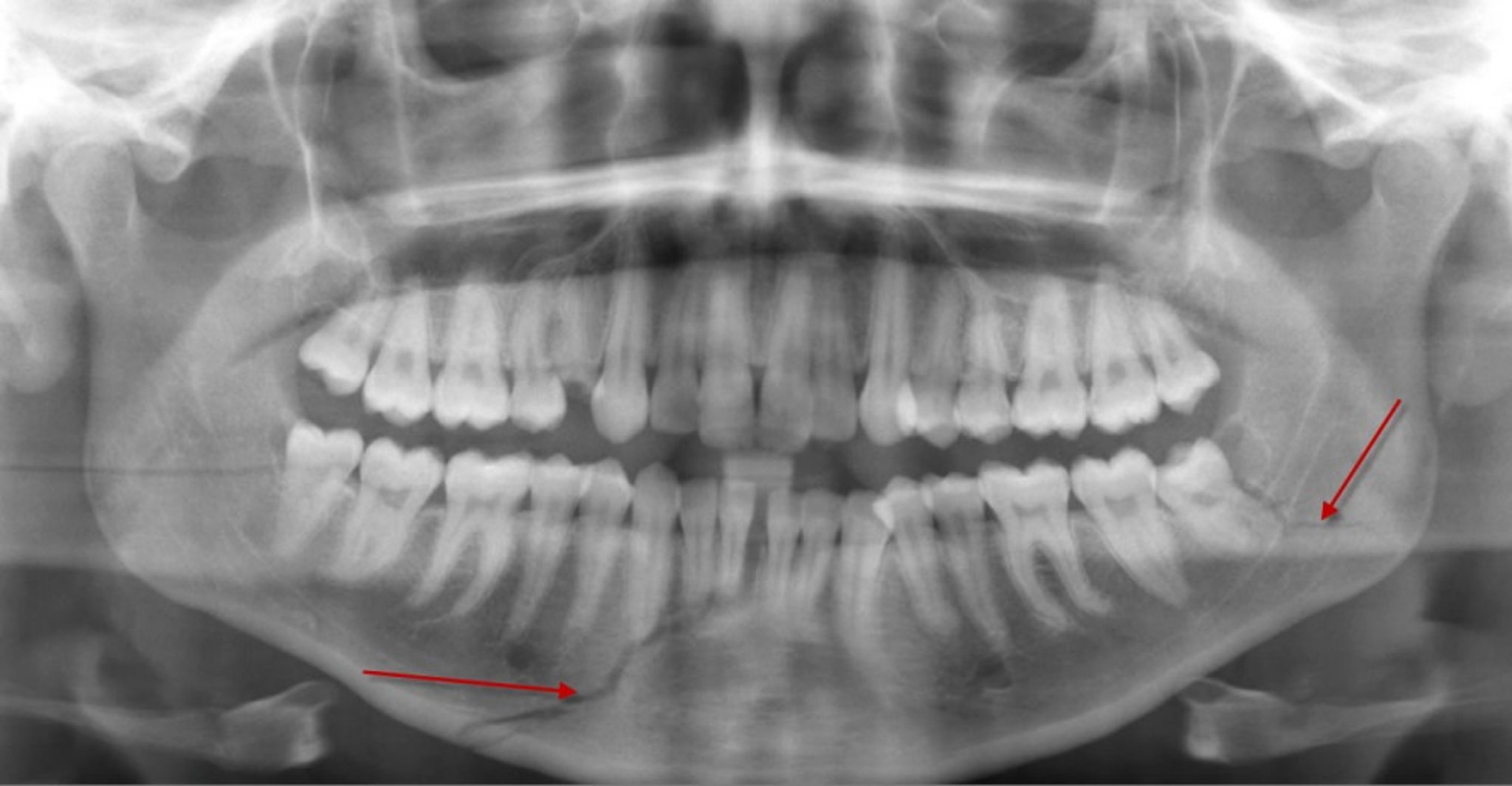
This panoramic radiograph shows a lucency (bottom arrow) in the body of the mandible, representing a fracture, that reaches to the roots of some lower anterior teeth. There is also a fracture of the left mandibular angle (arrow).
DU CANE MEDICAL IMAGING LTD/SCIENCE PHOTO LIBRARY
A panoramic radiograph may be used as an initial imaging study to screen for isolated mandibular fracture. Fine-cut CT (1-mm slices) in the axial and coronal planes is the preferred imaging modality to evaluate for facial fractures.
General reference
1. Gómez Roselló E, Quiles Granado AM, Artajona Garcia M, et al. Facial fractures: classification and highlights for a useful report. Insights Imaging. 2020;11(1):49. Published 2020 Mar 19. doi:10.1186/s13244-020-00847-w
Treatment of Mandibular and Midface Fractures
Endotracheal intubation, if necessary
Fracture management
Sometimes antibiotics
Maintaining a patent airway takes precedence over fracture management. If fractures cause significant tissue disruption, hemorrhage, or edema within the airway, an oral endotracheal airway may be required to maintain airway patency.
Definitive facial fracture management is complex and may include internal fixation.
Tooth socket fractures
Fractures through a tooth socket are considered open fractures because they communicate with the oral cavity. Antibiotic prophylaxis is recommended with antibiotics that cover aerobic and anaerobic oral flora (eg, clindamycin or ampicillin-sulbactam) and administered orally as a liquid or parenterally (1). Concurrent tooth avulsion and mandibular or maxillary fracture may require reimplantation, stabilization of the tooth, and fracture management; coordinated care is required by dental and oral and maxillofacial surgical specialists.
Mandible fractures
For a fractured mandible, treatment ranges from soft diet alone to maxillomandibular fixation (MMF, sometimes referred to as wiring the jaw shut; immobilization of the upper and lower jaws with arch bars, wires, or screws) and/or open reduction and internal fixation (ORIF; surgical procedure in which the bone is exposed and bone fragments are realigned and secured with metal plates) (2). If fixation is performed within the first few hours after injury, closure of any lip or oral lacerations should be delayed until the fracture has been reduced.
Eating is restricted to liquids, pureed foods, and nutritional supplements. Clinicians sometimes advise patients with maxillomandibular fixation to carry wire cutters in case of vomiting.
Because only buccal surfaces of the teeth can be brushed during maxillomandibular fixation, clinicians often recommend oral chlorhexidine rinses (eg, 60-second rinse with 30 mL of chlorhexidine 0.12% every morning and evening) to control plaque formation, infection, and halitosis. Physical therapy, including jaw-opening exercises (eg, maximum mouth opening, lateral movements), usually helps restore function after fixation is discontinued.
Duration of fixation may be up to 6 weeks and depends on location and severity of injury and patient (eg, age or any factor impacting healing time); combined MMF and ORIF usually allows earlier mobilization. Condylar fractures may require only 2 to 3 weeks of maxillomandibular fixation, followed by a soft diet. However, severely displaced, bilaterally fractured condyles may require ORIF. Condylar fractures in children should not be rigidly immobilized because ankylosis and abnormal facial development may result (3). Flexible (elastic) fixation for 5 to 10 days is usually sufficient.
Midface fractures
Fractures of the midface are treated surgically if they cause malocclusion, enophthalmos, diplopia, infraorbital nerve anesthesia, or unacceptable cosmetic deformity. Surgical treatment usually consists of internal stabilization using fine screws and plates. Surgery can often be delayed until swelling subsides to determine the extent of deformity. However, if surgery is required, it is best done within approximately 14 days of injury because after this time, bone callus can make reduction difficult.
Treatment references
1. Appelbaum RD, Farrell MS, Gelbard RB, et al. Antibiotic prophylaxis in injury: an American Association for the Surgery of Trauma Critical Care Committee clinical consensus document. Trauma Surg Acute Care Open. 2024;9(1):e001304. Published 2024 Jun 3. doi:10.1136/tsaco-2023-001304
2. Ma Y, Hao X, Zhao Y. Open Reduction and Internal Fixation Strategy For Treatment of Comminuted Mandibular Fracture. J Craniofac Surg. 2021;32(1):e90-e92. doi:10.1097/SCS.0000000000006778
3. Hajibandeh J, Peacock ZS. Pediatric Mandible Fractures. Oral Maxillofac Surg Clin North Am. 2023;35(4):555-562. doi:10.1016/j.coms.2023.05.001
Drug Information for the Topic


