


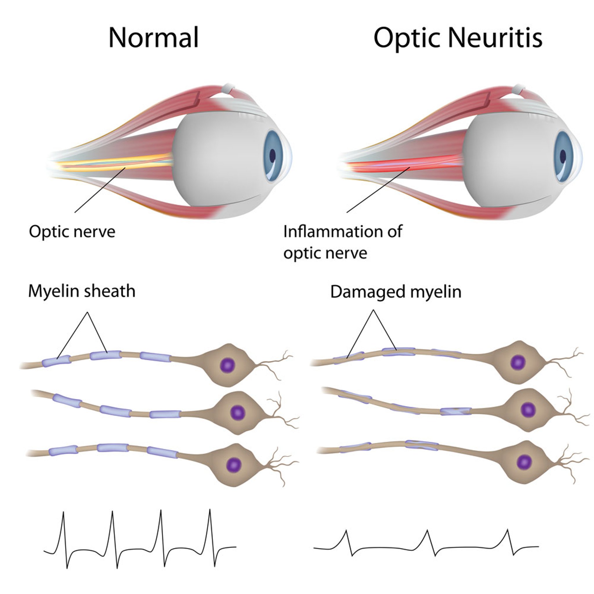
Optic Neuritis
In this illustration, the images in the left column show a healthy optic nerve and healthy myelin sheaths.
The images in the right column show an inflamed optic nerve and damaged myelin.
The bottom line graphics show visual evoked potentials (VEP). VEPs measure how quickly and strongly visual signals travel from the eye to the brain. In optic neuritis, VEPs often show delayed latency (slower signal) and lower amplitude (weaker signal).
Alila Medical Media/stock.adobe.com
In these topics