

Arthrocentesis of the metacarpophalangeal joints and interphalangeal joints of the hand is the process of puncturing the finger joints with a needle to withdraw synovial fluid. The procedure described is applicable to any of these joints.
(See also Evaluation of the Patient with Joint Symptoms and Evaluation of the Hand.)
Indications for MCP and IP Joint Arthrocentesis
Diagnosis of the cause of a synovial effusion (eg, infection, crystal-induced arthritis)
Removal of a synovial effusion and/or injection of medications as part of treatment and for pain relief
Contraindications to MCP and IP Joint Arthrocentesis
Absolute contraindications
Infection of skin or deeper tissues at the anticipated site of needle insertion
If possible, an alternate, uninfected puncture site should be used. However, acutely inflamed joints may be generally warm, tender, and erythematous, thus mimicking extra-articular infection and making it hard to find an uninvolved insertion site. Ultrasound may be helpful; visualization of a joint effusion by ultrasound can reinforce the decision to perform arthrocentesis despite surrounding erythema. NOTE: If infectious arthritis is strongly suspected, arthrocentesis should be performed regardless of erythema or negative ultrasound results because joint infection must not be missed.
Complications of MCP and IP Joint Arthrocentesis
Complications are uncommon and include:
Infection
Damage to tendon, nerve, or blood vessels (traumatic tap)
Equipment for MCP and IP Joint Arthrocentesis
Antiseptic solution (eg, chlorhexidine, povidone iodine, isopropyl alcohol), sterile gauze, and glovesAntiseptic solution (eg, chlorhexidine, povidone iodine, isopropyl alcohol), sterile gauze, and gloves
Nonsterile underpads
Local anesthetic (eg, 1% lidocaine, 25- to 30-gauge needle, 3-mL syringe) Local anesthetic (eg, 1% lidocaine, 25- to 30-gauge needle, 3-mL syringe)
For joint aspiration, a 25-mm (1-inch) 25-gauge needle and a 3-mL syringe
Appropriate containers for collection of fluid for laboratory tests (eg, cell count, crystals, cultures)
For intra-articular therapeutic injection, a syringe containing a glucocorticoid (eg, triamcinolone acetonide 10 mg or methylprednisolone acetate 20 mg) and/or a long-acting anesthetic (eg, 0.25% bupivacaine), a 25-gauge needle, and a hemostat to help switch syringes, if neededFor intra-articular therapeutic injection, a syringe containing a glucocorticoid (eg, triamcinolone acetonide 10 mg or methylprednisolone acetate 20 mg) and/or a long-acting anesthetic (eg, 0.25% bupivacaine), a 25-gauge needle, and a hemostat to help switch syringes, if needed
Additional Considerations for MCP and IP Joint Arthrocentesis
Enlist an assistant to provide flexion and traction to the finger or thumb.
Synovial fluid is usually not obtainable from a metacarpophalangeal (MCP) or interphalangeal (IP) joint that is not infected or inflamed.
Standard precautions, including the use of a sterile technique, is necessary to prevent microbial contamination of both the joint space and the aspirated synovial fluid.
Relevant Anatomy for MCP and IP Joint Arthrocentesis
The aspirating needle is inserted into the joint line along the dorsal portion of the joint, just medial or lateral to the extensor tendon.
Positioning for MCP and IP Joint Arthrocentesis
Position the patient sitting or supine with the forearm resting on a bedside table and the hand pronated.
The fingers are flexed; for the MCP joint, the fingers are flexed at the level of the MCPs, and for the interphalangeal joints, the patient slightly flexes the fingers.
Step-by-Step Description of MCP and IP Joint Arthrocentesis
First, have the patient extend the finger. In this position, palpate the dorsal aspect of the joint and the extensor tendon. Then have the patient relax the finger. Apply traction to the relaxed finger, which expands the joint space slightly and makes the joint line visible as skin depressions—the areas for needle insertion—on either side of the extensor tendon. These landmarks are most apparent over the MCP joints.
Rest the hand on an underpad. Prepare the area with a skin-cleansing agent, such as chlorhexidine or povidone iodine, then use an alcohol wipe to remove the agent.Rest the hand on an underpad. Prepare the area with a skin-cleansing agent, such as chlorhexidine or povidone iodine, then use an alcohol wipe to remove the agent.
Place a wheal of local anesthetic over the needle entry site using a 25- to 30-gauge needle.
Have the patient flex the fingers to the appropriate angle for the joint being punctured.
Have an assistant apply gentle axial traction to the finger to facilitate entry of the aspirating needle into the joint space.
Aspirate the joint using a 25-gauge needle. Enter the skin perpendicularly from above, at the level of the joint line, just medial or lateral to the extensor tendon or from the side at a 90° angle from above. Direct the needle toward the center of the joint space (see figure and figure ). Gently pull back on the plunger as you advance. Synovial fluid will enter the syringe when the joint is entered.
If the needle hits bone, retract almost to skin surface and then redirect at a different angle.
Drain all fluid from the joint (usually ≤ 1 mL).
If intra-articular medications (eg, anesthetic, glucocorticoid) are to be given, use a hemostat to hold the hub of the needle motionless while removing the synovial fluid-containing syringe and replace it with the medication-containing syringe. If the needle has remained in place in the joint space, there will be no resistance to medication injection. Injections into the MCP and IP joints should not exceed 0.5 mL in volume.
After injecting a glucocorticoid, move the joint through full range of motion to distribute the medication throughout the joint.
Transfer synovial fluid to tubes and other transport media for synovial fluid analysis. Inspect the fluid for blood and fat.
Apply an adhesive bandage or sterile dressing.
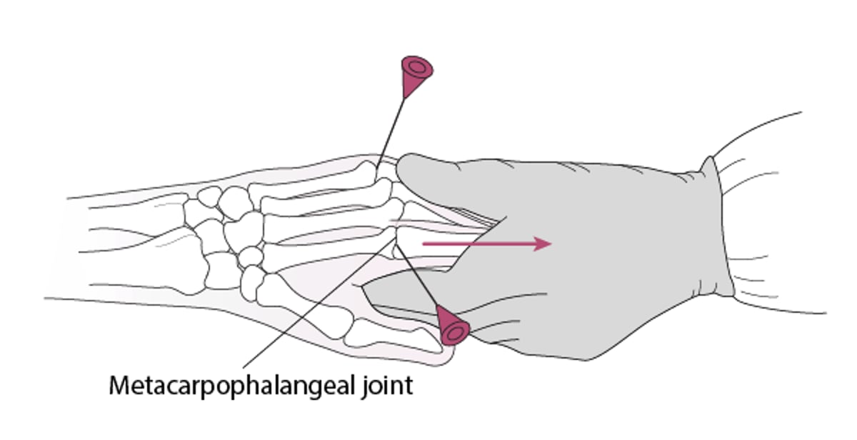
Arthrocentesis of the Metacarpophalangeal Joint
For arthrocentesis of the metacarpophalangeal joint, insert a 25-gauge needle at either side of the extensor tendon from above or at a 90° angle from above, while gentle traction is applied to the finger. |

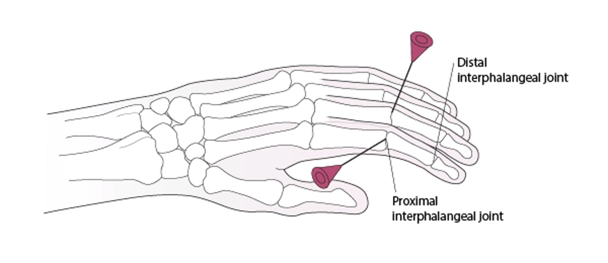
Arthrocentesis of the Proximal Interphalangeal Joint
For arthrocentesis of the proximal interphalangeal, insert a 25-gauge needle at either side of the extensor tendon from above or at a 90° angle from above, while gentle traction is applied distal to the joint. |

Aftercare for MCP and IP Joint Arthrocentesis
Ice, elevation, and oral nonsteroidal anti-inflammatory drugs (NSAIDs) may help relieve pain.
If an intra-articular anesthetic has been given, limited joint activity should be prescribed for 4 to 8 hours.
If an intra-articular glucocorticoid has been given, the joint should be rested for approximately 24 to 48 hours.
If the patient has increased erythema, pain, and/or swelling > 12 hours after the procedure, the joint should be examined for possible infection.
Warnings and Common Errors for MCP and IP Joint Arthrocentesis
Carefully ensure optimal positioning before joint puncture.
Allow adequate time for local anesthesia to take effect before proceeding.
To avoid damaging the synovium and articular cartilage, do not advance the needle against resistance and do not move the needle once it has begun draining synovial fluid.
If the needle tip must be relocated, first withdraw it almost to the skin surface and then redirect; do not try to change the angle of insertion while a needle is embedded in tissue.
Tips and Tricks for MCP and IP Joint Arthrocentesis
Note also that warmth, tenderness, and erythema may overlie an acutely inflamed arthritic joint, mimicking extra-articular infection.
When trying to differentiate infectious arthritis from infection of the overlying structures (a contraindication to arthrocentesis), infectious arthritis is more likely with the following:
Joint effusion
Circumferential joint pain and capsule tenderness
Pain with both gentle, passive motion and with active joint motion
When inspecting fluid, consider the following:
The hemarthrosis of a traumatic tap tends to be nonuniformly bloody and tends to clot.
There may be no visible aspirated fluid from small joints. However, the syringe should still be used to express even a trivial drop of fluid through the needle on to a slide for microscopic evaluation. This may be sufficient to document crystal-associated arthritis or increase suspicion for infection.
Drugs Mentioned In This Article


