

Paget disease of bone is a chronic disorder of the adult skeleton in which bone turnover is accelerated in localized areas. Normal matrix is replaced with softened and enlarged bone. The disease may be asymptomatic or cause gradual onset of bone pain or deformity. Diagnosis is by radiograph. Treatment includes symptomatic measures and often medications, usually bisphosphonates.
Approximately 2 to 3% of adults > 55 years old in the United States have Paget disease, with a slight male predominance (1). Prevalence increases with age; however, overall prevalence seems to be decreasing. The disease is most common in the United Kingdom and European countries including Spain, France, and Italy, as well as in countries settled by European immigrants (eg, Australia, New Zealand, the United States, and Canada) (2, 3). Paget disease is rare in Scandinavian countries and Asia.
General references
1. Ralston SH, Corral-Gudino L, Cooper C, et al: Diagnosis and management of Paget's disease of bone in adults: a clinical guideline. J Bone Miner Res 34(4):579-604, 2019. doi: 10.1002/jbmr.3657
2. Reid IR: Recent advances in understanding and managing Paget's disease. F1000Res. 2019;8:F1000 Faculty Rev-1485. Published 2019 Aug 22. doi:10.12688/f1000research.19676.1
3. Singer FR, Bone HG 3rd, Hosking DJ, et al: Paget's disease of bone: an Endocrine Society clinical practice guideline. J Clin Endocrinol Metab 99(12):4408-22, 2014. doi: 10.1210/jc.2014-2910
Etiology of Paget Disease of Bone
Approximately 10% of patients with Paget disease have mutations of the SQSTM1 (sequestosome-1) gene, resulting in increased nuclear factor kappa-B activity, which increases osteoclast activity (1, 2). Several other mutations affecting the RANK (receptor activator of nuclear factor kappa-B) signaling pathway, which is critical for osteoclast generation and activity, have been associated with Paget disease, including mutations in RANK itself (3).
A viral etiology, such as measles, has been proposed because nuclear inclusions in diseased osteoclasts that are similar to those seen in paramyxovirus-infected cells have been seen on electron microscopy. Although a viral cause remains controversial, it is hypothesized that in genetically predisposed patients an as yet unidentified virus triggers abnormal osteoclast activity.
Etiology references
1. Laurin N, Brown JP, Morissette J, Raymond V: Recurrent mutation of the gene encoding sequestosome 1 (SQSTM1/p62) in Paget disease of bone. Am J Hum Genet 70:1582–1588, 2002. doi: 10.1086/340731
2. Rea SL, Walsh JP, Layfield R, et al: New insights into the role of sequestosome 1/p62 mutant proteins in the pathogenesis of Paget's disease of bone. Endocr Rev 34(4):501-24, 2013. doi: 10.1210/er.2012-1034
3. Albagha OM, Visconti MR, Alonso N, et al: Genome-wide association study identifies variants at CSF1, OPTN and TNFRSF11A as genetic risk factors for Paget's disease of bone. Nat Genet 42(6):520-524, 2010. doi:10.1038/ng.562
Pathophysiology of Paget Disease of Bone
Any bone can be involved in Paget disease. The bones most commonly affected are the pelvis, femur, and skull. Other less commonly involved bones are the tibia, vertebrae, clavicle, and humerus.
Bone turnover is accelerated at involved sites. Pagetic lesions are metabolically active and highly vascular. Excessively active osteoclasts are often large and contain many nuclei. Osteoblastic repair is also hyperactive, causing coarsely woven, thickened lamellae and trabeculae. This abnormal structure weakens the bone, despite bone enlargement and areas of bone sclerosis.
Symptoms and Signs of Paget Disease of Bone
Paget disease of the bone is usually asymptomatic. If symptoms occur, they develop insidiously, with pain, stiffness, fatigue, and bone deformity. Bone pain is aching, deep, and occasionally severe, sometimes worse at night. Pain also may arise from compression neuropathy or osteoarthritis.
If the skull is involved, signs may include skull enlargement bitemporally and frontally (frontal bossing), dilated scalp veins, and nerve deafness in one or both ears. Symptoms may include vertigo, headaches, and hearing impairment. Deformities may develop from bowing of the long bones or osteoarthritis. Pathologic fractures may be the presenting manifestation. New onset or severe pain at a site of pagetic bone should prompt consideration of osteosarcoma.

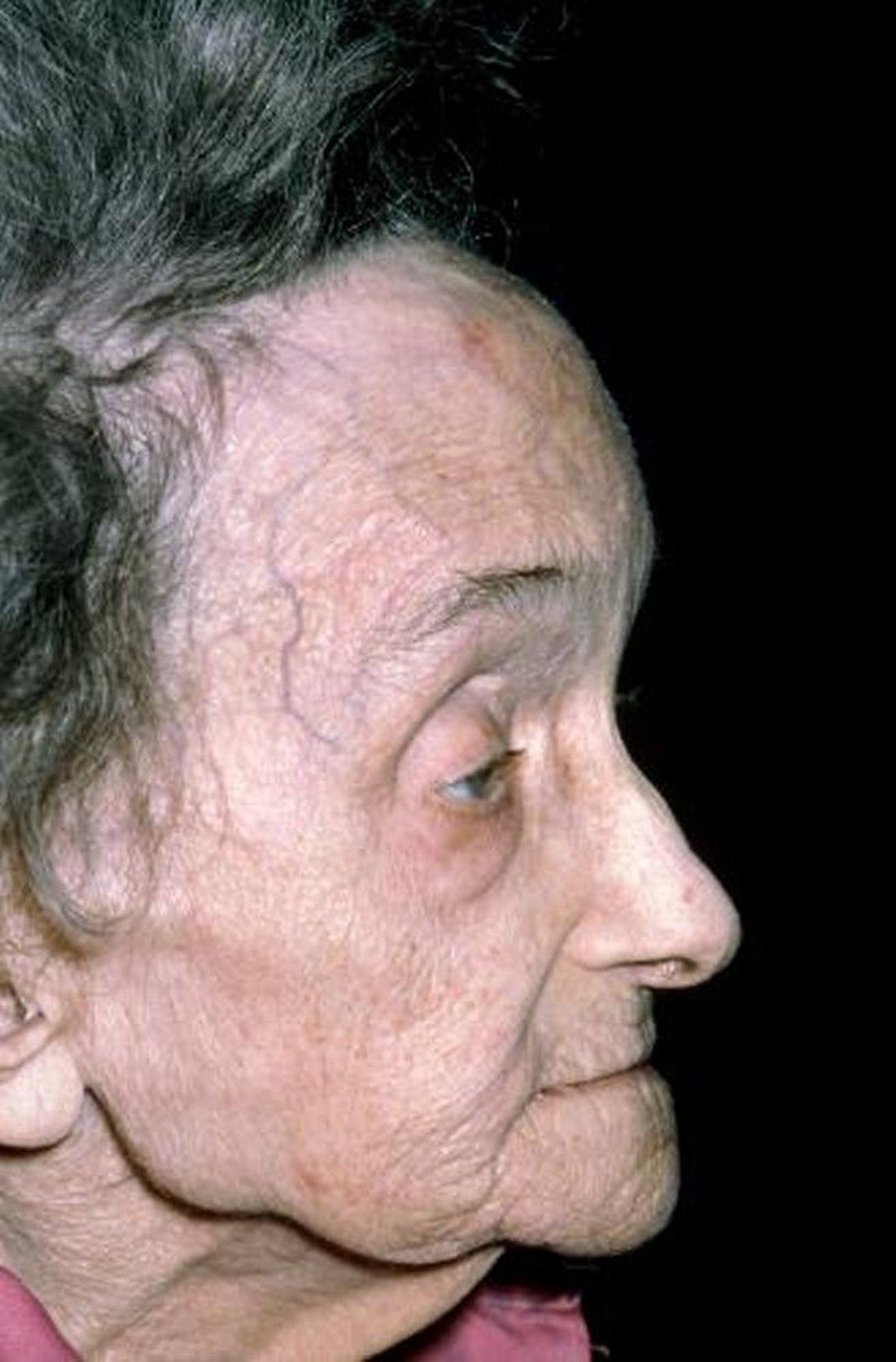
This photo shows an abnormally prominent forehead (frontal bossing) in a patient with Paget disease.
DR P. MARAZZI/SCIENCE PHOTO LIBRARY

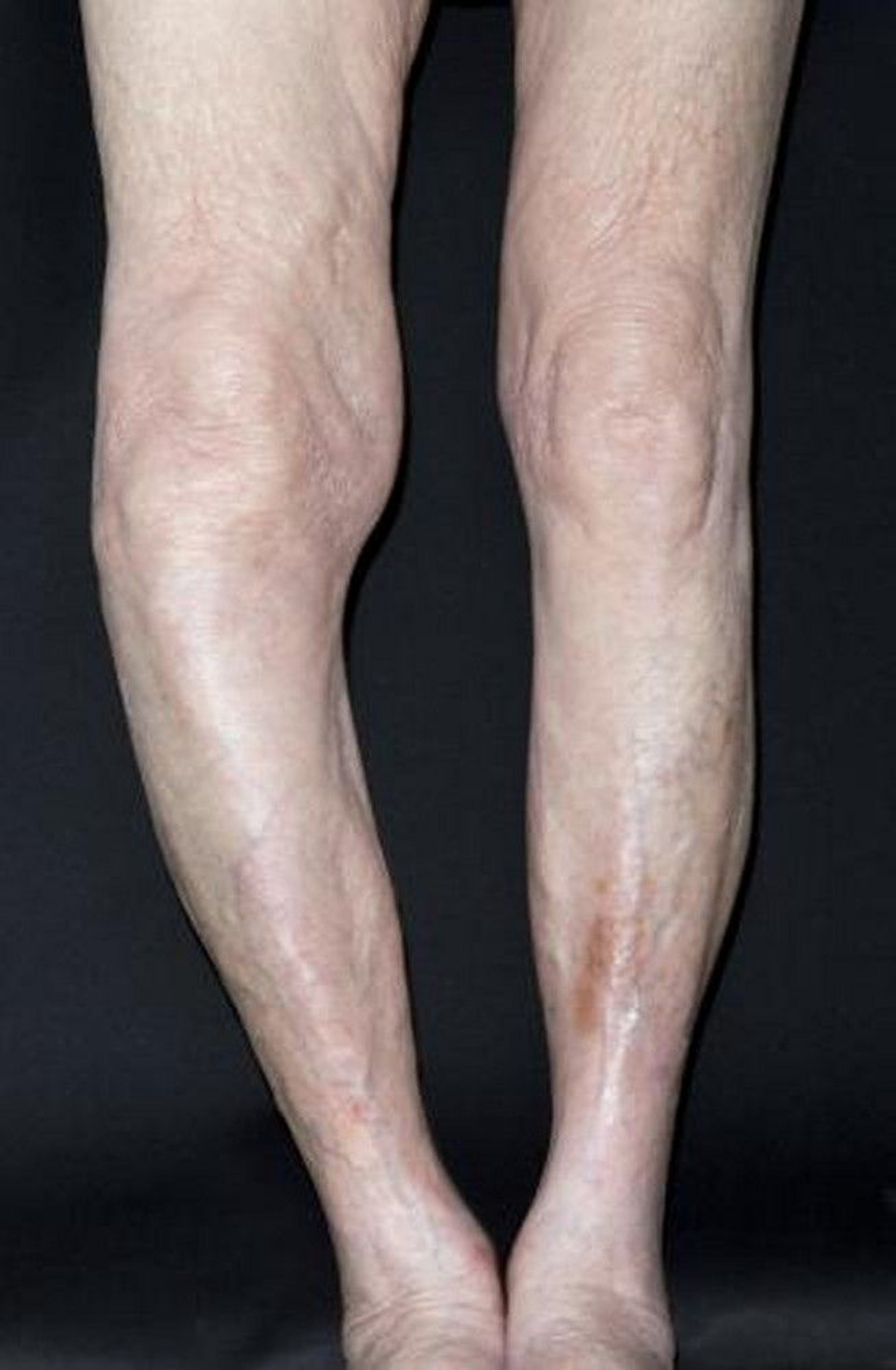
This photo shows bowing of the tibia of the right leg (seen at left) in a patient with Paget disease.
DR P. MARAZZI/SCIENCE PHOTO LIBRARY
Complications
The most common complication of Paget disease of bone is
Osteoarthritis occurs in up to 50% of patients and develops in joints adjacent to involved bone (1). Pathologic fracture is also common due to focal areas of weakened bone.
Overgrown bone may compress nerves and other structures passing through small foramina. Spinal stenosis or spinal cord compression may develop.
Rare complications include transformation to osteosarcoma in < 1% of patients. Highly vascular bones may bleed excessively during orthopedic surgery. Very rarely, hypercalcemia develops in patients who are immobile; however, hypercalcemia in ambulatory patients suggests the coexistence of hyperparathyroidism. High-output heart failure due to large or numerous hypervascular lesions has been reported.
Symptoms and signs reference
1. Berg K, Dockrell D, Colvin L, et al: Causes of Musculoskeletal Pain in Paget's Disease of Bone. Calcif Tissue Int 115(5):533-541, 2024. doi:10.1007/s00223-024-01279-0
Diagnosis of Paget Disease of Bone
Radiographs
Elevated serum alkaline phosphatase or other markers of increased bone turnover
Bone scan to establish the extent and location of disease
Paget disease should be suspected in patients with the following:
Unexplained bone pain or deformity
Suggestive findings on radiographs
Unexplained elevation of serum alkaline phosphatase on laboratory tests done for other reasons, particularly if liver-sourced 5'-nucleotidase or gamma-glutamyl-transpeptidase (GGT) is normal
If Paget disease is suspected, radiographs and serum alkaline phosphatase, 25-hydroxyvitamin D, creatinine, calcium, and phosphate levels should be obtained (1, 2, 3).
Characteristic laboratory findings include elevated serum alkaline phosphatase (increased anabolic activity of bone) but usually normal serum gamma-glutamyl-transpeptidase (GGT), 5'-nucleotidase, and serum phosphate levels. Serum calcium is usually normal, though it may be increased in patients who are immobilized. If hypercalcemia is present, the patient should be evaluated for concurrent hyperparathyroidism. If alkaline phosphatase is not elevated or it is unclear whether the increased serum alkaline phosphatase is of bony origin (ie, if GGT is increased in proportion to alkaline phosphatase), bone-specific alkaline phosphatase can be measured. Serum markers of bone turnover, such as procollagen type I intact N-terminal propeptide (PINP) and C-telopeptide cross-links (CTX), may be elevated. Serum 25-hydroxyvitamin D level and creatinine should be checked before treating symptomatic patients with bisphosphonates because bisphosphonates are not recommended for patients with a creatinine clearance < 35 mL/min/1.73m2 (0.58 mL/s/m2) and vitamin D deficiency increases the risk of hypocalcemia after bisphosphonate administration.
Confirmation on radiographs is required to establish the diagnosis. Characteristic radiographic findings include abnormal architecture with coarse trabeculae and cortical thickening and sclerosis. Involved bone may be enlarged, and pagetic lesions in the tibia or femur may result in bowing. When Paget disease is suspected based on imaging findings or elevated alkaline phosphatase of bone origin, a radionuclide bone scan using technetium-labeled phosphonates should be done to determine the extent of bone involvement. In cases of diagnostic uncertainty, a biopsy should be performed to exclude possible metastatic disease. Repeat imaging with CT or MRI and/or biopsy is indicated for patients with known pagetic bone involvement who have worsening symptoms at the affected site to evaluate for osteosarcoma or giant cell tumor of bone, which may rarely arise in pagetic bone.

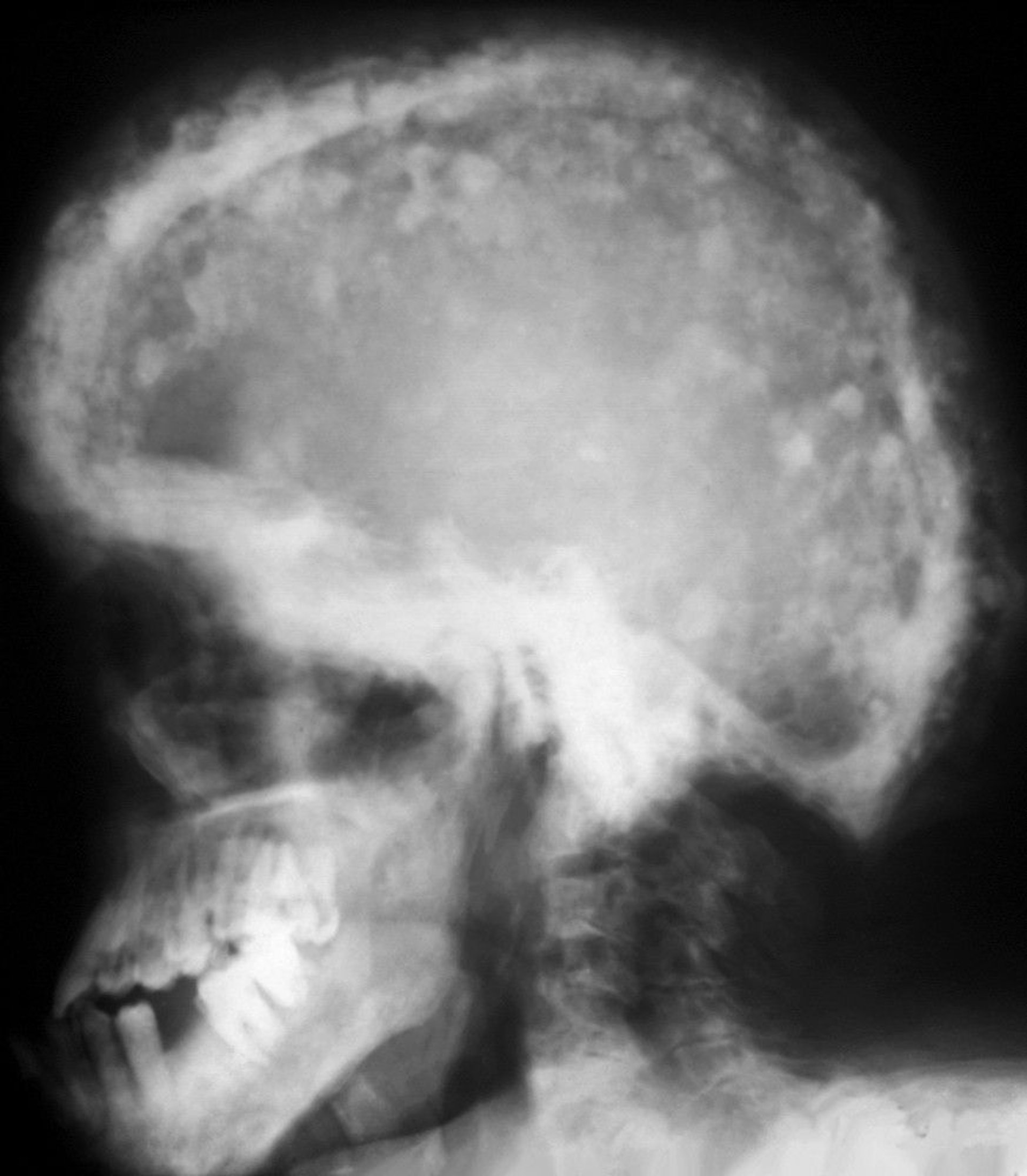
A typical mosaic pattern of areas of increased sclerosis and lucency is seen in this radiograph. These cotton-wool patches result from thickening of the calvarium.
ZEPHYR/SCIENCE PHOTO LIBRARY

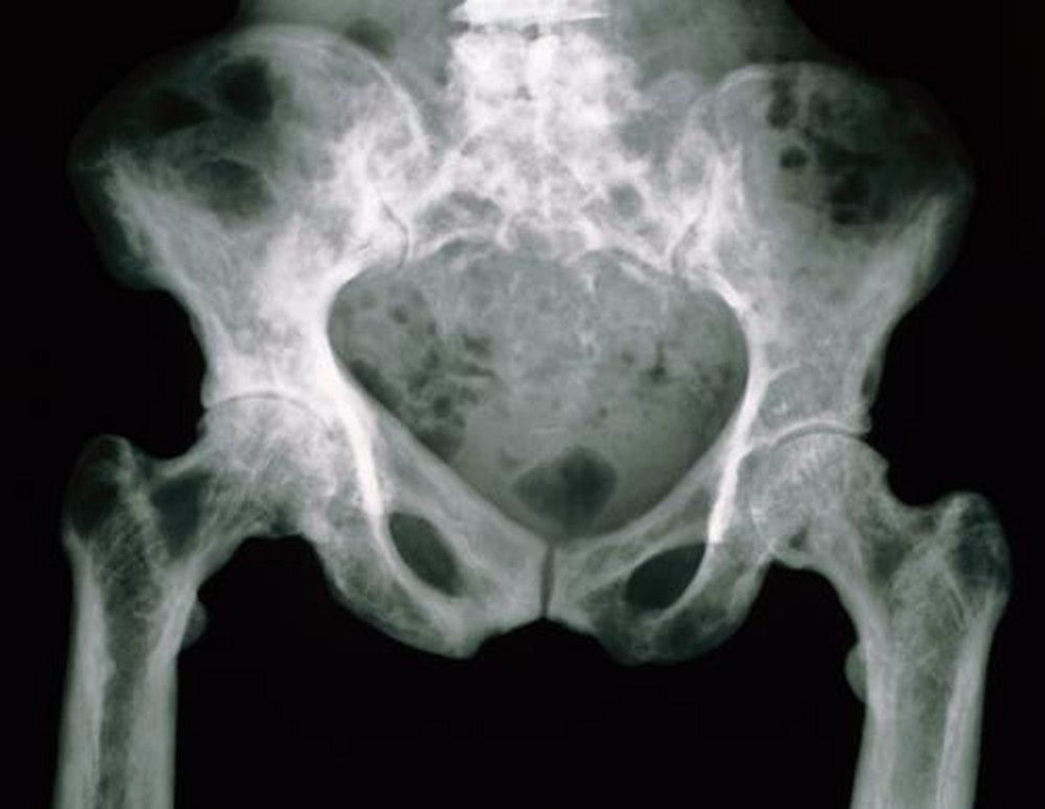
The pelvic bones in this radiograph have a mottled appearance due to their increased porosity.
SCIENCE PHOTO LIBRARY
Pearls & Pitfalls
|
Diagnosis references
1. Singer FR, Bone HG 3rd, Hosking DJ, et al. Paget's disease of bone: an endocrine society clinical practice guideline. J Clin Endocrinol Metab 2014;99(12):4408-4422. doi:10.1210/jc.2014-2910
2. Ralston SH, Corral-Gudino L, Cooper C, et al: Diagnosis and Management of Paget's Disease of Bone in Adults: A Clinical Guideline. J Bone Miner Res 34(4):579-604, 2019. doi:10.1002/jbmr.3657
3. Rendina D, Falchetti A, Diacinti D, et al: Diagnosis and treatment of Paget's disease of bone: position paper from the Italian Society of Osteoporosis, Mineral Metabolism and Skeletal Diseases (SIOMMMS). J Endocrinol Invest 47(6):1335-1360, 2024. doi:10.1007/s40618-024-02318-1
Treatment of Paget Disease of Bone
Supportive care for symptoms and complications
Bisphosphonates if bone disease is symptomatic or active in bones at risk of complications
Supportive treatment of Paget disease of bone includes analgesics or nonsteroidal anti-inflammatory drugs (NSAIDs) for pain. Orthotics help correct abnormal gait caused by bowed lower extremities. Some patients require orthopedic surgery (eg, hip or knee replacement, decompression of the spinal cord). Weight bearing should be encouraged, and bed rest should be avoided.
Localized, asymptomatic disease may not require treatment.
Pharmacologic therapy
Pharmacologic therapy is indicated to treat pain clearly related to the pagetic process and not to another source (eg, osteoarthritis). In addition, some guidelines (1, 2, 3) recommend treatment to:
Prevent or slow progression of complications (eg, hearing loss, deformity, osteoarthritis, paraparesis or paraplegia related to vertebral Paget disease, or other neurologic deficits), particularly in a patient for whom surgery is not appropriate
Prevent or minimize bleeding if orthopedic surgery is planned at a site with pagetic involvement
Suppress excessive osteoclast activity when serum alkaline phosphatase (of bony origin) is > 2 times the normal level, even in the absence of symptoms
Although disease progression can be slowed by medications, existing deficits (eg, deformity, osteoarthritis, hearing loss, neural impingement) are not reversed.
Several antiresorptive agents that suppress osteoclast activity and decrease bone pain are available for treating Paget disease.
Bisphosphonates are the medication class of choice. The amino-bisphosphonates (bisphosphonates with an extra nitrogen atom) more effectively suppress markers of disease activity and provide more prolonged response. Among these, zoledronic acid is recommended as first-line therapy for Paget disease of bone in professional guidelines (Bisphosphonates are the medication class of choice. The amino-bisphosphonates (bisphosphonates with an extra nitrogen atom) more effectively suppress markers of disease activity and provide more prolonged response. Among these, zoledronic acid is recommended as first-line therapy for Paget disease of bone in professional guidelines (1, 2), whereas other amino-bisphosphonates (such as alendronate, risedronate, and pamidronate) are second-line, and the simple bisphosphonates (bisphosphonates without an extra nitrogen atom, such as tiludronate and etidronate) are third-line therapy. Zoledronic acid and pamidronate are administered intravenously, while all other bisphosphonates are oral medications. Hypocalcemia after zoledronic acid administration has been reported in patients with serum 25-hydroxy), whereas other amino-bisphosphonates (such as alendronate, risedronate, and pamidronate) are second-line, and the simple bisphosphonates (bisphosphonates without an extra nitrogen atom, such as tiludronate and etidronate) are third-line therapy. Zoledronic acid and pamidronate are administered intravenously, while all other bisphosphonates are oral medications. Hypocalcemia after zoledronic acid administration has been reported in patients with serum 25-hydroxyvitamin D levels ≤ 10 ng/mL (24.96 nmol/L), and a level of > 25 ng/ml (62.4 nmol/L) is suggested to reduce the risk of hypocalcemia.
Synthetic salmon calcitonin is an alternative to bisphosphonates for patients intolerant of or resistant to them. Case reports suggest that denosumab may also be an alternative to bisphosphonates (Synthetic salmon calcitonin is an alternative to bisphosphonates for patients intolerant of or resistant to them. Case reports suggest that denosumab may also be an alternative to bisphosphonates (3); however, there are insufficient data to support its routine use.
Because bone turnover is increased, patients should ensure adequate intake of calcium and vitamin D, and supplements are often needed.
Treatment references
1. Singer FR, Bone HG 3rd, Hosking DJ, et al: Paget's disease of bone: an Endocrine Society clinical practice guideline. J Clin Endocrinol Metab 99(12):4408-22, 2014. doi: 10.1210/jc.2014-2910
2. Ralston SH, Corral-Gudino L, Cooper C, et al: Diagnosis and Management of Paget's Disease of Bone in Adults: A Clinical Guideline. J Bone Miner Res 34(4):579-604, 2019. doi:10.1002/jbmr.3657
3. Rendina D, Falchetti A, Diacinti D, et al: Diagnosis and treatment of Paget's disease of bone: position paper from the Italian Society of Osteoporosis, Mineral Metabolism and Skeletal Diseases (SIOMMMS). J Endocrinol Invest 47(6):1335-1360, 2024. doi:10.1007/s40618-024-02318-1
Prognosis of Paget Disease of Bone
The prognosis of Paget disease is generally favorable, and life expectancy is not affected by the presence of Paget's disease (1), though osteoarthritis, bone deformity, and bone pain are common (1, 2). Bone pain is relieved by zoledronic acid treatment in the majority of patients (). Bone pain is relieved by zoledronic acid treatment in the majority of patients (3), and biochemical markers of bone turnover are normalized in > 90% of patients. While studies conclusively demonstrating that treatment prevents long-term complications such as deformity and osteoarthritis are lacking, it is clear that progression of osteolytic lesions and worsening deformity occur in untreated patients (4).
Prognosis references
1. Wermers RA, Tiegs RD, Atkinson EJ, Achenbach SJ, Melton LJ 3rd. Morbidity and mortality associated with Paget's disease of bone: a population-based study. J Bone Miner Res 2008;23(6):819-825. doi:10.1359/jbmr.080215
2. Seton M, Moses AM, Bode RK, Schwartz C. Paget's disease of bone: the skeletal distribution, complications and quality of life as perceived by patients. Bone 2011;48(2):281-285. doi:10.1016/j.bone.2010.09.021
3. Reid IR, Miller P, Lyles K, et al. Comparison of a single infusion of zoledronic acid with risedronate for Paget's disease. N Engl J Med 2005;353(9):898-908. doi:10.1056/NEJMoa044241
4. Siris ES, Feldman F. Natural history of untreated Paget's disease of the tibia. J Bone Miner Res 1997;12(4):691-692. doi:10.1359/jbmr.1997.12.4.691
Key Points
Paget disease of bone is an often asymptomatic abnormality of bone metabolism, particularly among older adults.
Complications can include osteoarthritis, fractures, neural compression, osteosarcoma, and rarely hypercalcemia.
Confirmation is usually by radiographs showing findings such as bone sclerosis, coarse cortical trabeculation or cortical thickening, and bone bowing or enlargement.
First-line treatment is with bisphosphonates, preferably zoledronic acid.First-line treatment is with bisphosphonates, preferably zoledronic acid.
Drug Information for the Topic


