


Horner syndrome is ptosis, miosis, and anhidrosis due to dysfunction of cervical sympathetic output. Diagnosis is confirmed with pharmacologic testing using cocaine or apraclonidine eye drops. Imaging (MRI or CT) of the brain, spinal cord, chest, or neck may be needed to identify the cause. Treatment depends on the cause.
(See also Overview of the Autonomic Nervous System.)
Etiology of Horner Syndrome
Horner syndrome results when the cervical sympathetic pathway running from the hypothalamus to the eye is disrupted. The causative lesion may be primary (including congenital) or secondary to another disorder.
Lesions are usually classified as either:
Central (eg, brainstem ischemia, syringomyelia, brain tumor, spinal cord tumor)
Peripheral (eg, Pancoast tumor, cervical adenopathy, neck and skull injuries, aortic or carotid dissection, thoracic aortic aneurysm)
Peripheral lesions may be preganglionic or postganglionic in origin.
Symptoms and Signs of Horner Syndrome
Symptoms of Horner syndrome include ptosis (drooping eyelid), miosis (constricted pupil), anhidrosis, and hyperemia of the affected side.

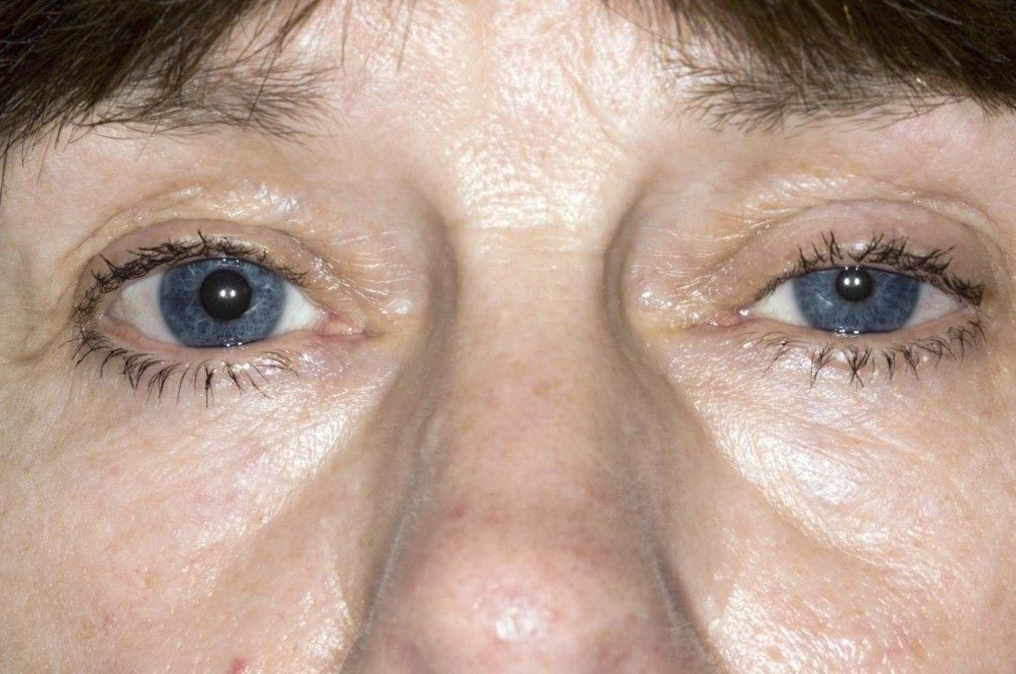
This photo shows ptosis (drooping of the eyelid) and miosis (constricted pupil) of the left eye of a patient with Horner syndrome.
DR P. MARAZZI/SCIENCE PHOTO LIBRARY
In the congenital form, the iris does not become pigmented and remains blue-gray.
Diagnosis of Horner Syndrome
Cocaine or apraclonidine eye drop test
MRI or CT to diagnose cause
Instilling eyedrops can help confirm and characterize Horner syndrome.
First, cocaine (4% or 10%) or apraclonidine (0.5%) drops are put in both eyes:
Cocaine:Cocaine blocks the synaptic reuptake of norepinephrine and causes the pupil of the unaffected eye to dilate. If a postganglionic lesion (peripheral Horner syndrome) is present, the pupil of the affected eye does not dilate because the postganglionic nerve terminals have degenerated; the result is increased anisocoria (unequal pupil size). If the lesion is above the superior cervical ganglion (preganglionic or central Horner syndrome) and the postganglionic fibers are intact, the pupil of the affected eye also dilates, and anisocoria decreases.
Apraclonidine:Apraclonidine is a weak alpha-adrenergic agonist that minimally dilates the pupil of a normal eye. If a postganglionic lesion is present (peripheral Horner syndrome), the pupil of the affected eye dilates more than that of the unaffected eye because the iris dilator muscle of the affected eye has lost its sympathetic innervation and has developed adrenergic supersensitivity. As a result, anisocoria decreases. (However, results may be falsely normal if the causative lesion is acute.) If the lesion is preganglionic (or a central Horner syndrome), the pupil of the affected eye does not dilate because the iris dilator muscle does not develop adrenergic supersensitivity; as a result, anisocoria increases.
If results suggest Horner syndrome, further testing is done to distinguish a postganglionic lesion from a central or preganglionic lesion.
Patients with Horner syndrome require MRI or CT of the brain, spinal cord, chest, or neck (depending on clinical suspicion) to localize the abnormality.
Treatment of Horner Syndrome
Treatment of the cause
The cause of Horner syndrome, if identified, is treated; there is no treatment for primary Horner syndrome.
Key Points
Horner syndrome causes ptosis, miosis, and anhidrosis.
It results from a central or peripheral lesion (preganglionic or postganglionic) that disrupts the cervical sympathetic pathway, which runs from the hypothalamus to the eye.
Instill cocaine or apraclonidine in both eyes to confirm the diagnosis of Horner syndrome and help locate the lesion (preganglionic or postganglionic).
Do MRI or CT of the brain, spinal cord, chest, or neck, depending on clinical suspicion.
Treat the cause, if identified; there is no treatment for primary Horner syndrome.
Drug Information for the Topic


