

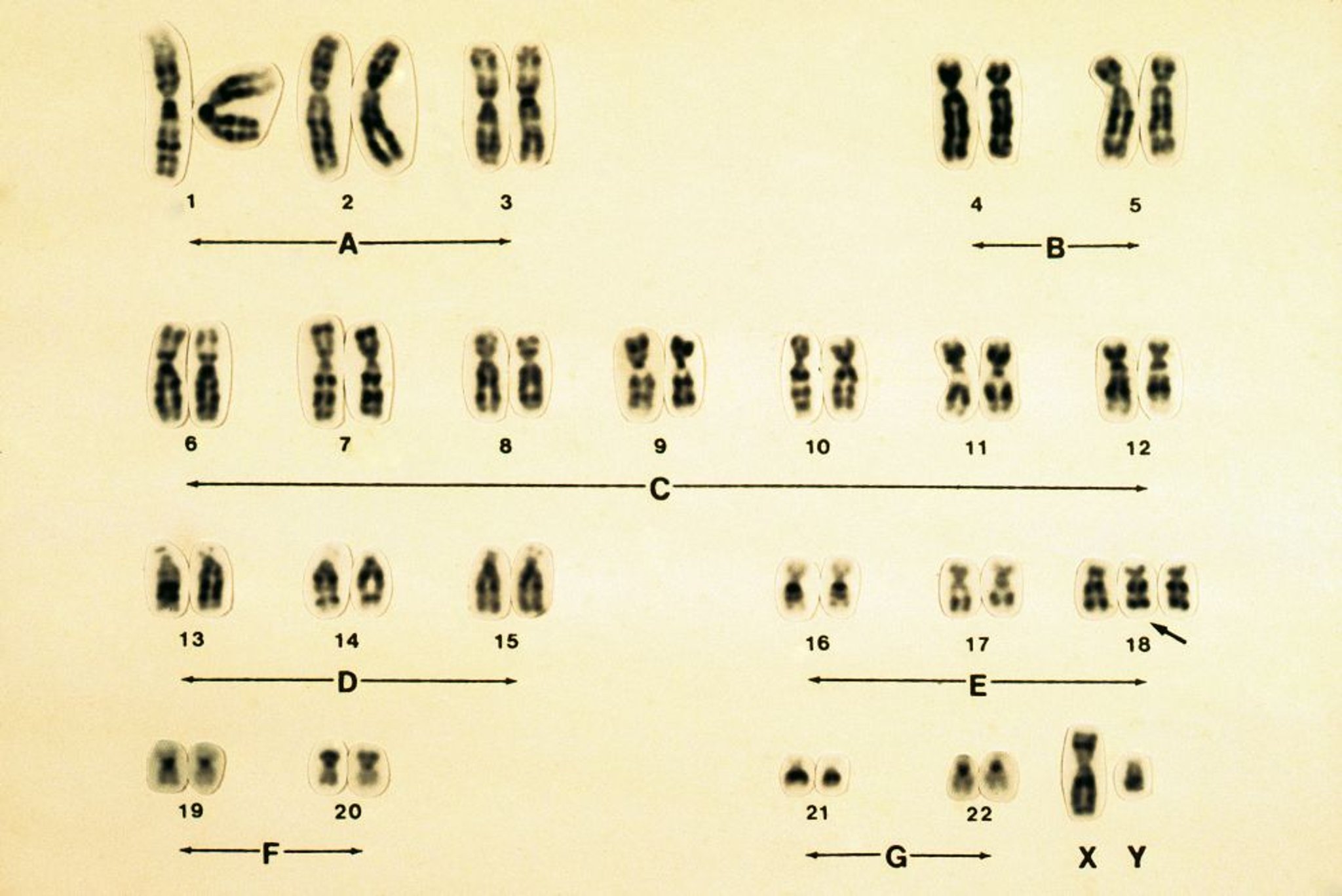
Trisomy 18 is caused by an extra chromosome 18 and is usually associated with intellectual disability, small birth size, and various congenital anomalies, including severe microcephaly, heart defects, prominent occiput, low-set malformed ears, and a characteristic pinched facial appearance. Prenatal diagnosis is with cytogenetic testing; postnatal diagnosis is with peripheral blood testing. Treatment is supportive.
(See also Overview of Chromosomal Abnormalities.)
Trisomy 18, also known as Edwards syndrome, is a chromosomal disorder caused by the presence of an extra chromosome 18 and is characterized by multiple congenital anomalies, severe developmental delays, and a high rate of perinatal mortality.
Trisomy 18 occurs in approximately 4.1/10,000 pregnancies (based on data from induced abortion for fetal anomalies, stillbirths, and live births) (1). More than 95% of affected children have complete trisomy 18 (2). The extra chromosome is almost always maternally derived, and advanced maternal age increases risk. Although most cases are due to nondisjunction, extra chromosome 18 material may rarely be present as a result of a translocation. The female:male ratio is 3:1.
Fewer than 10% of affected infants survive beyond the first year of life (3).
General references
1. Goel N, Morris JK, Tucker D, et al. Trisomy 13 and 18-Prevalence and mortality-A multi-registry population based analysis. Am J Med Genet A. 2019;179(12):2382-2392. doi:10.1002/ajmg.a.61365
2. Alberman E, Mutton D, Morris JK. Cytological and epidemiological findings in trisomies 13, 18, and 21: England and Wales 2004-2009. Am J Med Genet A. 2012;158A(5):1145-1150. doi:10.1002/ajmg.a.35337
3. Cereda A, Carey JC. The trisomy 18 syndrome. Orphanet J Rare Dis. 2012;7:81. Published 2012 Oct 23. doi:10.1186/1750-1172-7-81
Symptoms and Signs of Trisomy 18
Typical findings on prenatal ultrasound and other fetal testing include polyhydramnios, small placenta, a single umbilical artery, and fetal growth restriction.
Birth weight is low, and there is hypotonia and marked hypoplasia of skeletal muscle and subcutaneous fat.
The cry is weak, and response to sound is decreased. The orbital ridges are hypoplastic, the palpebral fissures are short, and the mouth and jaw are small; all of these characteristics give the face a pinched appearance. Microcephaly, prominent occiput, low-set malformed ears, narrow pelvis, and a short sternum are common.
A clenched fist with the index finger overlapping the third and fourth fingers often occurs. The distal crease on the fifth finger is often absent. Redundant skinfolds, especially over the back of the neck, are common. The fingernails are hypoplastic, and the big toe is shortened and frequently dorsiflexed. Clubfeet and rocker-bottom feet are common.
Severe congenital heart disease is common, especially patent ductus arteriosus and ventricular septal defects. Anomalies of lungs, diaphragm, gastrointestinal tract, abdominal wall, kidneys, and ureters are frequent. Boys may have undescended testes.
Approximately 30% of patients with trisomy 18 have seizures (generalized, focal, or mixed) (1).
Common muscular manifestations include hernias, separation of the rectus muscles of the abdominal wall, or both.

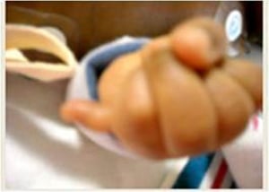
This photo shows the hand of an infant with trisomy 18. Note the clenched fist and postaxial polydactyly.
This photo shows the hand of an infant with trisomy 18. Note the clenched fist and postaxial polydactyly.
Image courtesy of Nina Powell-Hamilton, MD, FAAP, FACMG.

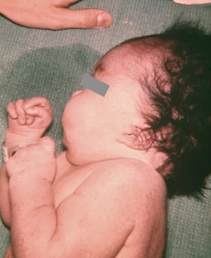
Image courtesy of the Centers for Disease Control and Prevention Public Health Image Library.


Image courtesy of the Centers for Disease Control and Prevention Public Health Image Library.

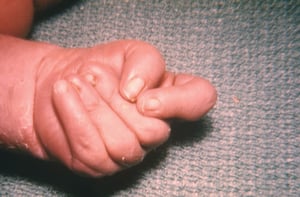
Image courtesy of the Centers for Disease Control and Prevention Public Health Image Library.
This photo shows the hand of an infant with trisomy 18. Note the clenched fist and postaxial polydactyly.
This photo shows the hand of an infant with trisomy 18. Note the clenched fist and postaxial polydactyly.
Image courtesy of Nina Powell-Hamilton, MD, FAAP, FACMG.
Image courtesy of the Centers for Disease Control and Prevention Public Health Image Library.
Image courtesy of the Centers for Disease Control and Prevention Public Health Image Library.
Image courtesy of the Centers for Disease Control and Prevention Public Health Image Library.
Symptoms and signs reference
1. Jaspersen SL, Bruns DA, Candee MS, Battaglia A, Carey JC, Fishler KP. Seizures in trisomy 18: Prevalence, description, and treatment. Am J Med Genet A. 2023;191(4):1026-1037. doi:10.1002/ajmg.a.63113
Diagnosis of Trisomy 18
Prenatal chorionic villus sampling and/or amniocentesis with cytogenetic testing by karyotype analysis, fluorescent in situ hybridization (FISH), and/or chromosomal microarray analysis (CMA)
(See also Next-generation sequencing technologies.)
Diagnosis of trisomy 18 may be suspected postnatally by appearance, or prenatally on ultrasound (eg, with abnormalities of extremities and fetal growth restriction), or by multiple marker screening or noninvasive prenatal screening (NIPS) using cell-free fetal DNA analysis on a maternal blood sample (1). The sensitivity and specificity of NIPS for trisomy 18 is relatively low, compared to trisomy 21. Management decisions should not be based only on the NIPS result.
Confirmation prenatally is by cytogenetic testing (karyotyping, FISH analysis, and/or chromosomal microarray analysis) of samples obtained by amniocentesis or chorionic villus sampling, or postnatally by testing peripheral neonatal blood for patients who did not wish to have additional procedures during pregnancy. Trisomy 18 detected on chorionic villus sampling may warrant further investigation either by amniocentesis or postnatal testing because the trisomy may represent mosaicism that is confined to the placenta, in which aneuploidy is present in the placenta but undetectable in the fetus.

RICHARD J. GREEN/SCIENCE PHOTO LIBRARY
Diagnosis reference
1. American College of Obstetricians and Gynecologists’ Committee on Practice Bulletins—Obstetrics; Committee on Genetics; Society for Maternal-Fetal Medicine: Screening for fetal chromosomal abnormalities: ACOG Practice Bulletin, Number 226. Obstet Gynecol 136(4):e48-e69, 2020. doi: 10.1097/AOG.0000000000004084
Treatment of Trisomy 18
Supportive care
The underlying genetic abnormality cannot be cured.
Children with trisomy 18 have marked developmental delay and disability, and mortality within the first year of life is high, so there is controversy about doing multiple, invasive procedures to correct various associated anomalies.
Multidisciplinary care is often needed. Recommended treatment options for trisomy 18 generally include both palliative care and life-prolonging interventions, such as noninvasive support for feeding difficulties (nasogastric or gastrostomy tube feeding), management of respiratory distress, and treatment of pain or seizures, that are tailored to the individual patient and family preferences.
Early referral for physical, speech, and feeding therapies is important. Support for the family is critical.
Screening for complications of trisomy 18
Treatment of some of the associated anomalies has increased survival for certain people with trisomy 18, which has led to the recognition of an increased risk of solid organ tumors (eg, hepatoblastoma, Wilms tumor). Because early recognition of these tumors and other anomalies is important for successful treatment (if desired), regular surveillance is recommended. Although the specifics are based mainly on expert opinion at one center, there is a published surveillance protocol for tumors and other complications that many specialists find reasonable (1). (See table Surveillance Protocol for Trisomy 18.)
If screening detects abnormalities, children should be referred to the appropriate specialists.
Surveillance Protocol for Trisomy 18
Time Period | Surveillance Recommendations |
|---|---|
Prenatal | Ultrasound at 19 weeks gestation Fetal echocardiogram considered if known trisomy 18 diagnosis or abnormal ultrasound |
Postnatal | Physical examination for external anomalies Complete blood count with differential Echocardiogram Total abdominal ultrasound, including urinary system (48–72 hours after birth)*,† Cranial ultrasound and/or MRI Early screening by pediatric ophthalmologist Airway assessment, possibly including sleep study Baseline serum AFP level |
0–12 months of age | Total abdominal ultrasound at 3, 6, 9, and 12 months of age Serum AFP levels at 3, 6, 9, and 12 months of age Frequent feeding assessments Audiology examination at 6–8 months of age Dental screening |
1–4 years of age | Annual ophthalmologic evaluation After 2 years of age, annual orthopedic examination and spinal radiographs Serum AFP levels every 3–4 months Abdominal ultrasound every 3 months until 4 years of age Dental evaluation every 6 months |
4–7 years of age | Annual ophthalmologic evaluation Annual orthopedic examination and spinal radiographs Abdominal ultrasound every 6 months Renal ultrasound every 3 months until 7 years of age Dental evaluation every 6 months |
7–12 years of age | Annual ophthalmologic evaluation Annual orthopedic examination Abdominal ultrasound every 6 months until 12 years of age |
Puberty | Clinical evaluation for seizures, behavioral changes, and normal sexual development, including menses in females‡ |
≥ 12 years of age | Annual ophthalmologic evaluation Annual orthopedic examination |
* If prenatal ultrasound showed or suggested hydronephrosis but postnatal ultrasound was normal, renal ultrasound is done at 4 to 6 weeks of age. | |
† If urinary tract anomalies are present on initial or subsequent ultrasound, voiding cystourethrogram is typically done. | |
‡ Risk of primary or secondary amenorrhea. | |
AFP = alpha-fetoprotein. | |
Data from Kepple JW, Fishler KP, Peeples ES: Surveillance guidelines for children with trisomy 18. Am J Med Genet A 185(4):1294–1303, 2021. doi: 10.1002/ajmg.a.62097 | |
Treatment reference
1. Kepple JW, Fishler KP, Peeples ES: Surveillance guidelines for children with trisomy 18. Am J Med Genet A 185(4):1294–1303, 2021. doi: 10.1002/ajmg.a.62097
Prognosis for Trisomy 18
More than 50% of children die within the first week of life; fewer than 10% survive the first year (1). However, there are now reports of adults with trisomy 18 (2).
Prognosis references
1. Rasmussen SA, Wong LY, Yang Q, et al. Population-based analyses of mortality in trisomy 13 and trisomy 18. Pediatrics. 2003;111(4 Pt 1):777-784. doi:10.1542/peds.111.4.777
2. Cereda A, Carey JC. The trisomy 18 syndrome. Orphanet J Rare Dis. 2012;7:81. Published 2012 Oct 23. doi:10.1186/1750-1172-7-81


