

Cryptorchidism is failure of one or both testes to descend into the scrotum; in younger children, it is typically accompanied by inguinal hernia. Diagnosis is by testicular examination, sometimes followed by laparoscopy to look for testes that cannot be palpated on examination. Imaging studies are rarely indicated. Treatment is surgical orchiopexy.
Cryptorchidism occurs in approximately 3% of term infants and in 30% of preterm infants. About 7% of cases are bilateral. Three-fourths of undescended testes spontaneously descend within the first 12 months of life (1).
Eighty percent of undescended testes are diagnosed at birth. The remainder are diagnosed during childhood or early adolescence; these are usually caused by an ectopic gubernacular attachment and become apparent after a somatic growth spurt.
Reference
1. Ghirri P, Ciulli C, Vuerich M, et al. Incidence at birth and natural history of cryptorchidism: a study of 10,730 consecutive male infants. J Endocrinol Invest. 2002;25(8):709-715. doi:10.1007/BF03345105
Pathophysiology of Cryptorchidism
Normally, the testes develop at 7 to 8 weeks of gestation and remain cephalad to the internal inguinal ring until about 28 weeks, when they begin their descent into the scrotum guided by condensed mesenchyme (the gubernaculum). Onset of descent is mediated by hormonal (eg, androgens, mullerian-inhibiting factor), physical (eg, gubernacular regression, intra-abdominal pressure), and environmental (eg, maternal exposure to estrogenic or antiandrogenic substances) factors.
A true undescended testis remains in the inguinal canal along the path of descent or is less commonly present in the abdominal cavity or retroperitoneum. An ectopic testis is one that descends normally through the external ring but diverts to an abnormal location and lies outside the normal course of descent (eg, suprapubically, in the superficial inguinal pouch, within the perineum, or along the inner aspect of the thigh).
Complications of cryptorchidism
Undescended testes may cause subfertility and are associated with testicular carcinoma, mainly in the undescended testis and particularly with intra-abdominal malposition. However, in patients with one undescended testis, 10% of cancers develop on the normal side. In untreated cases of intra-abdominal testes, testicular torsion may occur, manifesting as an acute abdomen. Almost all neonates who present with an undescended testis at birth also have an inguinal hernia (due to a patent processus vaginalis [channel left open when testicle descended]).
Etiology of Cryptorchidism
Undescended testes are almost always idiopathic. In phenotypic boys with bilateral, nonpalpable testes at birth suspicion should be high for female virilization caused by congenital adrenal hyperplasia (especially if there is also hypospadias).
Symptoms and Signs of Cryptorchidism
In about 80% of cases, the scrotum on the affected side is empty at birth; in the remainder of cases, a testis is palpable in the scrotum at birth but appears to ascend with linear growth because of an ectopic gubernacular attachment that restrains it from following the normal “descent” of the scrotum. Inguinal hernia associated with cryptorchidism is rarely symptomatic, but the patent processus is often detectable, especially in infants (but less commonly in those with ectopic undescended testes). Rarely, an undescended testis manifests acutely because of testicular torsion.

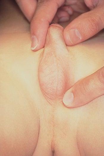
In this photo, the patient has cryptorchidism of the left testis, which is not palpable on physical examination. The left hemiscrotum is hypoplastic.
Diagnosis of Cryptorchidism
Clinical evaluation
Sometimes laparoscopy
Rarely ultrasound or MRI
All boys should have a testicular examination at birth and annually thereafter to assess testicular location and growth.
Undescended and ectopic testes must be distinguished from hypermobile (retractile) testes, which are present in the scrotum but easily retract into the inguinal canal via the cremasteric reflex. Diagnosis of cryptorchidism is by physical examination; a warm environment, warm examiner’s hands, and a relaxed patient are important to avoid stimulating testicular retraction. If needed, the child can be placed in a frog-leg position to better examine the testes. Lubricating the child's groin or the examiner's gloved hands with soap before the examination can help decrease friction and aid in the localization of the testis.
In patients with a unilateral nonpalpable testis, a descended testis that is larger than expected suggests an atrophic undescended testis; confirmation requires surgical intervention typically via diagnostic laparoscopy to seek an intra-abdominal testis or confirm testicular agenesis. However, scrotal or inguinal exploration is sometimes done if a testicular remnant distal to the internal inguinal ring is suspected.
For bilateral nonpalpable testes, patients in the immediate neonatal period should be evaluated for a possible disorder of sexual differentiation (consultation with a pediatric endocrinologist should be considered). If a disorder of sexual differentiation has been ruled out, laparoscopy is often necessary to identify testes located in the abdomen and then bilateral orchiopexy may be done.
(See also the American Urological Association's 2018 Evaluation and Treatment of Cryptorchidism guidelines.)
Treatment of Cryptorchidism
Surgical repair
For a palpable undescended testis, treatment is surgical orchiopexy, in which the testis is brought into the scrotum and sutured into place; an associated inguinal hernia is repaired if present.
For a nonpalpable undescended testis, abdominal laparoscopy is done; if the testis is present, it is moved into the scrotum. If it is atrophic (usually the result of prenatal testicular torsion), the tissue is removed.
Surgery should be done at about 6 months of age in term infants and at 1 year of age in preterm infants because early intervention improves fertility potential and may reduce cancer risk. Also, the shorter the child, the shorter the distance necessary to place the testis into the scrotum.
No intervention is needed for a retractile testis as long as the spermatic cord length is sufficient to allow the testis to rest in a dependent scrotal position without traction when the cremasteric reflex is not stimulated. Hypermobility usually resolves without treatment by puberty when increased testicular size makes retraction more difficult.
(See also the American Urological Association's 2018 Evaluation and Treatment of Cryptorchidism guidelines.)
Key Points
Cryptorchidism affects about 3% of term infants and up to 30% of preterm infants; two-thirds of undescended testes descend spontaneously.
Undescended testes may cause subfertility and increase risk of testicular carcinoma (including in the descended testis).
Clinical evaluation is usually adequate; imaging is rarely indicated.
Treatment is surgical repair.


