


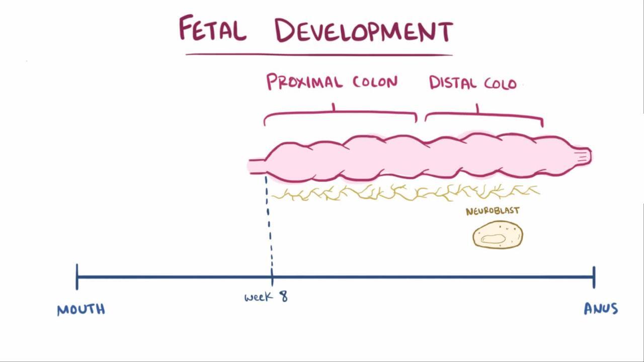
Hirschsprung disease is a congenital anomaly consisting of a failure of neuronal colonization (and thus a failure of innervation) of the lower intestine, usually limited to the colon, resulting in partial or total functional obstruction. Symptoms are obstipation and distention. The diagnosis may be suggested by evaluation with a barium enema; definitive diagnosis requires a rectal biopsy (suction biopsy or surgical biopsy). Anal manometry can help in the evaluation and reveals lack of relaxation of the internal anal sphincter upon rectal balloon distention. Treatment is surgical.
(Also see Overview of Congenital Gastrointestinal Anomalies.)
Hirschsprung disease, sometimes abbreviated HSCR, is caused by congenital absence of the Meissner and Auerbach autonomic plexus (aganglionosis) in the intestinal wall. The etiology of the aganglionosis is thought to be the failure of migration of neural progenitors from the neural crest.
The estimated incidence of Hirschsprung disease is 1 in 5000 live births (1, 2).
Disease is usually limited to the distal colon (75% of cases) but can involve the entire colon (3 to 10% of cases) or even the entire large and small bowels; the denervated area is always contiguous (3). Males are more commonly affected (male:female ratio 4:1) unless the entire colon is involved, in which case there is no gender difference.

General references
1. Mueller JL, Goldstein AM. The science of Hirschsprung disease: What we know and where we are headed. Semin Pediatr Surg. 2022;31(2):151157. doi:10.1016/j.sempedsurg.2022.151157
2. Montalva L, Cheng LS, Kapur R, et al. Hirschsprung disease. Nat Rev Dis Primers. 2023;9(1):54. Published 2023 Oct 12. doi:10.1038/s41572-023-00465-y
3. Urla C, Lieber J, Obermayr F, et al. Surgical treatment of children with total colonic aganglionosis: functional and metabolic long-term outcome. BMC Surg. 2018;18(1):58. Published 2018 Aug 15. doi:10.1186/s12893-018-0383-6
Pathogenesis of Hirschsprung Disease
There is a significant and complex genetic component to Hirschsprung disease, and > 40 different genes are associated with the pathogenesis (1). In 20% of cases, the disease is believed to be caused by a single gene mutation, whereas in the remainder of cases, multiple gene mutations are thought to be the cause. In 12% of cases, Hirschsprung disease is associated with a chromosome abnormality such as Down syndrome.
Approximately 20 to 30% of patients with Hirschsprung disease have another congenital anomaly. For example, approximately 20% of patients with congenital central hypoventilation syndrome also have Hirschsprung disease; the combination is referred to as Haddad syndrome. Other disorders associated with Hirschsprung disease include Waardenburg syndrome, Bardet-Biedl syndrome, Goldberg-Shprintzen syndrome, and cartilage-hair hypoplasia.
The likelihood of disease recurrence among family members increases with increasing length of the involved gut—from 4% overall to 50% for disease involving the entire colon.
Pearls & Pitfalls
|
Peristalsis in the involved segment is absent or abnormal, resulting in continuous smooth muscle spasm and partial or complete obstruction with accumulation of intestinal contents and massive dilation of the more proximal, normally innervated intestine. Skip lesions almost never occur.
Pathogenesis reference
1. Montalva L, Cheng LS, Kapur R, et al. Hirschsprung disease. Nat Rev Dis Primers. 2023;9(1):54. Published 2023 Oct 12. doi:10.1038/s41572-023-00465-y
Symptoms and Signs of Hirschsprung Disease
Patients most commonly present early in life, but some do not present until childhood or even adulthood.
Normally, almost all neonates pass meconium in the first 24 hours of life. The majority of neonates with Hirschsprung disease fail to pass meconium in the first 48 hours of life (1). Infants present with obstipation, abdominal distention, and, finally, vomiting as in other forms of distal bowel obstruction. Occasionally, infants with ultrashort segment aganglionosis have only mild or intermittent constipation, often with intervening bouts of mild diarrhea, resulting in delay in diagnosis.
Infants may also have growth and weight faltering (formerly known as failure to thrive). Less commonly, infants may present with Hirschsprung-associated enterocolitis.
In older infants and children, symptoms and signs may include anorexia, constipation, lack of a physiologic urge to defecate, and, on digital rectal examination, an empty rectum with stool palpable higher up in the colon and an explosive passage of stool upon withdrawal of the examining finger (blast sign).
Symptoms and signs reference
1. Bradnock TJ, Knight M, Kenny S, Nair M, Walker GM; British Association of Paediatric Surgeons Congenital Anomalies Surveillance System. Hirschsprung's disease in the UK and Ireland: incidence and anomalies. Arch Dis Child. 2017;102(8):722-727. doi:10.1136/archdischild-2016-311872
Diagnosis of Hirschsprung Disease
Barium enema
Rectal suction or surgical biopsy
Rectal manometry
Diagnosis of Hirschsprung disease should be made as soon as possible. The longer the disease goes untreated, the greater the chance of developing Hirschsprung-associated enterocolitis, a complication that may be fulminant and fatal. Most patients can be diagnosed in early infancy.

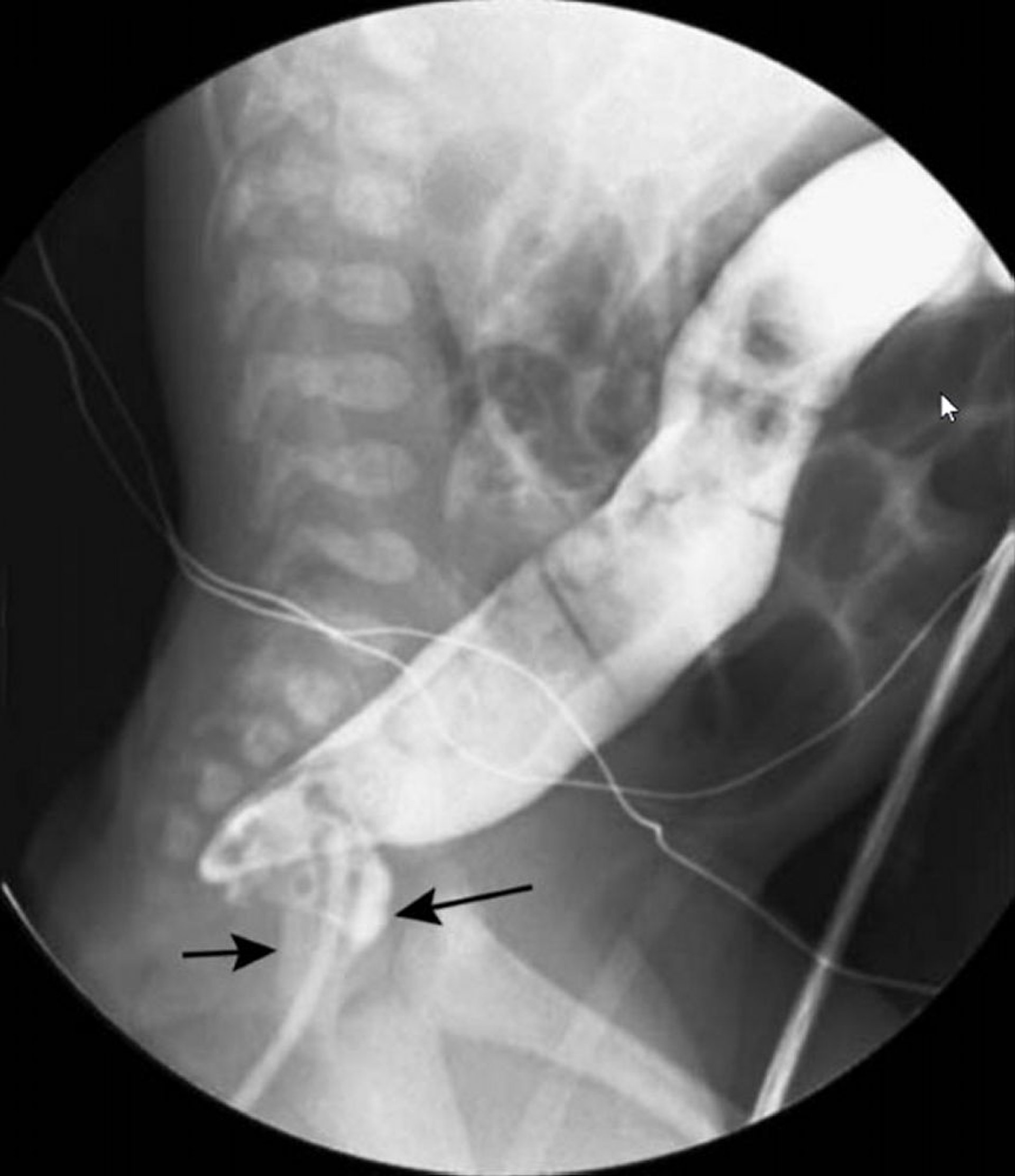
In this image, barium enema shows a narrowed rectum (black arrows) and a dilated colon (white arrow) more proximally.
© Springer Science+Business Media
Initial approach is typically with barium enema and/or rectal suction biopsy. (NOTE: A barium enema should not be performed in patients suspected of having Hirschsprung-associated enterocolitis because of the risk of perforation.) Barium enema may show a transition in diameter between the dilated, normally innervated colon proximal to the narrowed distal segment (which lacks normal innervation). Barium enema should be performed without prior preparation, which can dilate the abnormal segment and decompress the proximal colon, rendering the test nondiagnostic. Because characteristic findings may not be present in the neonatal period, a 24-hour post-evacuation radiograph should be taken; if the colon is still filled with barium, Hirschsprung disease is likely.
A rectal suction biopsy can disclose the absence of ganglion cells. Acetylcholinesterase staining can be performed to highlight the enlarged nerve trunks; calretinin staining can be performed to detect the absence of mucosal innervation (1).
Some centers also can do rectal manometry, which typically reveals a lack of relaxation of the internal anal sphincter upon balloon insufflation of the rectum, which is characteristic of the abnormal innervation.
Definitive diagnosis requires a surgical or suction biopsy of the rectum and then surgical biopsies to map the extent of disease and thus plan surgical treatment.
Genetic testing is not routine but may be performed if evaluation shows manifestations of a genetic syndrome or in cases of total colonic aganglionosis.

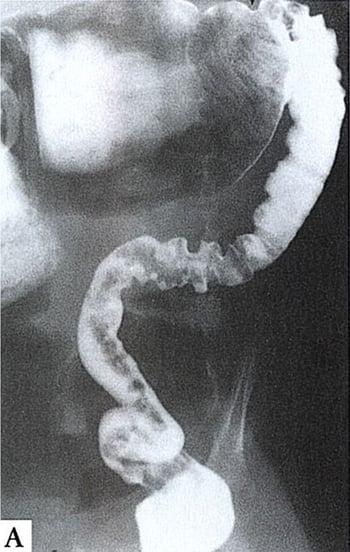
This barium enema shows aganglionosis up to the splenic flexure. The aganglionic bowel is narrowed and spastic (right), whereas the normally innervated proximal bowel (top) is dilated and filled with fecal material.
Pearls & Pitfalls
|
Diagnosis reference
1. Claxton HL, Lounis SA, Stanton M, Hall NJ, Aldeiri B. The Diagnostic Value of Immunohistochemistry Markers in Hirschsprung Disease; A Systematic Review and Meta-analysis. J Pediatr Surg. 2025;60(2):162010. doi:10.1016/j.jpedsurg.2024.162010
Treatment of Hirschsprung Disease
Surgical repair
Treatment of Hirschsprung disease is surgical repair by bringing normally innervated bowel to the anus with preservation of the anal sphincters. In the neonate with long-segment disease, this typically requires a 2-stage procedure beginning with a colostomy proximal to the aganglionic segment to decompress the colon (1). Then the neonate is allowed to grow before the second stage of the procedure, in which the entire aganglionic portion of the colon is resected and a pull-through procedure is performed. In the neonate with short-segment disease, a 1-stage procedure is performed at diagnosis or after a 1- to 3-month delay. Results using laparoscopic technique are similar to those of the open method and are associated with shorter hospitalizations, earlier initiation of feeding, and less pain.
After definitive repair, the prognosis is good, although a number of infants have chronic dysmotility with constipation, obstructive problems, or both.
Treatment reference
1. Montalva L, Cheng LS, Kapur R, et al. Hirschsprung disease. Nat Rev Dis Primers. 2023;9(1):54. Published 2023 Oct 12. doi:10.1038/s41572-023-00465-y
Key Points
Congenital denervation affects the distal colon and less often larger regions of the colon and sometimes even the small bowel.
Infants typically present with findings of distal bowel obstruction, such as obstipation, abdominal distention, and vomiting.
Barium enema findings (done without prior preparation) and rectal manometry are highly suggestive; diagnosis is confirmed by rectal biopsy.
The affected segment is resected surgically, and the normal segment is pulled through to the anus.
Hirschsprung-Associated Enterocolitis (Toxic Megacolon)
Hirschsprung-associated enterocolitis is a life-threatening complication of Hirschsprung disease resulting in a grossly enlarged colon, often followed by sepsis and shock.
The etiology of Hirschsprung-associated enterocolitis (HAEC), seems to be marked proximal dilation secondary to obstruction, with thinning of the colonic wall, bacterial overgrowth, and translocation of gut bacteria. Sepsis or shock can develop (more often when the entire colon is affected by Hirschsprung disease), and death can follow rapidly; however, with treatment mortality is rare (1–3). Up to 20% of full-term infants with Hirschsprung disease may develop HAEC, so close monitoring of infants with Hirschsprung disease is essential.
HAEC occurs most commonly in the first several months of life before surgical correction but can occur postoperatively, typically in the first year after surgery. Infants with Down syndrome are at increased risk of developing this complication postoperatively. Infants present with fever (neonates with temperature instability), abdominal distention, diarrhea (which may be bloody or explosive), and, subsequently, obstipation.
A barium enema should not be performed in patients suspected of having HAEC because of the risk of perforation.
Initial treatment of HAEC is supportive with bowel rest, fluid resuscitation, decompression with nasogastric and rectal tubes, and broad-spectrum antibiotics to include anaerobic coverage (eg, a metronidazole with either ampicillin and gentamicin, aztreonam, or piperacillin/tazobactam) (4). Some experts advocate rectal irrigation in infants who are not critically ill to clean out the colon, but this must be performed carefully so as not to increase colonic pressure and cause perforation.
Surgery is the definitive treatment for infants who have not yet undergone surgical repair, as well as for those with perforation or necrotic gut. For infants who can be stabilized, definitive surgical therapy can be performed instead of the previous standard approach of a diverting ileostomy or colostomy. The previous standard approach should be performed in infants with severe disease.
References
1. Pruitt LCC, Skarda DE, Rollins MD, Bucher BT. Hirschsprung-associated enterocolitis in children treated at US children's hospitals. J Pediatr Surg. 2020;55(3):535-540. doi:10.1016/j.jpedsurg.2019.10.060
2. Chantakhow S, Tepmalai K, Singhavejsakul J, Tantraworasin A, Khorana J. Prognostic factors of postoperative Hirschsprung-associated enterocolitis: a cohort study. Pediatr Surg Int. 2023;39(1):77. Published 2023 Jan 9. doi:10.1007/s00383-023-05364-7
3. Montalva L, Cheng LS, Kapur R, et al. Hirschsprung disease. Nat Rev Dis Primers. 2023;9(1):54. Published 2023 Oct 12. doi:10.1038/s41572-023-00465-y
4. Gosain A, Frykman PK, Cowles RA, et al. Guidelines for the diagnosis and management of Hirschsprung-associated enterocolitis. Pediatr Surg Int. 2017;33(5):517-521. doi:10.1007/s00383-017-4065-8
Drug Information for the Topic


