Wernicke encephalopathy is characterized by acute onset of confusion, nystagmus, partial ophthalmoplegia, and ataxia due to thiamine deficiency. Diagnosis is primarily clinical. The disorder may remit with treatment, persist, or degenerate into Korsakoff psychosis. Treatment consists of thiamine and supportive measures.
Wernicke encephalopathy results from inadequate intake or absorption of thiamine plus continued carbohydrate ingestion. Severe alcoholism is a common underlying condition. Excessive alcohol intake interferes with thiamine absorption from the gastrointestinal tract and hepatic storage of thiamine; the poor nutrition associated with alcoholism often precludes adequate thiamine intake.
Wernicke encephalopathy may also result from other conditions that cause prolonged undernutrition or vitamin deficiency (eg, recurrent dialysis, hyperemesis, starvation, gastric plication, cancer, advanced HIV infection). Giving a carbohydrate load to patients with thiamine deficiency (ie, refeeding after starvation or giving IV dextrose-containing solutions to high-risk patients) can trigger Wernicke encephalopathy, although in practice this is rarely seen except with prolonged glucose infusions prior to thiamine administration (1, 2).
Not all people who misuse alcohol and are thiamine-deficient develop Wernicke encephalopathy, suggesting that other factors may be involved. Genetic abnormalities that result in a defective form of transketolase, an enzyme that processes thiamine, may be involved.
Characteristically, central nervous system lesions are symmetrically distributed around the third ventricle, aqueduct, and fourth ventricle. Changes in the mamillary bodies, dorsomedial thalamus, locus ceruleus, periaqueductal gray matter, ocular motor nuclei, and vestibular nuclei are common.
General references
1. Jasti J, Sherman K, Gummin D, Farkas A. Prevalence of Wernicke's Encephalopathy When Receiving Dextrose Before Thiamine: A National Study of Veterans. Acad Emerg Med. 2025;32(11):1197-1202. doi:10.1111/acem.70131
2. Schabelman E, Kuo D. Glucose before thiamine for Wernicke encephalopathy: a literature review. J Emerg Med. 2012;42(4):488-494. doi:10.1016/j.jemermed.2011.05.076
Symptoms and Signs of Wernicke Encephalopathy
In patients with Wernicke encephalopathy, clinical changes occur suddenly.
Oculomotor abnormalities, including horizontal and vertical nystagmus and partial ophthalmoplegias (eg, lateral rectus palsy, conjugate gaze palsies), are common. Pupils may be abnormal; they are usually sluggish or unequal.
Vestibular dysfunction without hearing loss is common, and the oculovestibular reflex may be impaired. Gait ataxia may result from vestibular disturbances, cerebellar dysfunction, and/or polyneuropathy; gait is wide-based and slow, with short-spaced steps.
Global confusion is often present; it is characterized by profound disorientation, indifference, inattention, drowsiness, or stupor. Peripheral nerve pain thresholds are often elevated, and many patients develop severe autonomic dysfunction characterized by sympathetic hyperactivity (eg, tremor, agitation) or hypoactivity (eg, hypothermia, postural hypotension, syncope). In untreated patients, stupor may progress to coma, then to death.
Diagnosis of Wernicke Encephalopathy
Usually a clinical diagnosis
Whole blood high-performance liquid chromatography (HPLC) thiamine assay prior to administration of thiamine, where available
Sometimes MRI
There are no specific diagnostic studies. Diagnosis of Wernicke encephalopathy is clinical and depends on recognition of underlying undernutrition or vitamin deficiency in a patient with typical signs and symptoms.

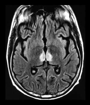
This axial T2-FLAIR MRI scan shows the classical findings of Wernicke encephalopathy with increased signal within the medial thalami.
This axial T2-FLAIR MRI scan shows the classical findings of Wernicke encephalopathy with increased signal within the m
Living Art Enterprises, LLC/Science Source/SCIENCE PHOTO LIBRARY

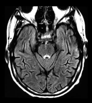
This axial T2-FLAIR MRI scan shows the classical findings of Wernicke encephalopathy with increased signal within the periaqueductal gray matter.
This axial T2-FLAIR MRI scan shows the classical findings of Wernicke encephalopathy with increased signal within the p
Living Art Enterprises, LLC/Science Source/SCIENCE PHOTO LIBRARY
This axial T2-FLAIR MRI scan shows the classical findings of Wernicke encephalopathy with increased signal within the medial thalami.
This axial T2-FLAIR MRI scan shows the classical findings of Wernicke encephalopathy with increased signal within the m
Living Art Enterprises, LLC/Science Source/SCIENCE PHOTO LIBRARY
This axial T2-FLAIR MRI scan shows the classical findings of Wernicke encephalopathy with increased signal within the periaqueductal gray matter.
This axial T2-FLAIR MRI scan shows the classical findings of Wernicke encephalopathy with increased signal within the p
Living Art Enterprises, LLC/Science Source/SCIENCE PHOTO LIBRARY
Brain MRI can be useful to confirm or support the diagnosis, particularly in cases not associated with alcohol use (1, 2). Lesions are typically seen in the thalami, mamillary bodies, tectal plate, and periaqueductal tissue.
A whole blood HPLC thiamine assay should be drawn prior to administration of thiamine, where available (2).
There are no characteristic abnormalities in cerebrospinal fluid, evoked potentials, brain imaging, or electroencephalogram. However, these tests, as well as laboratory tests (eg, blood tests, glucose, complete blood count, liver tests, arterial blood gas measurements, toxicology screening), should typically be performed to exclude other etiologies.
Diagnosis references
1. Manzo G, De Gennaro A, Cozzolino A, Serino A, Fenza G, Manto A. MR imaging findings in alcoholic and nonalcoholic acute Wernicke's encephalopathy: a review. Biomed Res Int. 2014;2014:503596. doi:10.1155/2014/503596
2. Galvin R, Bråthen G, Ivashynka A, et al. EFNS guidelines for diagnosis, therapy and prevention of Wernicke encephalopathy. Eur J Neurol. 2010;17(12):1408-1418. doi:10.1111/j.1468-1331.2010.03153.x
Treatment of Wernicke Encephalopathy
Parenteral thiamine
Sometimes parenteral magnesium
Supportive care
Treatment of Wernicke encephalopathy consists of immediate administration of thiamine, in conjunction with other B vitamins, including folate (1). A recommended thiamine regimen is 500 mg of thiamine IV infused over 30 minutes 3 times daily for 2 consecutive days and 250 mg IV or IM once daily for an additional 5 days (2). Magnesium is a necessary cofactor in thiamine-dependent metabolism, and hypomagnesemia should be corrected. Supportive treatment includes rehydration, correction of electrolyte abnormalities, and general nutritional therapy, including multivitamins. Patients with advanced disease require hospitalization. Alcohol cessation is mandatory.
Because Wernicke encephalopathy is preventable, all undernourished patients should be treated with parenteral thiamine plus vitamin B12 and folate, particularly if IV dextrose is necessary (1). Thiamine is also prudent before any treatment is begun in patients who present with a reduced level of consciousness. Patients who are undernourished, including patients who undergo obesity surgery (gastric bypass), should continue to receive thiamine as outpatients for 6 months.
Treatment references
1. Galvin R, Bråthen G, Ivashynka A, et al. EFNS guidelines for diagnosis, therapy and prevention of Wernicke encephalopathy. Eur J Neurol. 2010;17(12):1408-1418. doi:10.1111/j.1468-1331.2010.03153.x
2. Cook CC, Hallwood PM, Thomson AD. B Vitamin deficiency and neuropsychiatric syndromes in alcohol misuse. Alcohol Alcohol. 1998;33(4):317-336. doi:10.1093/oxfordjournals.alcalc.a008400
Prognosis for Wernicke Encephalopathy
Prognosis depends on timely diagnosis of Wernicke encephalopathy. If begun in time, treatment may correct all abnormalities. Ocular symptoms usually begin to abate within 24 hours after early thiamine administration. Ataxia and confusion may persist for days to months. Memory and learning impairment may not resolve completely. Untreated, mortality is approximately 20%. Of surviving patients, 80% develop Korsakoff psychosis (1, 2); the combination is called Wernicke–Korsakoff syndrome. Even when treated, 1 study estimated mortality of alcohol-related Wernicke encephalopathy at approximately 3%, and the rate of progression to Korsakoff psychosis at approximately 17% (3).
Prognosis references
1. Sinha S, Kataria A, Kolla BP, Thusius N, Loukianova LL. Wernicke Encephalopathy-Clinical Pearls. Mayo Clin Proc. 2019;94(6):1065-1072. doi:10.1016/j.mayocp.2019.02.018
2. Galvin R, Bråthen G, Ivashynka A, et al. EFNS guidelines for diagnosis, therapy and prevention of Wernicke encephalopathy. Eur J Neurol. 2010;17(12):1408-1418. doi:10.1111/j.1468-1331.2010.03153.x
3. Rasiah R, Gregoriano C, Mueller B, Kutz A, Schuetz P. Hospital Outcomes in Medical Patients With Alcohol-Related and Non-Alcohol-Related Wernicke Encephalopathy. Mayo Clin Proc. 2024;99(5):740-753. doi:10.1016/j.mayocp.2023.07.021
Drug Information for the Topic