

Leukemia is a malignant condition involving the excess production of immature or abnormal leukocytes, which eventually suppresses the production of normal blood cells and results in symptoms related to the cytopenias.
Malignant transformation usually occurs at the pluripotent stem cell level, although it sometimes involves a committed stem cell with more limited capacity for self-renewal. Abnormal proliferation, clonal expansion, aberrant differentiation, and diminished apoptosis (programmed cell death) lead to replacement of normal blood elements with malignant cells.
In the United States in 2025 there were approximately 66,890 new cases of leukemia (of all types) in adults and children, and approximately 23,540 deaths (1).
General reference
1. American Cancer Society: Cancer Statistics Center. Accessed February 6, 2026.
Classification of Leukemia
The current approach to classifying leukemia is based on the 2016 World Health Organization (WHO) system (1). The WHO classification is based on a combination of clinical features and morphology, immunophenotype, and genetic factors. Other less commonly used classification systems include the French-American-British (FAB) system, which is based on the morphology of the abnormal leukocytes (2).
Leukemias are commonly also categorized as:
Acute or chronic: Based on the percentage of blasts or leukemia cells in bone marrow or blood
Myeloid or lymphoid: Based on the predominant lineage of the malignant cells
The 4 most common leukemias and their distinguishing features are summarized in the table .
For 2025, the American Cancer Society estimates the distribution of new cases in the United States by leukemia type as follows (3):
Acute myeloid leukemia (AML): 33%
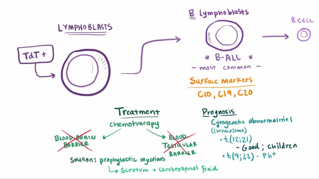
Acute lymphoblastic leukemia (ALL): 9%
Chronic myeloid leukemia (CML): 14%
Chronic lymphocytic leukemia (CLL): 35%
Other leukemias (eg, mixed phenotype acute leukemia [MPAL], hairy cell leukemia): 8%
Findings at Diagnosis in the Most Common Leukemias
Feature | ||||
|---|---|---|---|---|
Peak age of incidence | Childhood | Any age | Middle and old age | Adulthood |
White blood cell count | High in 50% Normal or low in 50% | High in 60% Normal or low in 40% | High in 98% Normal or low in 2% | High in 100% |
Differential white blood cell count | Many lymphoblasts | Many myeloblasts | Small lymphocytes | Entire myeloid series |
Anemia | Severe in > 90% | Severe in > 90% | Mild in approximately 50% | Mild in 80% |
Platelets | Low in > 80% | Low in > 90% | Low in 20 to 30% | High in 60% Low in 10% |
Lymphadenopathy | Common | Occasional | Common | Infrequent |
Splenomegaly | In 60% | In 50% | Usual and moderate | Usual and severe |
Other features | Without prophylaxis, central nervous system commonly involved | Central nervous system rarely involved Sometimes Auer rods in myeloblasts | Occasionally hemolytic anemia and hypogammaglobulinemia | Low leukocyte alkaline phosphatase level Philadelphia chromosome–positive in > 90% |
Acute leukemias
Acute leukemias consist of predominantly immature, poorly differentiated cells (usually blast forms). Acute leukemias are divided into:

Chronic leukemias
Chronic leukemias have more mature cells than do acute leukemias. They usually manifest as leukocytosis with or without cytopenias in otherwise asymptomatic patients. Findings and management differ significantly between:
Myelodysplastic syndromes
Myelodysplastic syndromes are a group of clonal hematopoietic stem cell disorders unified by the presence of distinct mutations of hematopoietic stem cells. They involve progressive bone marrow failure but with an insufficient proportion of blast cells (< 20%) for making a definite diagnosis of acute myeloid leukemia; 40 to 60% of cases evolve into acute myeloid leukemia.
Leukemoid reaction
A leukemoid reaction is a neutrophil count > 50,000/mcL (> 50 × 109/L) not caused by malignant transformation of a hematopoietic stem cell. It can result from a variety of causes, particularly other cancers or systemic infection. Usually the cause is apparent, but apparent benign neutrophilia can be mimicked by chronic neutrophilic leukemia or chronic myeloid leukemia.
Classification references
1. Swerdlow SH, Campo E, Pileri SA, et al. The 2016 revision of the World Health Organization classification of lymphoid neoplasms. Blood. 2016;127(20):2375-2390. doi:10.1182/blood-2016-01-643569
2. Bennett JM, Catovsky D, Daniel MT, et al. Proposals for the classification of the acute leukaemias. French-American-British (FAB) co-operative group. Br J Haematol. 1976;33(4):451-458. doi: 10.1111/j.1365-2141.1976.tb03563.x
3. American Cancer Society. Cancer Facts and Statistics. Accessed February 6, 2026.
Risk Factors for Leukemia
Risk of developing leukemia is increased in patients with:
History of exposure to ionizing radiation (eg, post–atomic bomb in Nagasaki and Hiroshima) or to chemicals (eg, benzene, some pesticides, polyaromatic hydrocarbons in tobacco smoke); exposure can lead to acute leukemias
Prior treatment with certain antineoplastic medications, including alkylating agents, topoisomerase II inhibitors, hydroxyurea, and maintenance lenalidomide after autologous stem cell transplantation with melphalan-containing conditioning regimens for multiple myeloma; can lead to therapy-related acute myeloid leukemia (AML)Prior treatment with certain antineoplastic medications, including alkylating agents, topoisomerase II inhibitors, hydroxyurea, and maintenance lenalidomide after autologous stem cell transplantation with melphalan-containing conditioning regimens for multiple myeloma; can lead to therapy-related acute myeloid leukemia (AML)
Infection with a virus (eg, human T lymphotropic virus [HTLV] 1, Epstein Barr virus) can rarely cause certain forms of leukemia and lymphoma, mainly in regions where such infections are common, such as Asia and Africa
History of antecedent hematologic disorders, including myelodysplastic syndromes and myeloproliferative neoplasms, which can lead to AML
Preexisting genetic conditions (eg, Fanconi anemia, Bloom syndrome, ataxia-telangiectasia, Down syndrome, xeroderma pigmentosum, Li-Fraumeni syndrome), which predispose to AML or ALL
More Information
The following English-language resource may be useful. Please note that The Manual is not responsible for the content of this resource.
Drug Information for the Topic


