

Streptococci are gram-positive aerobic organisms that cause many disorders, including pharyngitis, pneumonia, wound and skin infections, sepsis, and endocarditis. Symptoms vary with the organ infected. Sequelae of infections due to group A beta-hemolytic streptococci may include acute rheumatic fever and poststreptococcal glomerulonephritis. Most strains are sensitive to penicillin; macrolide-resistant strains have emerged.
(See also Pneumococcal Infections, Rheumatic Fever, Tonsillopharyngitis, Postinfectious Glomerulonephritis, and Toxic Shock Syndrome.)
Classification of Streptococci
Three different types of streptococci are initially differentiated by their appearance when they are grown in culture on sheep blood agar:
Beta-hemolytic streptococci produce zones of clear hemolysis around each colony.
Alpha-hemolytic streptococci (commonly called viridans streptococci) are surrounded by green discoloration resulting from incomplete hemolysis.
Gamma-hemolytic streptococci are nonhemolytic.
Subsequent classification, based on carbohydrates in the cell wall, divides streptococci into 20 Lancefield groups A through H and K through V (see table Lancefield Classification). In the Lancefield classification, enterococci were initially included among the group D streptococci but are now classified as a separate genus even though they do express Lancefield group D antigens. Lancefield groups K through V are streptococcal species of limited virulence that can cause infections in people who are immunocompromised.
Viridans streptococci form a separate group that is considered difficult to classify. Some streptococci such as Streptococcus pneumoniae are alpha-hemolytic, ie, they are a type of viridans streptococci but do not express Lancefield antigens.
Lancefield Classification*
Lancefield Group | Species | Hemolysis | Associated Diseases | Treatment |
|---|---|---|---|---|
A | Streptococcus pyogenes | Beta | Pharyngitis, tonsillitis, wound and skin infections, septicemia, scarlet fever, pneumonia, rheumatic fever, glomerulonephritis, endocarditis (rare) | Penicillin Erythromycin, clindamycin (resistance increasing in the United States)Erythromycin, clindamycin (resistance increasing in the United States) |
Necrotizing fasciitis | Expeditious surgical management Beta-lactam (usually broad spectrum until etiology is identified; if the presence of GABHS is confirmed, penicillin or cefazolin can be used) plus clindamycinBeta-lactam (usually broad spectrum until etiology is identified; if the presence of GABHS is confirmed, penicillin or cefazolin can be used) plus clindamycin | |||
B | S. agalactiae | Beta | Sepsis, postpartum or neonatal sepsis, meningitis, skin infections, endocarditis, septic arthritis, urinary tract infection | Penicillin or ampicillin, cephalosporins, vancomycinPenicillin or ampicillin, cephalosporins, vancomycin |
C and G | S. equi, S. equimilis, S. zooepidemicus, S. canis | Beta | Pharyngitis, pneumonia, cellulitis, pyoderma, erysipelas, impetigo, wound infections, puerperal sepsis, neonatal sepsis, endocarditis, septic arthritis Human infection with group C implies a zoonotic origin | Penicillin, vancomycin, cephalosporins, macrolides (variable susceptibility)Penicillin, vancomycin, cephalosporins, macrolides (variable susceptibility) |
D | Enterococcal:† Enterococcus faecalis, E. faecium Nonenterococcal: S. gallolyticus , S. equinus | Alpha or gamma | Endocarditis, urinary tract infection, intra-abdominal infection, cellulitis, wound infection as well as concurrent bacteremia | Penicillin, ampicillin, vancomycin (plus an aminoglycoside for serious infection)Penicillin, ampicillin, vancomycin (plus an aminoglycoside for serious infection) Vancomycin-resistant enterococci: , oxazolidinones (linezolid), lipopeptide (daptomycin)Vancomycin-resistant enterococci: , oxazolidinones (linezolid), lipopeptide (daptomycin) |
S. gallolyticus (formerly S. bovis biotype I) | Colonic adenomas or carcinomas, endocarditis | |||
Viridans‡ | S. mutans, S. sanguis, S. salivarius, S. mitis, S. anginosus , S. constellatus, S. intermedius | Alpha or gamma | Endocarditis, bacteremia, meningitis, localized infection, abscesses (particularly S. anginosus) | Penicillin, ampicillin, vancomycin (plus an aminoglycoside for serious infection), other antibiotics based on in vitro susceptibilityPenicillin, ampicillin, vancomycin (plus an aminoglycoside for serious infection), other antibiotics based on in vitro susceptibility |
S. suis | Meningitis, sometimes toxic shock syndrome | |||
S. iniae | Cellulitis, invasive infections from fish | Penicillin | ||
* This table includes only the most clinically relevant of the 20 Lancefield groups. | ||||
† Enterococci were initially included among the group D streptococci but are now classified as a separate genus even though they do express Lancefield group D antigens. | ||||
‡ Do not conform to specific Lancefield serogroups. | ||||
GABHS = group A beta-hemolytic streptococci. | ||||
Virulence Factors
Many streptococci elaborate virulence factors, including streptolysins, DNAases, and hyaluronidase, which contribute to tissue destruction and spread of infection. A few strains release exotoxins that activate certain T cells, triggering release of cytokines, including tumor necrosis factor-alpha, interleukins, and other immunomodulators. These cytokines activate the complement, coagulation, and fibrinolytic systems, leading to shock, organ failure, and death.
Diseases Caused by Streptococci
The most significant streptococcal pathogen is S. pyogenes, which is beta-hemolytic and in Lancefield group A and is thus denoted as group A beta-hemolytic streptococci (GABHS).
The most common acute diseases due to GABHS are:
In addition, delayed, nonsuppurative complications (rheumatic fever, acute glomerulonephritis) sometimes occur ≥ 2 weeks after infection.
GABHS can spread through the affected tissues and along lymphatic channels (causing lymphangitis) to regional lymph nodes (causing lymphadenitis). GABHS can also cause local suppurative complications, such as peritonsillar abscess, otitis media, sinusitis, and bacteremia. Suppuration depends on the severity of infection and the susceptibility of tissue.
Other serious GABHS infections include septicemia, puerperal sepsis, endocarditis, pneumonia, and empyema.
Disease caused by other streptococcal species is less prevalent and usually involves soft-tissue infection or endocarditis (see table Lancefield Classification). Some non-GABHS infections occur predominantly in certain populations (eg, group B streptococci in neonates and postpartum women).
Streptococcal pharyngitis
GABHS are the most common cause of acute-onset bacterial pharyngitis. Other bacterial causes include groups C and G streptococci and Fusobacterium necrophorum. Most patients with GABHS pharyngitis are children between 3 years and 14 years of age who present with acute onset of sore throat, fever, beefy-red pharynx, and purulent tonsillar exudate. GABHS pharyngitis is rare in children younger than 3 years of age.
The cervical and submaxillary nodes may enlarge and become tender. Streptococcal pharyngitis can lead to peritonsillar abscess. Cough, laryngitis, and stuffy nose are not characteristic of streptococcal pharyngeal infection; their presence suggests another cause (usually viral or allergic).
Humans are the primary reservoir for group A streptococci, which spread person-to-person via saliva or nasal secretions from an infected person. An asymptomatic carrier state may exist in as many as 20% of people, especially school-age children, in the winter and spring (1).

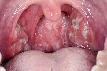
In this image, the pharynx is erythematous, and the tonsils have purulent exudates.
Scarlet fever
Scarlet fever (sometimes called scarlatina), a predominantly childhood disease, usually follows a pharyngeal streptococcal infection; less commonly, it follows streptococcal infections at other sites (eg, the skin). Transmission of scarlet fever is enhanced in environments that result in close contact among people (eg, in schools or day care centers).
Scarlet fever is caused by group A streptococcal strains that produce an erythrogenic toxin, leading to a diffuse pink-red cutaneous flush that commonly blanches with pressure. The rash is seen best on the abdomen or lateral chest and as dark red lines in skinfolds (Pastia lines) or as circumoral pallor. The rash consists of characteristic numerous small (1- to 2-mm) papular elevations, giving a sandpaper quality to the skin. The upper layer of the previously reddened skin often desquamates after fever subsides. The rash usually lasts 2 to 5 days. Scarlet fever occurs more frequently among children aged > 5 years who presented with a sore throat but experienced delay in diagnosis of GABHS infection (2).
Scarlet fever is not as common as it was in the past, but in recent years there have been a number of significant outbreaks in the United States, Europe, and China. For example, there has been a persistent increase in scarlet fever in England with the highest incidence for nearly 50 years. Potential reasons for the increased incidence include macrolide resistance, weakened herd immunity, environmental factors, and the absence of a vaccine for the bacteria that cause the disease (group A streptococci). Over a century ago, scarlet fever was feared as a potentially lethal disease. Its mortality and morbidity waned, however, well before antibiotics became available. Since the advent of antibiotic therapy, the mortality rate of treated scarlet fever has been < 1% (3).
To address some of these concerns, the Strep A Vaccine Global Consortium (SAVAC), in conjunction with the World Health Organization, support the development and availability of a safe and effective vaccine targeting streptococcal variants causing scarlet and rheumatic fevers (4).

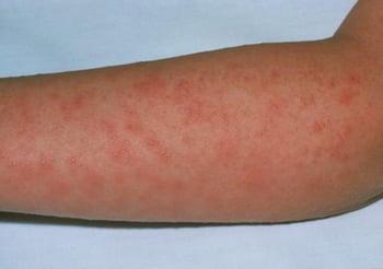
The classic scarlet fever rash initially appears as tiny red papules on the chest and abdomen. Papules may then spread over the body. The rash resembles sunburn, feels like rough sandpaper, and lasts about 2 to 5 days.
A strawberry tongue (inflamed papillae protruding through a bright red coating) also occurs and must be differentiated from that seen in toxic shock syndrome and Kawasaki disease.


This tongue is erythematous with prominent papillation.
Other symptoms are similar to those in streptococcal pharyngitis, and the course and management of scarlet fever are the same as those of other group A infections.
Streptococcal skin infections
Skin infections include:
Impetigo is a superficial skin infection that causes crusting or bullae.

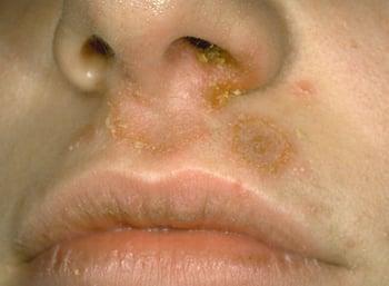
In impetigo, clusters of vesicopustular or bullous lesions form, rupture, and develop a honey-colored crust.
Erysipelas is a superficial cellulitis that also involves the lymphatics. Patients have shiny, red (violaceous in dark skin), raised, indurated lesions with distinct margins. It is most often caused by GABHS, but other streptococcal and nonstreptococcal organisms are sometimes involved.


CID/SCIENCE PHOTO LIBRARY
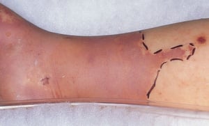
Cellulitis involves the deeper layers of skin and may spread rapidly because of the numerous lytic enzymes and toxins produced mainly by group A streptococci. Cellulitis typically presents with erythema, sometimes with a clear line of demarcation. Warmth and tenderness are often present.

This photo shows the focal erythema and swelling, usually accompanied by warmth and tenderness, characteristic of focal cellulitis. Note the clinician has marked the border of the cellulitis with a pen, to facilitate recognition of spread or resolution.
This photo shows the focal erythema and swelling, usually accompanied by warmth and tenderness, characteristic of focal
© Springer Science+Business Media

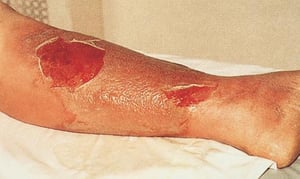
This photo shows the focal redness and swelling of the lower leg, usually accompanied by warmth and tenderness, characteristic of focal cellulitis. The clinician has marked the border of the cellulitis with a pen, to facilitate recognition of spread or resolution. Note the line of redness extending up the thigh due to lymphangitis.
This photo shows the focal redness and swelling of the lower leg, usually accompanied by warmth and tenderness, charact
© Springer Science+Business Media

© Springer Science+Business Media
This photo shows the focal erythema and swelling, usually accompanied by warmth and tenderness, characteristic of focal cellulitis. Note the clinician has marked the border of the cellulitis with a pen, to facilitate recognition of spread or resolution.
This photo shows the focal erythema and swelling, usually accompanied by warmth and tenderness, characteristic of focal
© Springer Science+Business Media
This photo shows the focal redness and swelling of the lower leg, usually accompanied by warmth and tenderness, characteristic of focal cellulitis. The clinician has marked the border of the cellulitis with a pen, to facilitate recognition of spread or resolution. Note the line of redness extending up the thigh due to lymphangitis.
This photo shows the focal redness and swelling of the lower leg, usually accompanied by warmth and tenderness, charact
© Springer Science+Business Media
© Springer Science+Business Media
Necrotizing fasciitis
Monomicrobial (type-2) necrotizing fasciitis due to S. pyogenes is a severe dermal (and sometimes muscle) infection that spreads along fascial planes (5). Inoculation originates through the skin or bowel.
Polymicrobial (type-1) necrotizing fasciitis is likely when the source is the bowel (eg, after intestinal surgery, bowel perforation, diverticulitis, or appendicitis) (5). Formerly known as streptococcal gangrene and popularized as the flesh-eating bacteria, polymicrobial necrotizing fasciitis may involve a host of aerobic and anaerobic flora, including Clostridium perfringens.
Necrotizing fasciitis is prevalent among people who use injection drugs.
Symptoms of necrotizing fasciitis begin with fever and exquisite localized pain out of proportion to physical findings; pain increases rapidly over time and is often the first (and sometimes only) manifestation. Diffuse or local erythema may be present. Thrombosis of the microvasculature causes ischemic necrosis, leading to rapid spread and disproportionally severe toxicity. In some patients, adjacent muscles are invaded. Shock and renal dysfunction are common. Mortality is high, even with treatment.

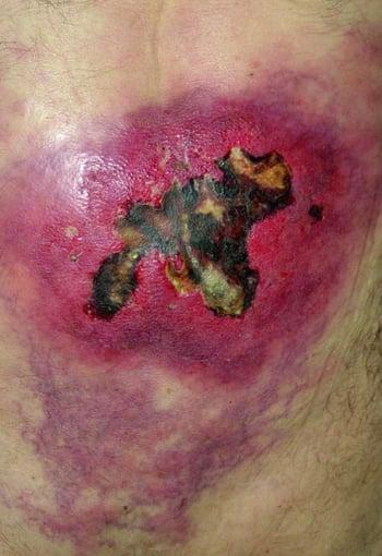
This photo shows life-threatening infection of the subcutaneous fat and muscles by streptococci (group A), causing widespread necrosis involving the lower back.
Streptococcal toxic shock syndrome
Streptococcal toxic shock syndrome (TSS), similar to that caused by S. aureus, may result from toxin-producing strains of GABHS and occasionally from other streptococci.
Delayed complications of streptococcal infection
The mechanism by which certain strains of GABHS cause delayed complications is unclear but may involve cross-reactivity of streptococcal antibodies against host tissue.
Rheumatic fever, an inflammatory disorder, occurs in approximately 3% of patients in the weeks after untreated GABHS (exudative) pharyngitis (6). It is much less common in regions where antibiotics are used to treat streptococcal infections, but incidence is still high in regions where antibiotics are not as widely used.
Diagnosis of a first episode is based on a combination of arthritis, carditis, chorea, specific cutaneous manifestations, and laboratory test results (Jones criteria—see table Modified Jones Criteria for a First Episode of Acute Rheumatic Fever).
One of the most important reasons for treating GABHS pharyngitis (strep throat) is to prevent rheumatic fever.
Poststreptococcal acute glomerulonephritis (PSGN) is an acute nephritic syndrome following pharyngitis or skin infection due to a certain limited number of nephritogenic strains of GABHS (eg, M protein serotypes 12 and 49). After a throat or skin infection with one of these strains, approximately 5 to 25% of patients develop acute glomerulonephritis (7). It is most common among children, occurring 1 to 3 weeks after infection (. Nearly all children, but somewhat fewer adults, recover without permanent renal damage. Antibiotic treatment of GABHS infection has little effect on the development of glomerulonephritis (8).
PANDAS syndrome (pediatric autoimmune neuropsychiatric disorder associated with streptococcal infections) refers to a subset of obsessive disorders in children or tic disorders in children known as pediatric acute-onset neuropsychiatric syndrome (PANS) that were thought to be exacerbated by GABHS infection; however, this association remains inconclusive until further high-quality data are available (9, 10). Importantly, prolonged antibiotic treatment or prophylaxis for PANDAS or PANS has not been shown to have a significant effect on symptom occurrence or resolution.
Certain forms of psoriasis (eg, guttate) may also be related to beta-hemolytic streptococcal infections.
Diseases caused by streptococci references
1. Shulman ST, Bisno AL, Clegg HW, et al. Clinical practice guideline for the diagnosis and management of group A streptococcal pharyngitis: 2012 update by the Infectious Diseases Society of America [published correction appears in Clin Infect Dis. 2014 May;58(10):1496. Dosage error in article text]. Clin Infect Dis. 2012;55(10):e86-e102. doi:10.1093/cid/cis629
2. Herdman MT, Cordery R, Karo B, et al. Clinical management and impact of scarlet fever in the modern era: findings from a cross-sectional study of cases in London, 2018-2019. BMJ Open. 2021;11(12):e057772. Published 2021 Dec 24. doi:10.1136/bmjopen-2021-057772
3. Lamagni T, Guy R, Chand M, et al. Resurgence of scarlet fever in England, 2014-16: a population-based surveillance study. Lancet Infect Dis. 2018;18(2):180-187. doi:10.1016/S1473-3099(17)30693-X
4. International Vaccine Institute: Strep A Vaccine Global Consortium (SAVAC). Accessed August 1, 2025.
5. Stevens DL, Bryant AE. Necrotizing Soft-Tissue Infections. N Engl J Med. 2017;377(23):2253-2265. doi:10.1056/NEJMra1600673
6. Hawkes MA, Ameriso SF. Neurologic complications of rheumatic fever. Handb Clin Neurol. 2021;177:23-31. doi:10.1016/B978-0-12-819814-8.00002-0
7. Anthony BF, Kaplan EL, Wannamaker LW, Briese FW, Chapman SS. Attack rates of acute nephritis after type 49 streptococcal infection of the skin and of the respiratory tract. J Clin Invest. 1969;48(9):1697-1704. doi:10.1172/JCI106135
8. Lasch EE, Frankel V, Vardy PA, Bergner-Rabinowitz S, Ofek I, Rabinowitz K. Epidemic glomerulonephritis in Israel. J Infect Dis. 1971;124(2):141-147. doi:10.1093/infdis/124.2.141
9. Board of Directors. Pediatric Acute-Onset Neuropsychiatric Syndrome (PANS): Clinical Report. Pediatrics. 2025;155(3):e2024070334. doi:10.1542/peds.2024-070334
10. Hardin H, Shao W, Bernstein JA. An updated review of pediatric autoimmune neuropsychiatric disorders associated with Streptococcus/pediatric acute-onset neuropsychiatric syndrome, also known as idiopathic autoimmune encephalitis: What the allergist should know. Ann Allergy Asthma Immunol. 2023;131(5):567-575. doi:10.1016/j.anai.2023.08.022
Diagnosis of Streptococcal Infections
Culture
Sometimes rapid antigen tests, nucleic acid amplification tests (NAATs)
Sometimes antistreptococcal antibody titers (commonly ASO, anti-DNase B; less commonly antihyaluronidase, antinicotinamide adenine dinucleotidase, antistreptokinase)
Streptococci are readily identified by culture on a sheep blood agar plate.
Rapid antigen-detection tests that can detect GABHS directly from throat swabs are available (ie, for point-of-care use) (1). Many tests use enzyme immunoassay, but tests that use optical immunoassay are also available. These rapid tests have high specificity (> 95%) but vary considerably in sensitivity (70% to 80 to 90% for the optical immunoassay test) (2). Thus, positive results can establish the diagnosis, but negative results, at least in children, should be confirmed by culture. Because streptococcal pharyngitis is less common among adults and adults are unlikely to have poststreptococcal complications, many clinicians do not confirm a negative rapid screening result in adults by culture unless use of a macrolide is being considered; in such cases, culture with susceptibility testing to detect macrolide resistance should be done.
Point-of-care NAATs (eg, polymerase chain reaction) can provide results in approximately 15 to 30 minutes to rapidly inform clinical decision-making; they offer improved sensitivity compared to rapid antigen-detection tests while maintaining specificity (3).
The Centor criteria can be used to guide decisions about testing for GABHS or selecting empiric antibiotic treatment for patients with pharyngitis. The criteria are age, presence of tonsillar exudate, anterior cervical lymphadenopathy, fever > 40° C, and absence of cough. The absence of cough often helps in differentiating streptococcal from other organisms implicated in pharyngitis.

Demonstrating antistreptococcal antibodies in serum during convalescence provides only indirect evidence of infection. Antistreptococcal antibody tests are not useful in diagnosing acute GABHS infection because antibodies first develop several weeks after GABHS infection begins and a single high antibody titer is more likely to reflect an infection that occurred in the distant past. Antibodies are most useful in diagnosis of poststreptococcal sequelae, such as rheumatic fever and glomerulonephritis.
Antistreptolysin O (ASO) and antideoxyribonuclease B (anti-DNase B) titers begin to increase about 1 week after the GABHS infection and peak about 1 to 2 months after the infection. Both titers may remain elevated for several months, even after uncomplicated infections. The ASO titer increases in only 75 to 80% of infections (suggesting that 20% or more of patients with infections may not mount an ASO titer response) (4). For completeness in difficult cases, any one of the other tests (antihyaluronidase, antinicotinamide adenine dinucleotidase, antistreptokinase) can also be used.
Titers are measured in the acute phase and in the convalescent phase 2 to 4 weeks later; a positive result is defined as a ≥ 2-fold increase in the titer. A single titer greater than the upper limit of normal suggests an antecedent streptococcal infection or the presence of high streptococcal endemicity in the community.
Penicillin given within the first 5 days for symptomatic streptococcal pharyngitis may delay the appearance and decrease the magnitude of the ASO response.
Patients with streptococcal pyoderma usually do not have a significant ASO response but may have a response to other antigens (ie, anti-DNAase, antihyaluronidase).
Diagnosis references
1. Mustafa Z, Ghaffari M: Diagnostic Methods, Clinical Guidelines, and Antibiotic Treatment for Group A Streptococcal Pharyngitis: A Narrative Review. Front Cell Infect Microbiol. 10:563627, 2020. Published 2020 Oct 15. doi:10.3389/fcimb.2020.563627
2. Plainvert C, Duquesne I, Touak G, et al: In vitro evaluation and comparison of 5 rapid antigen detection tests for the diagnosis of beta-hemolytic group A streptococcal pharyngitis. Diagn Microbiol Infect Dis 83(2):105–111, 2015. doi: 10.1016/j.diagmicrobio.2015.06.012
3. May L, Sickler J, Robbins EM, Tang S, Chugh K, Tran N: The Impact of Point-of-Care Polymerase Chain Reaction Testing on Prescribing Practices in Primary Care for Management of Strep A: A Retrospective Before-After Study. Open Forum Infect Dis. 9(5):ofac147, 2022. doi:10.1093/ofid/ofac147
4. Wannamaker LW, Ayoub EM: Antibody titers in acute rheumatic fever. Circulation. 21:598-614, 1960. doi:10.1161/01.cir.21.4.598
Treatment of Streptococcal Infections
Usually penicillin
Pharyngitis
Pharyngeal GABHS infections, including scarlet fever, are ordinarily self-limited (1). Antibiotics (especially penicillin) typically shorten the course in young children, especially those with scarlet fever, but have only modest effect on symptoms in adolescents and adults. However, antibiotics help prevent local suppurative complications (eg, peritonsillar abscess), otitis media, and rheumatic fever (2, 3).
Penicillin is preferred for pharyngeal GABHS infections. No isolate of GABHS has shown penicillin resistance clinically. However, some streptococcal strains appear to have in vitro tolerance to penicillin (ie, significantly decreased bactericidal effect of penicillin). The in vivo (ie, the clinical) significance of such resistance is still unclear. However, one Cochrane systematic review comparing the efficacy of different antibiotics concluded that there were no clinically relevant differences in symptom resolution when comparing cephalosporins and macrolides with penicillin in the treatment of GABHS tonsillopharyngitis (4). Insufficient evidence was available to draw conclusions regarding shortening disease duration, preventing relapse, and complications.
A single injection of penicillin G benzathine 600,000 units IM for small children (A single injection of penicillin G benzathine 600,000 units IM for small children (< 27 kg) or 1.2 million units IM for children weighing ≥ 27 kg, adolescents, and adults usually suffices.
Oral medications may be used if the patient can be trusted to maintain the regimen for the required 10 days. Choices include:
Penicillin V 500 mg (250 mg for children < 27 kg) orally every 8 to 12 hoursPenicillin V 500 mg (250 mg for children < 27 kg) orally every 8 to 12 hours
Amoxicillin 500 mg (maximum 1 g) once or twice a day for 10 days (which is an effective substitute for penicillin V and is often preferred for ease of administration)Amoxicillin 500 mg (maximum 1 g) once or twice a day for 10 days (which is an effective substitute for penicillin V and is often preferred for ease of administration)
Oral narrow-spectrum cephalosporins (eg, cephalexin, cefadroxil) are also effective and can be used, unless patients have an anaphylactic reaction to penicillin. Alternatives include macrolides (azithromycin, clarithromycin) and clindamycin. However, these non-beta-lactam alternative options have high rates of resistance in some communities. Local antibiotic susceptibility patterns should be confirmed before use, and confirmation by in vitro susceptibility testing is recommended.
Macrolides, 12 mg/kg once daily (maximum 500 mg), are also inactive against Fusobacterium necrophorum, a common cause of pharyngitis in adolescents and adults.. Delaying treatment 1 to 2 days until laboratory confirmation increases neither the duration of disease nor the incidence of GABHS pharyngitis complications.
Amoxicillin/clavulanic acid is also effective.Amoxicillin/clavulanic acid is also effective.
Sulfamethoxazole/trimethoprim (TMP/SMX), some of the fluoroquinolones, and tetracyclines are unreliable for treating GABHS infection.Sulfamethoxazole/trimethoprim (TMP/SMX), some of the fluoroquinolones, and tetracyclines are unreliable for treating GABHS infection.
Sore throat, headache, and fever can be treated with analgesics or antipyretics. Aspirin should be avoided in children. Bed rest and isolation are unnecessary. Close contacts who are symptomatic or have a history of poststreptococcal complications should be examined for streptococci.
Skin infection
Cellulitis is often treated without doing a culture because isolating organisms can be difficult. Thus, regimens effective against both streptococci and staphylococci are used; for example, one of the following may be used:
Dicloxacillin or cephalexin if methicillin-resistant Dicloxacillin or cephalexin if methicillin-resistantStaphylococcus aureus (MRSA) is not likely
TMP/SMX, linezolid, tedizolid, minocycline, or clindamycin if MRSA is suspected (see TMP/SMX, linezolid, tedizolid, minocycline, or clindamycin if MRSA is suspected (seetreatment of cellulitis)
Necrotizing fasciitis should be treated in an intensive care unit. Extensive (sometimes repeated) surgical debridement is required. Until etiology is confirmed by culture, a recommended initial antibiotic regimen is a broad-spectrum beta-lactam (eg, piperacillin/tazobactam) or a carbapenem (eg, meropenem, imipenem) plus clindamycin for antitoxin effects. If clindamycin resistance is known or suspected, linezolid or tedizolid can be used instead. Vancomycin should be added if infection with MRSA is suspected. The use of intravenous immunoglobulin (IVIG), especially when used concurrently with clindamycin, has also been associated with decreased mortality in GABHS necrotizing fasciitis (should be treated in an intensive care unit. Extensive (sometimes repeated) surgical debridement is required. Until etiology is confirmed by culture, a recommended initial antibiotic regimen is a broad-spectrum beta-lactam (eg, piperacillin/tazobactam) or a carbapenem (eg, meropenem, imipenem) plus clindamycin for antitoxin effects. If clindamycin resistance is known or suspected, linezolid or tedizolid can be used instead. Vancomycin should be added if infection with MRSA is suspected. The use of intravenous immunoglobulin (IVIG), especially when used concurrently with clindamycin, has also been associated with decreased mortality in GABHS necrotizing fasciitis (5).
Other streptococcal infections
For treating groups B, C, and G infections, antibiotics of choice are:
Penicillin
AmpicillinAmpicillin
VancomycinVancomycin
Cephalosporins or macrolides are usually effective, but susceptibility tests must guide therapy, especially in very ill, immunocompromised, or debilitated patients and in patients with foreign bodies at the infection site. Surgical wound drainage and debridement as adjuncts to antimicrobial therapy may be lifesaving.
S. gallolyticus is relatively susceptible to antibiotics. Although vancomycin-resistant is relatively susceptible to antibiotics. Although vancomycin-resistantS. gallolyticus isolates have been reported, the organism remains susceptible to penicillin and aminoglycosides.
Most viridans streptococci are susceptible to penicillin G and other beta-lactams. Resistance to penicillin is rare, and when present is relative resistance. Therapy for such strains should be dictated by the results of in vitro susceptibility tests or knowledge of local resistance patterns, and may require higher doses of penicillin. For some conditions, the addition of a second antimicrobial agent (ie, infective endocarditis and gentamicin) may be necessary.are susceptible to penicillin G and other beta-lactams. Resistance to penicillin is rare, and when present is relative resistance. Therapy for such strains should be dictated by the results of in vitro susceptibility tests or knowledge of local resistance patterns, and may require higher doses of penicillin. For some conditions, the addition of a second antimicrobial agent (ie, infective endocarditis and gentamicin) may be necessary.
Treatment references
1. Herdman MT, Cordery R, Karo B, et al. Clinical management and impact of scarlet fever in the modern era: findings from a cross-sectional study of cases in London, 2018-2019. BMJ Open. 2021;11(12):e057772. doi:10.1136/bmjopen-2021-057772
2. Shulman ST, Bisno AL, Clegg HW, et al. Clinical practice guideline for the diagnosis and management of group A streptococcal pharyngitis: 2012 update by the Infectious Diseases Society of America [published correction appears in Clin Infect Dis. 2014 May;58(10):1496. Dosage error in article text]. Clin Infect Dis. 2012;55(10):e86-e102. doi:10.1093/cid/cis629
3. Kumar RK, Antunes MJ, Beaton A, et al. Contemporary Diagnosis and Management of Rheumatic Heart Disease: Implications for Closing the Gap: A Scientific Statement From the American Heart Association [published correction appears in Circulation. 2021 Jun 8;143(23):e1025-e1026. doi: 10.1161/CIR.0000000000000984]. Circulation. 2020;142(20):e337-e357. doi:10.1161/CIR.0000000000000921
4. Hedin K, Thorning S, van Driel ML. Different antibiotic treatments for group A streptococcal pharyngitis. Cochrane Database Syst Rev. 2023;11(11):CD004406. doi:10.1002/14651858.CD004406.pub6
5. Carapetis JR, Jacoby P, Carville K, Ang SJ, Curtis N, Andrews R. Effectiveness of clindamycin and intravenous immunoglobulin, and risk of disease in contacts, in invasive group a streptococcal infections. Clin Infect Dis. 2014;59(3):358-365. doi:10.1093/cid/ciu304
Key Points
The most significant streptococcal pathogen is S. pyogenes, which is denoted as group A beta-hemolytic streptococci (GABHS).
The 2 most common acute diseases due to GABHS are pharyngitis and skin infections.
Delayed nonsuppurative complications, including rheumatic fever and poststreptococcal glomerulonephritis, can occur.
Rapid antigen tests (ie, for point-of-care use) are very specific but not highly sensitive; confirm negative results using culture, at least in children.
NAATs provide a more sensitive rapid testing option.
A penicillin or cephalosporin is preferred for pharyngitis; because macrolide resistance is increasing, susceptibility testing is recommended if that class of antibiotic is used.
For skin infection, use dicloxacillin or cephalexin if MRSA is not likely. TMP/SMX, linezolid, doxycycline, or clindamycin (if resistance rates are low in the community) are options if MRSA is suspected.For skin infection, use dicloxacillin or cephalexin if MRSA is not likely. TMP/SMX, linezolid, doxycycline, or clindamycin (if resistance rates are low in the community) are options if MRSA is suspected.
Treat groups B, C, and G streptococcal infections with the same antibiotics used for treating GABHS.
Drugs Mentioned In This Article


