

Pelvic fractures can involve the pubic symphysis, innominate bones, acetabulum, sacroiliac joint or sacrum. They range from minimally displaced stable injuries caused by low energy falls to dramatically displaced and unstable injures that can cause massive hemorrhage. Genitourinary, intestinal, and neurologic injuries may also occur. Diagnosis is by radiographs and usually CT. Minor stable fractures require only symptomatic treatment. Unstable fractures and fractures with significant hemorrhage usually require external fixation or open reduction with internal fixation (ORIF).
(See also Overview of Fractures.)
Pathophysiology of Pelvic Fractures
The pelvic bones, with the anterior and posterior sacroiliac ligaments and fibrous joints between bones (syndesmoses), form a ring. Pelvic fractures may or may not disrupt the ring; ring disruption results from fractures in ≥ 2 places and results in instability.
Complications
Many significant anatomic structures traverse the pelvis and are often damaged. Vascular injuries (eg, iliac vein injuries) may occur and cause significant hemorrhage, especially with posterior pelvic fractures. Hemorrhage may be external (indicating open fracture) or only internal; either can cause hemorrhagic shock.
Concomitant genitourinary injuries (eg, urethral or bladder tears) are common, particularly in anterior fractures. Intestinal injuries may occur, particularly in patients with posterior fractures. Nerve roots and plexuses near the sacral foramina may be damaged in posterior fractures.
Etiology of Pelvic Fractures
Most pelvic fractures result from high-energy injuries, most commonly caused by motor vehicle crashes (including motor vehicle–pedestrian collisions) or a fall from a height. Some (eg, symphyseal or pubic ramus fractures) result from minor or low-energy injuries (eg, falls at home), especially in patients with osteoporosis.
Some pelvic fractures, typically in adolescents with open growth plates, are small avulsion fractures of the anterior or inferior iliac spine or of the ischial tuberosity.
There are complex classification systems based on the mechanism, location, and/or stability of the injury
Symptoms and Signs of Pelvic Fractures
Most patients with a pelvic fracture have groin and/or lower back pain. Compression of the pubic symphysis or simultaneous compression of both anterior superior iliac spines is usually painful, particularly in severe fractures, and may indicate instability.
Depending on the severity of the fracture, patients may or may not be able to walk.
Signs of genitourinary and/or gynecologic (usually vulvovaginal) injuries include
Hematuria
Anuria
Blood at the urethral meatus
Scrotal or perineal hematoma
Abnormal location of the prostate on rectal examination (called a high-riding prostate)
Vaginal bleeding
Intestinal or rectal injuries can cause
Abdominal or pelvic pain
Rectal bleeding
Later development of peritonitis
Neurologic injuries can cause
Weakness or loss of sensation and reflexes in the lower extremities, rectum, or perineum
Incontinence
Urinary retention
Mortality rate is high when fractures are unstable or posterior or when they cause hemorrhagic shock.
Diagnosis of Pelvic Fractures
Radiographs
Usually noncontrast CT
Pelvic fractures should be considered if patients have pain in the pelvic region or hip or have had major trauma. An anteroposterior pelvis radiograph shows most fractures.

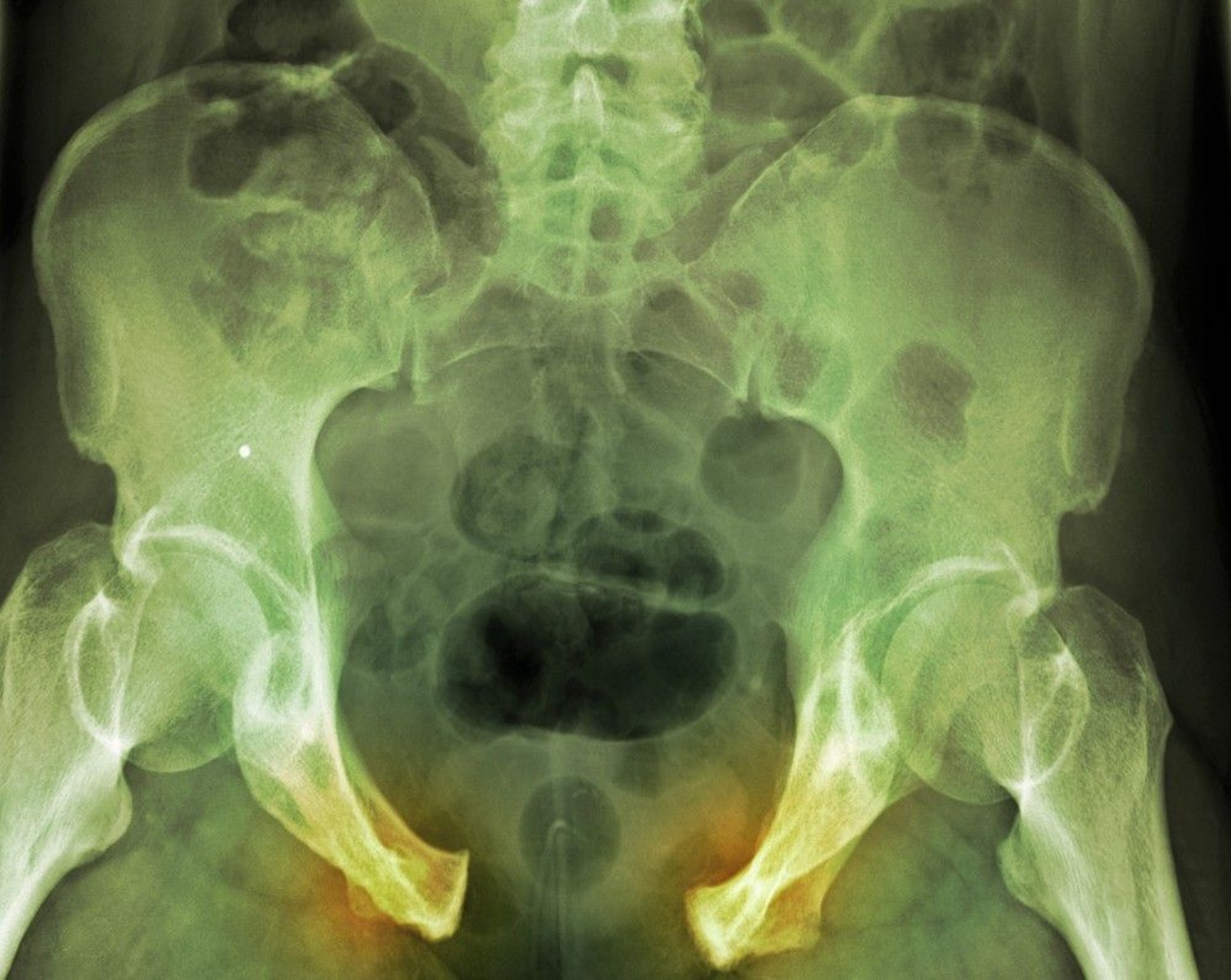
DU CANE MEDICAL IMAGING LTD/SCIENCE PHOTO LIBRARY
A displaced fracture indicates that the pelvic ring is disrupted, suggesting another fracture or syndesmotic or ligamentous disruption. Specialized radiograph views (eg, Judet views to visualize the acetabulum) may be necessary.
CT is more sensitive than radiographs and is usually done to identify all fracture fragments and certain associated injuries when the fracture is due to a high-energy injury. CT is often unnecessary when patients have an isolated pubic ramus fracture due to a low-energy injury or a small avulsion fracture.
Diagnosis and treatment of associated injuries takes precedence over full definition of the pelvic fracture. Bladder and urethral injuries should be considered and evaluated. Tests include
Urinalysis to check for hematuria
Neurologic examination
Pelvic examination in women to check for vulvovaginal injury
Retrograde urethrography to check for urethral injury, if indicated
Traditionally, digital rectal examination is done in men to check for a high-riding prostate, which suggests increased risk of posterior urethral injury. However, the usefulness of this examination is not clear. If urethral injury is suspected (eg, because blood is seen at the meatus), retrograde urethrography should be done.
Treatment of Pelvic Fractures
For stable fractures, usually only symptomatic treatment
For unstable fractures, external fixation or open reduction with internal fixation (ORIF)
For significant hemorrhage, external fixation or sometimes angiographic embolization or pelvic packing
For pelvic fractures, an orthopedic or trauma surgeon is consulted. Almost all patients with pelvic fractures (other than stable fractures, eg, minimally displaced fractures of the pubic rami) should be admitted for observation to watch for continued bleeding.
Stable pelvic fractures often require only symptomatic treatment, particularly when patients can walk unaided.
Acetabular fractures result from a high-energy injury (eg, a fall from height or a motor vehicle crash). Acetabular fractures are treated surgically if the fractures are displaced or instability persists after closed reduction. Acetabular fractures with posterior wall injuries are managed nonsurgically. Orthopedic consultation is recommended for these injuries.
Unstable pelvic fractures should be wrapped (eg, in sheets) or stabilized with a commercially available pelvic binder as soon as possible in the emergency department; such stabilization can often decrease or stop bleeding. Orthopedic consultation is needed when pelvic fractures are unstable to determine whether ORIF or external (percutaneous) screw fixation should be done. External screw fixation can be done in the emergency department by orthopedic surgeons.
Indications for external screw fixation include
Ongoing hemorrhage or hemodynamic instability, particularly in patients with large pelvic disruption
Multisystem trauma
Need for stabilization before transfer for definitive care
External screw fixation reduces morbidity and the length of stay in a hospital.
Pearls & Pitfalls
|
If bleeding persists, angiographic embolization or surgery for pelvic packing and/or internal pelvic fixation is required.
Unstable fractures without significant hemorrhage require a pelvic binder, applied in the emergency department; ORIF is the definitive treatment.
Other associated injuries are treated.
Key Points
Serious pelvic fractures due to high-energy injuries are often associated with genitourinary and vascular injuries.
Some (eg, symphyseal or pubic ramus fractures), particularly in patients with osteoporosis, result from minor injuries (eg, falls at home).
Do CT for high-energy injuries.
Stabilize fractures, control bleeding, and treat associated injuries and symptoms.


