


Progressive multifocal leukoencephalopathy (PML) is caused by reactivation of the JC virus. The disease usually occurs in patients with impaired cell-mediated immunity, particularly patients with HIV infection. PML results in subacute and progressive demyelination in the central nervous system, multifocal neurologic deficits, and death, usually within 9 months. Diagnosis is with MRI plus cerebrospinal fluid analysis using polymerase chain reaction testing. In AIDS patients, highly active antiretroviral therapy may slow down the progression, and patients taking immunosuppressants may improve when those drugs are withdrawn. Limited data suggest that treatment with immune checkpoint inhibitors, which activate the immune response to the JC virus, is sometimes helpful. Treatment is otherwise supportive.
(See also Introduction to Brain Infections.)
Etiology of PML
Progressive multifocal leukoencephalopathy is caused by reactivation of the JC virus, a ubiquitous human papovavirus that is typically acquired during childhood and remains latent in the kidneys and possibly other sites (eg, mononuclear cells, central nervous system [CNS]). The reactivated virus has a tropism for oligodendrocytes.
Most patients who develop PML have depressed cell-mediated immunity due to
AIDS (the most common risk factor)
Reticuloendothelial system disorders (eg, leukemia, lymphoma)
Other conditions (eg, Wiskott-Aldrich syndrome, organ transplantation)
The risk in AIDS increases with increasing HIV viral load; prevalence of PML has decreased because of widespread use of more effective antiretrovirals.
Increasingly, PML is occurring as a complication of immunomodulatory therapy. The drugs most frequently implicated include
Symptoms and Signs of PML
Clumsiness may be the first symptom of progressive multifocal leukoencephalopathy. Hemiparesis is the most common finding. Aphasia, dysarthria, and hemianopia are also common. Multifocal cortical damage produces cognitive impairment in two thirds of patients. Sensory, cerebellar, and brain stem deficits may be present.
Headaches and convulsive seizures are rare and occur most often in patients with AIDS.
Gradual, relentless progression culminates in death, usually 1 to 9 months after symptoms begin.
Diagnosis of PML
MRI
Cerebrospinal fluid (CSF) testing for JC viral DNA
Progressive multifocal leukoencephalopathy is suspected in patients with unexplained progressive brain dysfunction, particularly in those with depressed cell-mediated immunity.
Provisional diagnosis of PML is made by contrast-enhanced MRI, which shows single or multiple white matter lesions on T2-weighted images. A contrast agent enhances, usually faintly and peripherally, 5 to 15% of lesions. CT may show low-density, nonenhancing lesions but is significantly less sensitive than MRI.

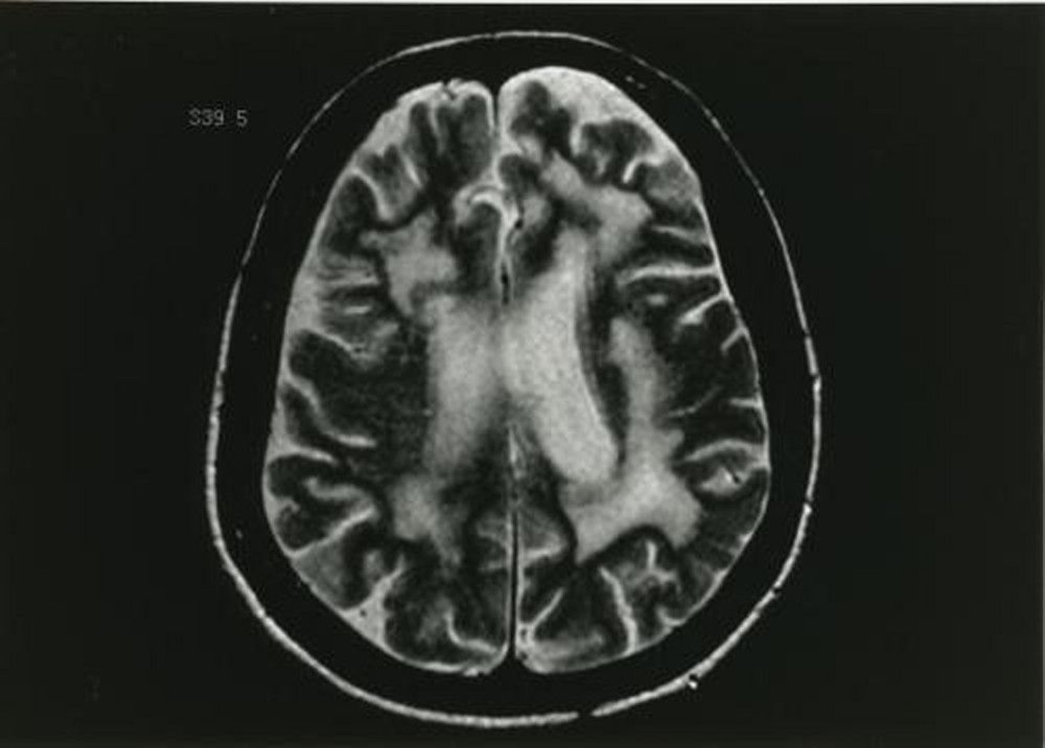
Image provided by John E. Greenlee, MD.
CSF is analyzed for JC viral DNA using PCR; a positive result with compatible neuroimaging findings is nearly pathognomonic. Routine CSF analysis is usually normal.
Serologic tests are not helpful. Stereotactic biopsy can provide a definitive diagnosis but is rarely warranted.
Treatment of PML
Supportive care
Treatment of progressive multifocal leukoencephalopathy is mainly supportive.
immune reconstitution inflammatory syndrome (IRIS). In IRIS, the recovering immune system produces an intense inflammatory response against the JC virus, thus worsening symptoms. Imaging done after IRIS develops shows greater contrast enhancement of the lesions and may show significant cerebral edema. Corticosteroids may be helpful. Depending on the severity of IRIS and of AIDS, clinicians may decide to withdraw ART.
Withdrawal of immunosuppressants may result in clinical improvement. However, patients who stop taking these drugs are also at risk of developing IRIS.
Treatment references
1. Cortese I, Muranski P, Enose-Akahata Y, et alN Engl J Med 380 (17):1597–1605, 2019. doi: 10.1056/NEJMoa1815039 Epub 2019 Apr 10.
2. Lambert N, El Moussaoui M, Maquet P: Immune checkpoint inhibitors for progressive multifocal leukoencephalopathy: Identifying relevant outcome factors. Eur J Neurol 2021 28 (11):3814–3819, 2021. doi: 10.1111/ene.15021 Epub 2021 Jul 26.
Key Points
Reactivation of the ubiquitous JC virus, usually due to impaired cell-mediated immunity, leads to PML.
PML commonly causes clumsiness, hemiparesis, aphasia, dysarthria, hemianopia, and cognitive impairment.
Do MRI and test CSF for JC virus DNA in patients who have impaired cell-mediated immunity and unexplained progressive brain dysfunction.


