


( See also Cytomegalovirus (CMV) Infection in adults and Overview of Neonatal Infections.)
Cytomegalovirus (CMV) is frequently isolated from neonates. Although most infants shedding this virus are asymptomatic, others have life-threatening illness and devastating long-term sequelae.
It is not known when a woman with primary CMV can safely conceive. Because risk to the fetus is difficult to assess, women who develop primary CMV during pregnancy should be counseled, but few experts recommend routine serologic testing for CMV before or during pregnancy in healthy women.
Etiology of Congenital and Perinatal CMV Infection
Congenital CMV infection, which occurs in 0.2 to 1% of live births worldwide, may result from transplacental acquisition of either a primary or recurrent maternal infection. Clinically apparent disease in the neonate is much more likely to occur after a primary maternal exposure, particularly in the first half of pregnancy. In some higher socioeconomic groups in the US, 50% of young women lack antibody to CMV, making them susceptible to primary infection.
Perinatal CMV infection is acquired by exposure to infected cervical secretions, breast milk, or blood products. Maternal antibody is thought to be protective, and most exposed term infants are asymptomatic or not infected. In contrast, preterm infants (who lack antibody to CMV) can develop serious infection or can die, particularly when transfused with CMV-positive blood. Efforts should be made to transfuse these infants with only CMV-negative blood or components or to use blood that has been filtered to remove leukocytes, which carry CMV. Such leukoreduced blood is considered by many experts to be CMV safe.
Symptoms and Signs of Congenital and Perinatal CMV Infection
Many women who become infected with CMV during pregnancy are asymptomatic, but some develop a mononucleosis-like illness.
About 10% of infants with congenital CMV infection are symptomatic at birth. Manifestations include the following:
Intrauterine growth restriction
Petechiae
Hepatosplenomegaly
Periventricular calcifications
Chorioretinitis
Hepatitis
Pneumonitis
Sensorineural hearing loss
Infants who acquire CMV during or after birth, especially if they are premature, may develop a sepsis-like syndrome, pneumonia, hepatosplenomegaly, hepatitis (which can lead to liver failure), thrombocytopenia, and atypical lymphocytosis. However, if transmission is via breast milk, the risk of severe symptomatic disease and long-term sequelae is low.
Diagnosis of Congenital and Perinatal CMV Infection
Viral culture using urine, saliva, or tissue
Polymerase chain reaction (PCR) testing using urine, saliva, blood, or tissue
Symptomatic congenital CMV infection must be distinguished from other congenital infections, including toxoplasmosis, rubella, lymphocytic choriomeningitis virus (LCMV), and syphilis.
In neonates, viral detection using culture or PCR of urine, saliva, or a tissue sample is the primary diagnostic tool; maternal diagnosis can also be made by serologic testing or PCR ( see Diagnosis). Culture specimens should be refrigerated until inoculation of fibroblast cells. Congenital CMV is diagnosed if the virus is identified in urine, saliva, or other body fluids obtained within the first 2 to 3 weeks of life; urine and saliva have the highest sensitivity. After 3 weeks, viral detection may indicate perinatal or congenital infection. Infants may shed CMV for several years after either type of infection.

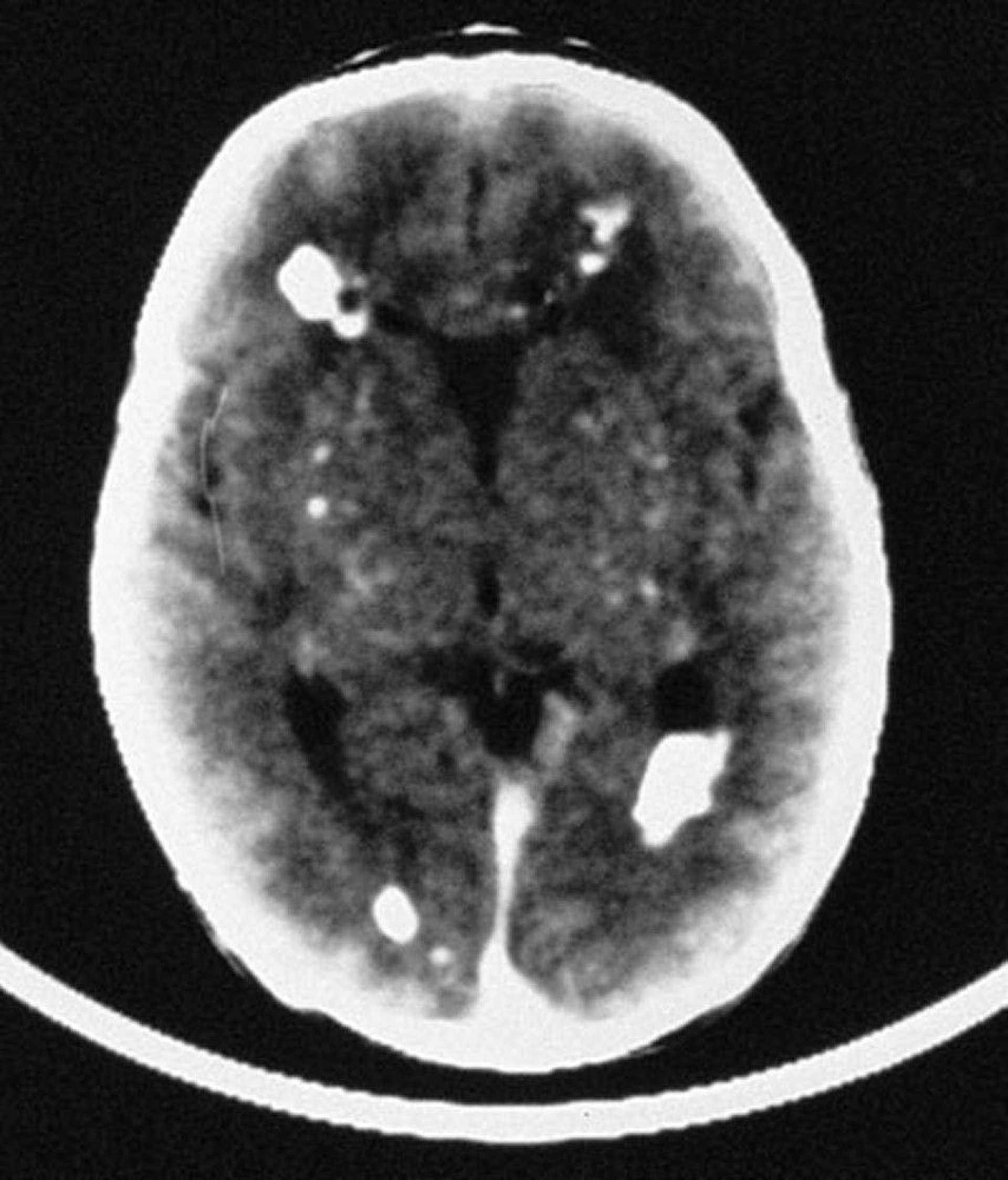
By permission of the publisher. From Demmler G: Congenital and perinatal infections. In Atlas of Infectious Diseases: Pediatric Infectious Diseases. Edited by CM Wilfert. Philadelphia, Current Medicine, 1998.
A complete blood count (CBC) with differential and liver tests may be helpful but are not specific. Cranial ultrasonography or CT and an ophthalmologic evaluation should also be done. Periventricular calcifications are commonly found on CT.
Hearing tests should be routinely done at birth in all infected neonates, and continued close monitoring is required because hearing loss may develop after the neonatal period and be progressive.
Prognosis for Congenital and Perinatal CMV Infection
Symptomatic neonates have a mortality rate of up to 30%, and 40 to 90% of survivors have some neurologic impairment, including
Visual disturbances
Among asymptomatic neonates, 5 to 15% eventually develop neurologic sequelae; hearing loss is the most common.
Treatment of Congenital and Perinatal CMV Infection
The main toxicity of treatment is neutropenia.
Treatment reference
1. Kimberlin DW, Jester PM, Sánchez PJ, et alN Engl J Med 372(10):933–943, 2015. doi: 10.1056/NEJMoa1404599
Prevention of Congenital and Perinatal CMV Infection
Nonimmune pregnant women should attempt to limit exposure to the virus. For instance, because CMV infection is common among children attending day care centers, pregnant women should always wash their hands thoroughly after exposure to urine and oral or respiratory secretions from children.
Transfusion-associated perinatal CMV disease can be avoided by giving preterm neonates blood products from CMV-seronegative donors or leukoreduced products.
A vaccine to prevent congenital CMV is under development. CMV hyperimmune globulin given to pregnant women with primary CMV infection did not show a reduction in congenital infection in a randomized, controlled trial (12).
Prevention references
1. Revello MG, Lazzarotto T, Guerra B, et al: A randomized trial of hyperimmune globulin to prevent congenital cytomegalovirus. N Engl J Med 370(14):1316–1326, 2014. doi: 10.1056/NEJMoa1310214
2. Hughes BL, Clifton RG, Rouse DJ, et al: A trial of hyperimmune globulin to prevent congenital cytomegalovirus infection. N Engl J Med 385(5):436–444, 2021. doi: 10.1056/NEJMoa1913569
Key Points
Cytomegalovirus (CMV) is the most common congenital viral infection and may be asymptomatic or symptomatic.
Multiple organs can be affected, and risk of premature birth increases.
Distinguish symptomatic congenital CMV infection from other congenital infections (eg, toxoplasmosis, rubella, lymphocytic choriomeningitis virus, syphilis) using polymerase chain reaction (PCR) testing or viral culture.


