

X-rays are a type of medical imaging that use very low-dose radiation waves to take pictures of bones and soft tissues. Chest x-rays are sometimes used in people who may have a heart disorder.
People thought to have a heart disorder have chest x-rays taken from the front and the side. Typically, the person is standing upright, but chest x-rays can be done with people lying in bed if they cannot stand. A machine is then used to beam x-rays through the person's body and record a picture on an x-ray film. The test is painless.
The x-rays show the shape and size of the heart and the outline of the large blood vessels in the lungs and chest. Abnormal heart shape or size and abnormalities, such as calcium deposits within blood vessels, are readily seen. Chest x-rays also can detect information about the condition of the lungs, particularly whether blood vessels in the lungs are abnormal and whether there is fluid in or around the lungs.

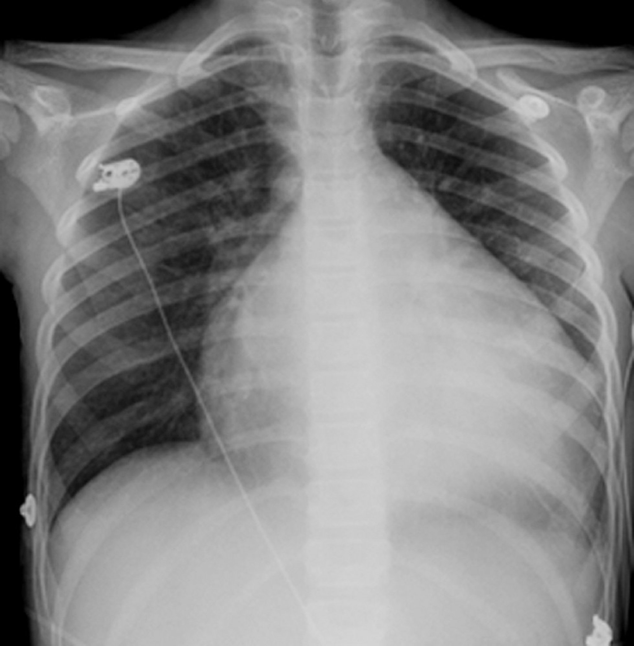
The heart is the globular, white structure in the middle of the chest. Usually it occupies less than half the chest but, in this person who has an enlarged heart (cardiomyopathy), the heart is almost 3/4 the dimension of the chest.
© Springer Science+Business Media
X-rays can detect enlargement of the heart, which is often due to heart failure or a heart valve disorder. X-rays can sometimes be helpful in diagnosis of constrictive pericarditis by detecting calcium deposits of the sac that envelops the heart (pericardium).
The appearance of blood vessels in the lungs is often more useful in making a diagnosis than the appearance of the heart itself. For instance, enlargement of the pulmonary arteries (the arteries that carry blood from the heart to the lungs) and narrowing of the arteries within the lung tissue suggest high blood pressure in the pulmonary arteries, which may lead to thickening of the muscle of the right ventricle (the lower heart chamber that pumps blood through the pulmonary arteries to the lungs).


