


Cardiac tumors may be primary (benign or malignant) or metastatic (malignant). Myxoma, a benign tumor, is the most common type of primary cardiac tumor. Cardiac tumors may occur in any cardiac tissue. They can cause valvular or inflow-outflow tract obstruction, thromboembolism, arrhythmias, or pericardial disorders. Diagnosis is by echocardiography and frequently cardiac MRI. Treatment of benign tumors is usually surgical resection; tumors may recur. Treatment of metastatic cancer depends on tumor type and origin; prognosis is generally poor.
Primary cardiac tumors have been found in < 1/5000 people at autopsy (1), with estimates of population prevalence ranging widely from approximately 1/300 to 1/60,000 (2). Metastatic tumors are much more common, occurring in series in approximately 8 to 10% of patients with cancer (3, 4, 5). Usually, primary cardiac tumors originate in the myocardium or endocardium; they may also originate in valve tissue, cardiac connective tissue, or the pericardium. Metastatic cardiac tumors originate most often from the lung. Other sources of cardiac metastases include breast and kidney carcinoma, melanoma, and lymphoma (5, 6).
General references
1. Reynen K. Frequency of primary tumors of the heart. Am J Cardiol. 1996;77(1):107. doi:10.1016/s0002-9149(97)89149-7
2. Petris AO, Alexandrescu DM, Costache II. Cardiac tumors. Rev Med Chir Soc Med Nat Iasi. 2014;118(2):289-292.
3. Maleszewski JJ, Bois MC, Bois JP, Young PM, Stulak JM, Klarich KW. Neoplasia and the Heart: Pathological Review of Effects With Clinical and Radiological Correlation. J Am Coll Cardiol. 2018;72(2):202-227. doi:10.1016/j.jacc.2018.05.026
4. Nova-Camacho LM, Gomez-Dorronsoro M, Guarch R, Cordoba A, Cevallos MI, Panizo-Santos A. Cardiac Metastasis From Solid Cancers: A 35-Year Single-Center Autopsy Study. Arch Pathol Lab Med. 2023;147(2):177-184. doi:10.5858/arpa.2021-0418-OA
5. Silvestri F, Bussani R, Pavletic N, Mannone T. Metastases of the heart and pericardium. G Ital Cardiol. 1997;27(12):1252-1255.
6. Klatt EC, Heitz DR. Cardiac metastases. Cancer. 1990;65(6):1456-1459. doi:10.1002/1097-0142(19900315)65:6<1456::aid-cncr2820650634>3.0.co;2-5
Classification of Cardiac Tumors
Some of the more common primary and secondary cardiac tumors are listed in table . Primary cardiac tumors may be:
Benign primary cardiac tumors
Examples are myxomas, papillary fibroelastomas, rhabdomyomas, fibromas, hemangiomas, teratomas, lipomas, paragangliomas, and pericardial cysts.
Myxoma is most common (2), accounting for 45 to 85% of all primary cardiac tumors (3, 4). Incidence in females is 2 to 3 times that in males (5, 6). In uncommon familial forms (Carney complex), males are affected more often. Most myxomas occur in the left atrium (3), and the rest occur in the other chambers as a solitary tumor or, less commonly, at several sites. Myxomas may be up to 15 cm in diameter. Most are pedunculated and may prolapse through the mitral valve and obstruct ventricular filling during diastole. The remainder of the tumors are broad-based and sessile. Myxomas may be myxoid and gelatinous; smooth, firm, and lobular; or friable and irregular. Friable, irregular myxomas increase risk of systemic embolism.
Carney complex is a familial, autosomal dominant syndrome of recurrent cardiac myxomas with some combination of cutaneous myxomas, myxoid mammary fibroadenomas, pigmented skin lesions (lentigines, ephelides, blue nevi), multiple endocrine neoplasia (primary pigmented nodular adrenocortical disease causing Cushing syndrome, growth hormone and prolactin-producing pituitary adenoma, testicular tumors, thyroid adenoma or carcinoma, and ovarian cysts), psammomatous melanotic schwannoma, breast ductal adenoma, and osteochondromyxoma. Patients are often young at presentation (median age, 25 to 29 years) (7), have multiple myxomas (particularly in the ventricles), and have a higher risk of myxoma recurrence.
Papillary fibroelastomas are avascular papillomas that occur on heart valves in approximately 80% of cases (8). The papillomas are more likely to occur on the left side of the heart, predominantly on the aortic and mitral valves. Males and females are affected equally. They have papillary fronds branching from a central core, resembling sea anemones. Most are pedunculated. They do not cause valvular dysfunction but increase the risk of embolism.
Rhabdomyomas affect mainly infants and children, and comprise the most common type of primary cardiac tumor in children (9). Up to 90% of children with rhabdomyomas (particularly multiple ones) also have tuberous sclerosis (10, 11). Rhabdomyomas are usually multiple and located intramurally in the septum or free wall of the left ventricle, where they affect the cardiac conduction system. They are firm, white lobules that typically regress with age. A minority of patients develop tachyarrhythmias and heart failure due to left ventricular outflow tract obstruction.
Fibromas occur mainly in children. They occur primarily on the left side of the heart (9), are often located within the ventricular myocardium, and may develop in response to inflammation. They can compress or invade the cardiac conduction system, causing arrhythmias and sudden death. Some fibromas occur as part of a syndrome with generalized body overgrowth, jaw keratocytes, skeletal abnormalities, ocular abnormalities, and various benign and malignant tumors (Gorlin, or basal cell nevus syndrome).
Cardiac hemangiomas may be incidentally detected during examinations performed for other reasons.
Teratomas of the pericardium affect mainly infants and children. They are often attached to the base of the great vessels (12). They are often associated with a pericardial effusion.
Lipomas can develop at a wide range of ages. They originate in the endocardium or epicardium and have a large pedunculated base. Most are not hemodynamically significant, but some obstruct flow or cause arrhythmias (1, 9).
Paragangliomas, including pheochromocytomas, rarely occur in the heart; when they do, they are most often localized to the left atrium near vagus nerve endings (2). They may manifest with symptoms due to catecholamine secretion (eg, increased heart rate and blood pressure, excessive sweating, tremor). Paragangliomas may be benign or malignant.
Pericardial cysts may resemble a cardiac tumor or pericardial effusion on chest radiograph. They are usually asymptomatic (12), although some cause compressive symptoms (eg, chest pain, dyspnea, cough).
Malignant primary cardiac tumors
Malignant primary tumors include sarcomas, pericardial mesothelioma, and primary lymphomas.
Sarcomas, which include undifferentiated pleomorphic sarcoma, angiosarcoma, leiomyosarcoma, malignant fibrous histiocytoma, rhabdomyosarcoma, liposarcoma, and osteosarcoma, are the most common type of malignant cardiac tumor (2). Sarcomas affect mainly middle-aged adults (mean, 44 to 47 years) (9). Undifferentiated pleomorphic sarcoma and angiosarcoma are the most common (13). Undifferentiated sarcoma usually originates in the posterior wall of the left atrium while angiosarcoma tends to originate in the right atrium and may involve the pericardium. Depending on location, sarcomas can cause mitral valve obstruction, heart failure, right ventricular inflow tract obstruction, and cardiac tamponade, and they may give rise to lung and other metastases.
Pericardial mesothelioma is rare (9). It affects all ages, males more than females (1, 14). It causes cardiac tamponade and constriction, and can metastasize to the spine, adjacent soft tissues, and brain.
Primary lymphoma is extremely rare. It most commonly occurs in immunocompromised individuals (2) and is usually located in the right atrium or pericardium. These tumors grow rapidly and cause heart failure, arrhythmias, cardiac tamponade, and superior vena cava (SVC) syndrome.
Metastatic tumors
Metastatic tumor from other organs can involve the heart through hematogenous spread or direct invasion. Metastatic tumors are more common than primary cardiac tumors (2).
Melanoma is a tumor with a high propensity for cardiac involvement (2). Lung and breast carcinoma, soft-tissue sarcoma, and renal cancer are also common sources of metastases to the heart (15, 16). Leukemia and lymphoma often involve the heart, but cardiac involvement may be clinically silent and detected incidentally.
Types of Cardiac Tumors
Type | Examples* |
|---|---|
Benign primary tumors | Myxomas Papillary fibroelastomas Rhabdomyomas Fibromas Hemangiomas Teratomas Lipomas Paraganglionomas† Pericardial cysts |
Malignant primary tumors | Sarcomas Pericardial mesothelioma Primary lymphomas |
Metastatic tumors | Soft-tissue sarcoma |
* Listed from most common to least common. | |
† Paragangliomas can also be malignant. | |
Classification references
1. Corradi D, Moreno PR, Rahouma M, et al. Cardiac tumors: Updated classifications and main clinico-pathologic findings. Trends Cardiovasc Med. 2025;35(5):297-308. doi:10.1016/j.tcm.2025.01.005
2. Tyebally S, Chen D, Bhattacharyya S, et al. Cardiac Tumors: JACC CardioOncology State-of-the-Art Review. JACC CardioOncol. 2020;2(2):293-311. Published 2020 Jun 16. doi:10.1016/j.jaccao.2020.05.009
3. Tazelaar HD, Locke TJ, McGregor CG. Pathology of surgically excised primary cardiac tumors. Mayo Clin Proc. 1992;67(10):957-965. doi:10.1016/s0025-6196(12)60926-4
4. Islam AKMM. Cardiac myxomas: A narrative review. World J Cardiol. 2022;14(4):206-219. doi:10.4330/wjc.v14.i4.206
5. Petris AO, Alexandrescu DM, Costache II. Cardiac tumors. Rev Med Chir Soc Med Nat Iasi. 2014;118(2):289-292.
6. Sido V, Volkwein A, Hartrumpf M, et al. Gender-Related Outcomes after Surgical Resection and Level of Satisfaction in Patients with Left Atrial Tumors. J Clin Med. 2023;12(5):2075. doi:10.3390/jcm12052075
7. Pitsava G, Zhu C, Sundaram R, Mills JL, Stratakis CA. Predicting the risk of cardiac myxoma in Carney complex. Genet Med. 2021;23(1):80-85. doi:10.1038/s41436-020-00956-3
8. Tamin SS, Maleszewski JJ, Scott CG, et al. Prognostic and Bioepidemiologic Implications of Papillary Fibroelastomas. J Am Coll Cardiol. 2015;65(22):2420-2429. doi:10.1016/j.jacc.2015.03.569
9. Maleszewski JJ, Bois MC, Bois JP, Young PM, Stulak JM, Klarich KW. Neoplasia and the Heart: Pathological Review of Effects With Clinical and Radiological Correlation. J Am Coll Cardiol. 2018;72(2):202-227. doi:10.1016/j.jacc.2018.05.026
10. Bosi G, Lintermans JP, Pellegrino PA, Svaluto-Moreolo G, Vliers A. The natural history of cardiac rhabdomyoma with and without tuberous sclerosis. Acta Paediatr. 1996;85(8):928-931. doi:10.1111/j.1651-2227.1996.tb14188.x
11. Sasongko TH, Kademane K, Chai Soon Hou S, Jocelyn TXY, Zabidi-Hussin Z. Rapamycin and rapalogs for tuberous sclerosis complex. Cochrane Database Syst Rev. 2023;7(7):CD011272. Published 2023 Jul 11. doi:10.1002/14651858.CD011272.pub3
12. Klein AL, Wang TKM, Cremer PC, et al. Pericardial Diseases: International Position Statement on New Concepts and Advances in Multimodality Cardiac Imaging. JACC Cardiovasc Imaging. 2024;17(8):937-988. doi:10.1016/j.jcmg.2024.04.010
13. Ramlawi B, Leja MJ, Abu Saleh WK, et al. Surgical Treatment of Primary Cardiac Sarcomas: Review of a Single-Institution Experience. Ann Thorac Surg. 2016;101(2):698-702. doi:10.1016/j.athoracsur.2015.07.087
14. Thomason R, Schlegel W, Lucca M, Cummings S, Lee S. Primary malignant mesothelioma of the pericardium. Case report and literature review. Tex Heart Inst J. 1994;21(2):170-174.
15. Agaimy A, Rösch J, Weyand M, Strecker T. Primary and metastatic cardiac sarcomas: a 12-year experience at a German heart center. Int J Clin Exp Pathol. 2012;5(9):928-938.
16. Klatt EC, Heitz DR. Cardiac metastases. Cancer. 1990;65(6):1456-1459. doi:10.1002/1097-0142(19900315)65:6<1456::aid-cncr2820650634>3.0.co;2-5
Symptoms and Signs of Cardiac Tumors
Symptoms and signs of all cardiac tumors depend on tumor type, location, size, and friability. Cardiac symptoms are often typical of much more common disorders (eg, heart failure, stroke, coronary artery disease). Malignant cardiac tumors (including cardiac metastatic disease originating elsewhere and extracardiac tumors metastasizing to the heart), and some benign cardiac tumors, may also cause extracardiac symptoms.
Types of symptoms and signs
Symptoms can be classified as:
Extracardiac
Intramyocardial
Intracavitary
Extracardiac symptoms and signs may be constitutional (fever, chills, lethargy, arthralgias) or mechanical. Petechiae and Raynaud phenomenon may also occur. These and other findings may suggest conditions such as bacterial endocarditis, systemic rheumatic disorders, or other cancers, making diagnosis challenging. With some tumors (especially gelatinous myxomas), thrombi or tumor fragments may embolize into the systemic circulation (eg, brain, coronary arteries, kidneys, spleen, extremities) or the lungs and cause manifestations specific to those organs. Mechanical symptoms (eg, dyspnea, chest discomfort) result from external compression of cardiac chambers or coronary arteries or from pericardial irritation or tamponade caused by growth or hemorrhage within the pericardium. Pericardial tumors may cause pericardial friction rubs.
Intramyocardial symptoms and signs are caused by arrhythmias, usually atrioventricular or intraventricular block or paroxysmal supraventricular or ventricular tachycardias due to compression or encroachment on the conduction system (notably rhabdomyomas and fibromas).
Intracavitary symptoms and signs are due to tumors that obstruct valvular function, blood flow, or both (causing valvular stenosis, valvular insufficiency, or heart failure). Intracavitary symptoms and signs may vary with body position, which can alter hemodynamics and physical forces associated with the tumor.
Symptoms and signs by tumor type
Myxomas may manifest with the triad of heart failure, embolic disease, and constitutional symptoms. Myxomas sometimes cause constitutional symptoms, perhaps as a result of cytokine (eg, interleukin-6) release (1, 2). Myxomas may cause a diastolic murmur that mimics the murmur of mitral stenosis but whose loudness and location vary from beat to beat with body position. Approximately 15% of pedunculated left atrial myxomas produce an audible “tumor plop” as they drop into the mitral orifice during diastole (1). Myxomas may also cause arrhythmias. Raynaud phenomenon and finger clubbing are less typical but may occur.
Fibroelastomas, often discovered incidentally at autopsy, are usually asymptomatic; however, they may be a source of systemic emboli, or, less commonly cause left-sided heart failure or sudden death (1).
Rhabdomyomas, when symptomatic, can present with ventricular arrhythmia or outflow tract obstruction (1, 3).
Fibromas cause arrhythmias, possibly causing sudden death, and obstructive symptoms (1).
Hemangiomas may cause any of the extracardiac, intramyocardial, or intracavitary symptoms.
Teratomas, particularly large ones in the anterior mediastinum, can cause respiratory distress and cyanosis due to compression of the great arteries or airway structures, or SVC syndrome due to compression of the superior vena cava.
Symptoms and signs of malignant cardiac tumors are often more acute in onset and progress more rapidly than those of benign tumors (1, 3, 4). Cardiac sarcomas, depending upon extent and location, cause symptoms of ventricular inflow tract obstruction and cardiac tamponade. Mesothelioma causes symptoms of pericarditis or tamponade. Primary lymphoma causes refractory progressive heart failure, tamponade, arrhythmias, and SVC syndrome. Metastatic cardiac tumors may manifest as sudden cardiac enlargement, tamponade (due to rapid accumulation of hemorrhagic pericardial effusion), heart block, other arrhythmias, or sudden unexplained heart failure. Fever, malaise, weight loss, night sweats, and loss of appetite may also be present.
Symptoms and signs references
1. Maleszewski JJ, Bois MC, Bois JP, Young PM, Stulak JM, Klarich KW. Neoplasia and the Heart: Pathological Review of Effects With Clinical and Radiological Correlation. J Am Coll Cardiol. 2018;72(2):202-227. doi:10.1016/j.jacc.2018.05.026
2. Pinede L, Duhaut P, Loire R: Clinical presentation of left atrial cardiac myxoma. A series of 112 consecutive cases. Medicine (Baltimore) 80(3):159–172, 2001. doi:10.1097/00005792-200105000-00002
3. Tyebally S, Chen D, Bhattacharyya S, et al. Cardiac Tumors: JACC CardioOncology State-of-the-Art Review. JACC CardioOncol. 2020;2(2):293-311. Published 2020 Jun 16. doi:10.1016/j.jaccao.2020.05.009
4. Corradi D, Moreno PR, Rahouma M, et al. Cardiac tumors: Updated classifications and main clinico-pathologic findings. Trends Cardiovasc Med. 2025;35(5):297-308. doi:10.1016/j.tcm.2025.01.005
Diagnosis of Cardiac Tumors
Echocardiography
Cardiac MRI
Cardiac CT
PET scan
Biopsy
Diagnosis is often delayed because symptoms and signs mimic those of much more common disorders. Symptoms are often initially evaluated with transthoracic or transesophageal echocardiography. Transesophageal echocardiography is better for visualizing atrial tumors, and transthoracic echocardiography is better for ventricular tumors. Due to its superior temporal resolution, echocardiography is the imaging modality of choice in visualizing very mobile cardiac tumors, particularly when they are small.
Cardiac MRI, CT, and positron emission tomography (PET) scanning are also used to further characterize the tumor (1, 2). The choice of advanced imaging modality depends upon the age of the patient, location and size of the tumor, and differential diagnosis. MRI provides information about tumor morphology, characterization, and infiltration into surrounding tissue, and it sometimes correlates with histopathology. CT, particularly with angiography, can also assess morphology particularly for calcified masses, vascular involvement and compression, and it can concomitantly evaluate for coronary artery disease. PET can help distinguish between benign and malignant tumors, stage tumors, evaluate for metastases, and guide biopsy or radiotherapy.


This image shows a large left atrial myxoma (indicated by the intersecting measurement lines ("+" and "x").
Belezapoy/stock.adobe.com
Cardiac MRI is the imaging modality of choice for tumor tissue characterization and provides the best clues to tumor types.

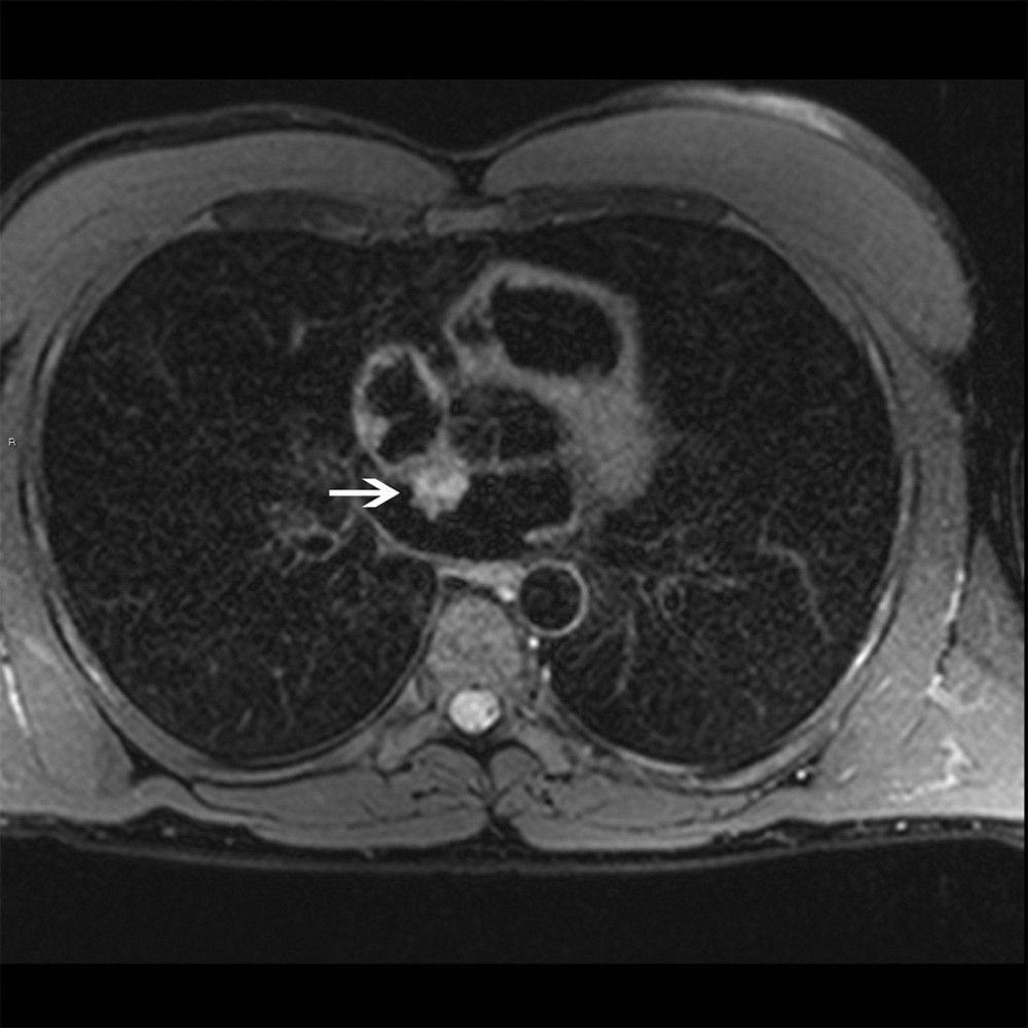
This cardiac MRI shows a left atrial myxoma (arrow) that is hyperintense on T2-weighted image, an appearance typical of myxoma.
© 2017 Elliot K. Fishman, MD.


This CTA shows a left atrial mass attached by a stalk to the interatrial septum, an appearance that is typical of atrial myxoma.
Lauren A. Baldassarre, MD, and Emmanuel Akintoye, MD, MPH
Positron emission tomography (PET) may also provide the first evidence suggesting cardiac metastasis in patients with extracardiac malignant tumors. However, PET may not be able to discriminate between physiologic and pathologic uptake in cardiac tissues and cannot provide tissue characterization of the tumor as well as MRI or CT do.
Biopsy is not usually performed because imaging studies can often distinguish benign from malignant tumors, and biopsy may inadvertently spread cancerous cells in patients with a malignant primary tumor. In carefully selected patients, an imaging-guided percutaneous transcatheter biopsy can be performed without the need for open heart surgery and provides information that affects management (3).
Extensive testing often precedes echocardiography in patients with myxomas because their symptoms are nonspecific. Anemia; thrombocytopenia; and elevation of white blood cell count, erythrocyte sedimentation rate, C-reactive protein, and gamma-globulins are common. ECG may show left atrial enlargement. Routine chest radiographs may show calcium deposits in right atrial myxomas or in teratomas seen as anterior mediastinal masses. Myxomas are sometimes diagnosed when tumor cells are found in a surgically removed embolus.
Arrhythmias and heart failure in patients with features of tuberous sclerosis suggest rhabdomyomas or fibromas. New cardiac symptoms and signs in a patient with a known extracardiac cancer suggest cardiac metastases. Chest radiographs may show changes in the cardiac silhouette.
Diagnosis references
1. Lyon AR, López-Fernández T, Couch LS, et al. 2022 ESC Guidelines on cardio-oncology developed in collaboration with the European Hematology Association (EHA), the European Society for Therapeutic Radiology and Oncology (ESTRO) and the International Cardio-Oncology Society (IC-OS). Eur Heart J. 2022;43(41):4229-4361. doi:10.1093/eurheartj/ehac244
2. Tyebally S, Chen D, Bhattacharyya S, et al. Cardiac Tumors: JACC CardioOncology State-of-the-Art Review. JACC CardioOncol . 2020;2(2):293-311. doi:10.1016/j.jaccao.2020.05.009
3. Reddy G, Maor E, Bois MC, et al. Percutaneous transcatheter biopsy for intracardiac mass diagnosis. EuroIntervention. 2017;13(12):e1436-e1443. doi:10.4244/EIJ-D-17-00707
Treatment of Cardiac Tumors
Benign primary: Usually excision
Malignant primary: Palliation
Metastatic: Depends on tumor origin
Treatment can involve excision, palliation, and/or other measures (1).
Treatment of benign primary tumors is generally surgical excision followed by serial echocardiography or other imaging to monitor for recurrence. Tumors are excised unless another disorder (eg, dementia) contraindicates surgery. Surgery is usually curative (eg, in one series, survival rates were 96% at 1 year and 75% at 10 years [2]). Exceptions are rhabdomyomas and pericardial teratomas. Most rhabdomyomas regress spontaneously; if symptomatic, rhabdomyomas may be treated with mTOR inhibitors (sirolimus and everolimus) (3). Pericardial teratomas may require urgent pericardiocentesis. Patients with fibroelastoma may also require valvular repair or replacement. Heart transplantation may be considered if the tumor is considered unresectable (4).
Chemotherapy and/or radiation therapy are seldom indicated for benign primary cardiac tumors. Reports have also highlighted the feasibility of autotransplantation (a procedure that involves explantation of the heart, tumor resection, heart reconstruction, and reimplantation of the reconstructed heart) in carefully selected patients with complex tumors (ie, not easily excised in situ) and in experienced centers (5).
Treatment of malignant primary tumors is usually palliative (eg, radiation therapy, chemotherapy, debulking surgery, management of complications); prognosis is poor (6).
Treatment of metastatic cardiac tumors depends on tumor origin. It may include systemic chemotherapy or palliation.
Pericardial tumors associated with effusion may require pericardiocentesis for symptom management. Intrapericardial cytostatic agents (chemotherapy or antimicrobials) are sometimes administered to prevent recurrence of malignant pericardial effusion after drainage (7).
Treatment references
1. Joshi M, Kumar S, Noshirwani A, Harky A. The Current Management of Cardiac Tumours: a Comprehensive Literature Review. Braz J Cardiovasc Surg. 2020;35(5):770-780. doi:10.21470/1678-9741-2019-0199
2. Mkalaluh S, Szczechowicz M, Torabi S, et al. Surgical Treatment of Cardiac Tumors: Insights from an 18-Year Single-Center Analysis. Med Sci Monit. 2017;23:6201-6209. doi:10.12659/msm.905451
3. Hurtado-Sierra D, Ramos Garzón JX, Romero-Guevara SL, Serrano-García AY, Rojas LZ. Everolimus and sirolimus in the treatment of cardiac rhabdomyomas in neonates. Pediatr Res. 2025;98(6):2045-2057. doi:10.1038/s41390-025-04043-8
4. Gowdamarajan A, Michler RE. Therapy for primary cardiac tumors: is there a role for heart transplantation? Curr Opin Cardiol. 2000;15(2):121-125. doi:10.1097/00001573-200003000-00010
5. Reardon MJ, Malaisrie SC, Walkes JC, et al. Cardiac autotransplantation for primary cardiac tumors. Ann Thorac Surg. 2006;82(2):645-650. doi:10.1016/j.athoracsur.2006.02.086
6. Tyebally S, Chen D, Bhattacharyya S, et al. Cardiac Tumors: JACC CardioOncology State-of-the-Art Review. JACC CardioOncol . 2020;2(2):293-311. doi:10.1016/j.jaccao.2020.05.009
7. Lorenzo-Esteller L, Ramos-Polo R, Pons Riverola A, et al. Pericardial Disease in Patients with Cancer: Clinical Insights on Diagnosis and Treatment. Cancers (Basel). 2024;16(20):3466. doi:10.3390/cancers16203466
Key Points
Most cardiac tumors are metastatic, most commonly from lung and breast carcinoma, melanoma, soft-tissue sarcoma, and renal carcinoma.
Primary cardiac tumors are much less common; most originate in the myocardium or endocardium, but they can develop in any cardiac tissue and be benign or malignant.
Manifestations depend on the location and type of tumor but include constitutional symptoms, valvular or inflow-outflow tract obstruction, thromboembolism, and arrhythmias.
Diagnosis is by echocardiography and often cardiac MRI, CT, or PET scan.
For benign tumors, treatment is usually surgical excision; for malignant primary and most metastatic tumors, treatment is usually palliative.
Drug Information for the Topic



