


Cellulitis is an acute bacterial infection of the skin and subcutaneous tissue most often caused by streptococci or staphylococci. Symptoms and signs are pain, warmth, rapidly spreading erythema, and swelling, typically without purulent drainage. Fever may occur, and regional lymph nodes may enlarge in more serious infections. Diagnosis is based on clinical appearance; cultures may help. Treatment with antibiotics should be initiated empirically; choice of antibiotic depends on the type of infection and antibiotic susceptibility pattern. Prognosis is excellent with timely treatment.
Cellulitis is a common, potentially severe bacterial skin and subcutaneous tissue infection. (See also Overview of Bacterial Skin Infections.)
Etiology of Cellulitis
The most common causes of cellulitis are:
Group A beta-hemolytic streptococci (eg, Streptococcus pyogenes)
Staphylococcus aureus
Streptococci cause diffuse, rapidly spreading infection because enzymes produced by the organism (streptokinase, DNase, hyaluronidase) break down cellular components that would otherwise contain and localize the inflammation.
Staphylococcal cellulitis is typically more localized and usually occurs in open wounds or cutaneous abscesses.
Infection involves bacterial entry through breaks in the skin barrier, such as those caused by prior fungal infections (eg, onychomycosis, tinea pedis), pressure injuries, venous leg ulcers, or other cutaneous disruptions (eg, psoriatic or eczematous lesions that are abraded by scratching).

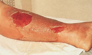
This photo shows the lower limb of a patient with the affected area showing discrete erythema, swelling, and desquamation of overlying skin.
This photo shows the lower limb of a patient with the affected area showing discrete erythema, swelling, and desquamati
© Springer Science+Business Media

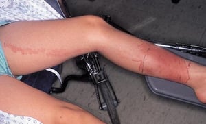
This photo shows focal erythema and swelling of the lower leg that is characteristic of focal cellulitis. The clinician has marked the border of the cellulitis with a pen to facilitate recognition of spread or resolution. Note the line of erythema extending up the thigh due to lymphangitic spread.
This photo shows focal erythema and swelling of the lower leg that is characteristic of focal cellulitis. The clinician
© Springer Science+Business Media
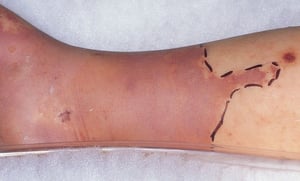
This photo shows focal erythema and swelling, usually accompanied by warmth and tenderness, characteristic of focal cellulitis. Erythema may be purplish and can mimic venous congestion. Note the clinician has marked the border of the cellulitis with a pen to facilitate recognition of spread or resolution.
This photo shows focal erythema and swelling, usually accompanied by warmth and tenderness, characteristic of focal cel
© Springer Science+Business Media
This photo shows the lower limb of a patient with the affected area showing discrete erythema, swelling, and desquamation of overlying skin.
This photo shows the lower limb of a patient with the affected area showing discrete erythema, swelling, and desquamati
© Springer Science+Business Media
This photo shows focal erythema and swelling of the lower leg that is characteristic of focal cellulitis. The clinician has marked the border of the cellulitis with a pen to facilitate recognition of spread or resolution. Note the line of erythema extending up the thigh due to lymphangitic spread.
This photo shows focal erythema and swelling of the lower leg that is characteristic of focal cellulitis. The clinician
© Springer Science+Business Media
This photo shows focal erythema and swelling, usually accompanied by warmth and tenderness, characteristic of focal cellulitis. Erythema may be purplish and can mimic venous congestion. Note the clinician has marked the border of the cellulitis with a pen to facilitate recognition of spread or resolution.
This photo shows focal erythema and swelling, usually accompanied by warmth and tenderness, characteristic of focal cel
© Springer Science+Business Media
While the overall prevalence of methicillin-resistant S. aureus (MRSA) has declined in recent years in the United States, it is responsible for about half of all community-associated S. aureus infections (1). Also, while strain USA300has become increasingly predominant in health care settings in the United States, traditional health care-associated MRSA strains (particularly USA100) persist in hospital and nursing facilities and exhibit different antibiotic resistance patterns compared to that of MRSA–USA300 (2, 3). (See also MRSA surveillance in health care institutions.)
Less common causes of cellulitis are:
Group B streptococci (eg, S. agalactiae) in older adults with diabetes
Gram-negative bacilli (eg, Haemophilus influenzae) in children
Pseudomonas aeruginosa in patients with diabetes or neutropenia, hot tub or spa users, and patients who are hospitalized
Anaerobic bacteria (eg, Bacteroides fragilis, Clostridium species)
Animal bites may result in cellulitis and are often polymicrobial. Pasteurella multocida is often the cause in cat bites, and Pasteurella or Capnocytophaga species are typically the cause in dog bites (4). Obligate anaerobes (ie, organisms that can survive only in the absence of oxygen), such as Fusobacterium, Bacteroides, Porphyromonas, and Prevotella species, can also cause cellulitis after an animal bite (5).
Exposure to contaminated water, particularly immersion of nonintact skin, may result in cellulitis caused by a variety of uncommon pathogens including Vibrio vulnificus, V. parahaemolyticus, Erysipelothrix rhusiopathiae, Mycobacterium marinum, Aeromonas species, Shewanella species, and Streptococcus iniae.
Immunocompromised patients may become infected by opportunistic organisms, including gram-negative bacteria (eg, Proteus, Serratia, Enterobacter, Citrobacter), anaerobic bacteria, and Helicobacter and Fusarium species. Mycobacteria are a rare cause of cellulitis.
Risk factors include skin abnormalities (eg, trauma, ulceration, fungal infection, other skin barrier compromise due to preexisting skin disease), which are common among patients with chronic venous insufficiency or lymphedema. Scars from saphenous vein removal for cardiac or vascular surgery are common sites for recurrent cellulitis, especially if tinea pedis is present. Frequently, no predisposing condition or site of entry is evident.
Etiology references
1. Burillo A, Bouza E. Community-acquired methicillin-resistant Staphylococcus aureus: is it still a significant pathogen for skin and soft tissue infections? A 30-year overview. Curr Opin Infect Dis. 2025;38(2):78-91. doi:10.1097/QCO.0000000000001086
2. Lakhundi S, Zhang K. Methicillin-Resistant Staphylococcus aureus: Molecular Characterization, Evolution, and Epidemiology. Clin Microbiol Rev. 2018, 31:e00020-18. Published 2018 Sep 12. doi:10.1128/CMR.00020-18
3. Centers for Disease Control and Prevention (CDC). Invasive Staphylococcus aureus Infection Surveillance. February 10, 2026. Accessed January 16, 2026.
4. Oehler RL, Velez AP, Mizrachi M, Lamarche J, Gompf S. Bite-related and septic syndromes caused by cats and dogs. Lancet Infect Dis. 2009;9(7):439-447. doi:10.1016/S1473-3099(09)70110-0
5. Talan DA, Citron DM, Abrahamian FM, Moran GJ, Goldstein EJ. Bacteriologic analysis of infected dog and cat bites. Emergency Medicine Animal Bite Infection Study Group. N Engl J Med. 1999;340(2):85-92. doi:10.1056/NEJM199901143400202
Symptoms and Signs of Cellulitis
Cellulitis is most common in the lower extremities. It is typically unilateral.
The major findings are local erythema and tenderness and, in more severe infections, often lymphangitis (inflammation of the lymphatic vessels presenting as streaks of erythema that extend proximally from a site of infection towards regional lymph notes) and regional lymphadenopathy. The skin in affected areas is warm, erythematous, and edematous, often with a surface appearance resembling the skin of an orange (peau d’orange). This appearance results from epidermal edema and tautness that surrounds a "dimpled" hair follicle (ie, the hair follicle remains tethered to the dermis, and the skin around it is swollen). The borders are usually indistinct, except in erysipelas (a type of cellulitis with sharply demarcated margins). Petechiae are common; large areas of ecchymosis are rare.
Vesicles and bullae may develop and rupture, occasionally with necrosis of the involved skin.
Most cellulitis is nonpurulent. However, cellulitis sometimes is accompanied by one or more pustules, furuncles, or abscesses with or without purulent drainage or exudate and is referred to as purulent.
Fever, chills, tachycardia, headache, hypotension, and delirium (usually indicating severe infection) may precede cutaneous findings by several hours, but many patients do not look ill. Leukocytosis is common. Cellulitis with rapid spread of infection, rapidly increasing pain, hypotension, delirium, or skin sloughing, particularly with bullae and fevers, suggests life-threatening infection.
Purulent cellulitis and other risk factors predispose to complicated/serious (ie, deeper, invasive, systemic) infection.
Risk factors for MRSA and complicated infection include the following:
Penetrating trauma
Surgical wounds
Recent hospitalization or nursing home exposure
Injection drug use
Proximity of infection to an implanted medical device such as a prosthetic joint
Previous MRSA infection
Known nasal colonization with MRSA
Clinical features suggestive of serious infection
Clinical features suggestive of complicated infection include the following:
Pain disproportionate to physical findings
Cutaneous hemorrhage
Bullae
Skin sloughing
Skin anesthesia
Rapid progression
Tissue gas
Signs of systemic toxicity (eg, fever or hypothermia, tachycardia, hypotension, delirium)

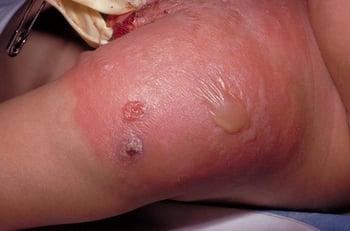
This example of cellulitis at the site of a previous vaccination shows warm, erythematous, edematous skin with formation of bullae.
Diagnosis of Cellulitis
Primarily history and physical examination
Blood cultures in selected cases
Tissue cultures in selected cases
The diagnosis of cellulitis is primarily based on clinical evaluation and the exclusion of differential diagnoses that may present similarly.
Skin and wound cultures (including cutaneous aspirates, biopsies, or swabs) as well as blood cultures are not routinely recommended in all patients with cellulitis (1).
In patients who are immunocompromised and in patients who have signs of systemic infection (eg, fever and leukocytosis) (1), blood cultures may be useful for detecting bacteremia. Culture of involved tissue may be required in immunocompromised patients if they are not responding to empiric therapy or if blood cultures do not isolate an organism and in patients with cellulitis at the site of certain injuries (eg, animal bite wounds, penetrating injuries).
Abscesses and necrotizing soft-tissue infection can develop as a complication of cellulitis. Abscesses should be identified primarily through clinical evaluation (eg, focal tenderness, fluctuance). Imaging modalities can effectively distinguish between cellulitis and abscess (eg, point of care ultrasound) or necrotizing soft-tissue infection (eg, MRI) and help guide management decisions (2, 3).
Differential diagnosis
Contact dermatitis, stasis dermatitis, and deep vein thrombosis (DVT) should be considered as potential alternative diagnoses (see table ). Cellulitis may also mimic stasis dermatitis; however, stasis dermatitis is usually bilateral. Stasis dermatitis can sometimes be differentiated by features of dermatitis itself (eg, scaling, eczematous findings, lichenification), evidence of venous stasis, and bilateral location. Contact dermatitis can often be differentiated by the presence of itching, limitation of lesions to the site of contact, absence of systemic signs, and sometimes unilateral location. Other disorders to consider include cutaneous T-cell lymphoma, nummular dermatitis, and tinea infection.
Differentiating Cellulitis and Deep Venous Thrombosis
Feature | Cellulitis | Deep Venous Thrombosis |
|---|---|---|
Skin temperature | Warm | Normal or warm (rarely cool unless limb ischemia is present due to extensive venous disease causing arterial insufficiency) |
Skin color | Erythematous | Normal or erythematous but blanchable (infrequently cyanotic) |
Skin surface | Peau d’orange | Smooth |
Lymphangitis and regional lymphadenopathy | Frequent | Uncommon |
Diagnosis references
1. Stevens DL, Bisno AL, Chambers HF, et al. Practice guidelines for the diagnosis and management of skin and soft tissue infections: 2014 update by the Infectious Diseases Society of America. Clin Infect Dis. 2014;59(2):e10-e52. doi:10.1093/cid/ciu444
2. Gottlieb M, Avila J, Chottiner M, Peksa GD. Point-of-Care Ultrasonography for the Diagnosis of Skin and Soft Tissue Abscesses: A Systematic Review and Meta-analysis. Ann Emerg Med. 2020;76(1):67-77. doi:10.1016/j.annemergmed.2020.01.004
3. Chun CW, Jung JY, Baik JS, Jee WH, Kim SK, Shin SH. Detection of soft-tissue abscess: Comparison of diffusion-weighted imaging to contrast-enhanced MRI. J Magn Reson Imaging. 2018;47(1):60-68. doi:10.1002/jmri.25743
Treatment of Cellulitis
Antibiotics
The first-line treatment of cellulitis is antibiotics, and selection is based on the presence or absence of purulence and other risk factors for serious and/or resistant infection (1). The Infectious Diseases Society of America typically recommends 5 days of treatment for patients who show clinical improvement; however, treatment should be extended if patients have not improved (2).
Nonpharmacologic modalities are important treatment adjuncts but often remain under-utilized. Immobilization and elevation of the affected area help reduce edema; cool and/or wet dressings help reduce pain. Compression therapy can help prevent repeat episodes of leg cellulitis in patients with recurrent cellulitis who have chronic lower extremity edema.
Nonpurulent, uncomplicated cellulitis
For most patients with nonpurulent cellulitis, empiric therapy effective against both group A streptococci and methicillin-susceptible S. aureus is used.
Oral therapy is usually adequate for mild infections, typically with dicloxacillin or cephalexin. In patients allergic to penicillin, clindamycin is an alternative.
Patients with mild cellulitis caused by animal bites can be treated as outpatients with amoxicillin/clavulanic acid. In patients allergic to penicillin, clindamycin plus either an oral fluoroquinolone (eg, ciprofloxacin 500 mg 2 times a day) or double-strength oral sulfamethoxazole/trimethoprim may be administered.
Cellulitis that develops after exposure to fresh or brackish water should be treated with a first-generation cephalosporin such as oral cephalexin or IV cefazolin in addition to a fluoroquinolone. If cellulitis develops after exposure to brackish or salt water, oral doxycycline should also be added.
Evaluation of skin integrity, including the interdigital toe spaces for fissures, cracks, or maceration, is important. Cellulitis can recur in patients with risk factors such as tinea pedis, obesity, venous insufficiency, edema, and atopic dermatitis. These disorders should be identified and treated to decrease the likelihood of recurrent cellulitis. Prophylactic antibiotics such as benzathine penicillin 1.2 million units IM monthly or penicillin V or erythromycin 250 mg orally 2 times a day for 1 to 12 months may be considered for patients who have 3 to 4 episodes of cellulitis per year despite treatment of risk factors. Patients taking prophylactic antibiotics should be assessed regularly to monitor for adverse effects and efficacy of treatment. Antibiotics should be continued until risk factors also have been managed. If these regimens prove unsuccessful, tissue culture may be required.
MRSA and purulent or complicated cellulitis
Patients with purulent cellulitis or risk factors for complicated or serious infection (ie, invasive, systemic) should receive antibiotics with activity against MRSA.
For patients with suspected MRSA without features suggesting complicated infection, empiric outpatient treatment is reasonable using double-strength sulfamethoxazole/trimethoprim (800 mg sulfamethoxazole/160 mg trimethoprim) orally 2 times a day, doxycycline 100 mg orally 2 times a day, linezolid 600 mg orally 2 times a day, or clindamycin 300 to 450 mg orally 3 times a day (however, resistance to clindamycin is becoming more prevalent).
Patients who have a more serious infection, with high-risk symptoms and suspected or confirmed MRSA, or whose oral therapy failed are hospitalized and typically treated with IV vancomycin. Alternative IV options include daptomycin, linezolid (although highly bioavailable oral option), and teicoplanin (outside of the United States).
The use of alternative medications for severe acute bacterial skin and skin structure infection (ABSSSI) with S. aureus (including MRSA) are based on availability, ease of administration, adverse effect profile, and cost. Alternatives include:
Linezolid or tedizolid (IV or oral)
Delafloxacin (IV or oral)
Omadacycline (IV or oral)
Ceftaroline and ceftobiprole (IV)
Dalbavancin, oritavancin, and telavancin (IV)
Cellulitis in a patient with neutropenia requires broad-spectrum antibiotic coverage. Vancomycin plus cefepime or meropenem is recommended until blood culture results are available to guide therapy. Tissue culture should be strongly considered for identification of the causative organism because of the increased risk of fungal infection. Culture should be considered for immunocompromised patients if they are not responding to empiric therapy or if blood cultures do not isolate an organism and for patients with cellulitis at the site of certain injuries (eg, animal bite wounds, penetrating injuries).
Treatment references
1. Brindle R, Williams OM, Barton E, Featherstone P. Assessment of antibiotic treatment of cellulitis and erysipelas: A systematic review and meta-analysis. JAMA Dermatol. 2019;155(9):1033–1040. doi:10.1001/jamadermatol.2019.0884
2. Stevens DL, Bisno AL, Chambers HF, et al. Practice guidelines for the diagnosis and management of skin and soft tissue infections: 2014 update by the Infectious Diseases Society of America. Clin Infect Dis. 2014;59(2):e10-e52. doi:10.1093/cid/ciu444
Prognosis for Cellulitis
Most cases of cellulitis resolve quickly with antibiotic therapy. Local abscesses occasionally form, requiring incision and drainage. Serious but rare complications include severe necrotizing subcutaneous infection and bacteremia with metastatic foci of infection.
Recurrences in the same area are common. For example, one large cohort study of severe lower limb cellulitis reported a cumulative incidence of 6.3% at 12 months after a first recurrence, and this increased substantially with each subsequent episode (17.2% at 12 months after a first recurrence, and 19.4% after a second recurrence). Recurrent cellulitis may cause serious damage to the lymphatics, chronic lymphatic obstruction, and lymphedema (1).
Prognosis reference
1. Stevens DL, Bisno AL, Chambers HF, et al. Practice guidelines for the diagnosis and management of skin and soft tissue infections: 2014 update by the Infectious Diseases Society of America. Clin Infect Dis. 2014;59(2):e10-e52. doi:10.1093/cid/ciu444
Key Points
The most common pathogens causing cellulitis overall are S. pyogenes and S. aureus.
Methicillin-resistant S. aureus (MRSA) should be considered in patients with certain risk factors (eg, purulent cellulitis, penetrating trauma, wound infection, nasal colonization), particularly if there is a known outbreak or local prevalence is high.
Differentiate leg cellulitis from deep vein thrombosis by the presence of skin warmth, erythema, peau d'orange quality, and lymphadenopathy.
Consider obtaining tissue culture in immunocompromised patients if they are not responding to empiric therapy or if blood cultures do not isolate any organism and in patients with cellulitis at the site of certain injuries (eg, animal bite wounds, penetrating injuries).
Direct antibiotic therapy against the most likely pathogens in specific clinical situations.
Drug Information for the Topic


