


Diverticulitis is inflammation with or without infection of a diverticulum, which can result in phlegmon of the bowel wall, peritonitis, perforation, fistula, or abscess. The primary symptom is abdominal pain. Diagnosis is by CT and ultrasound. Treatment is with bowel rest, sometimes antibiotics, and occasionally surgery.
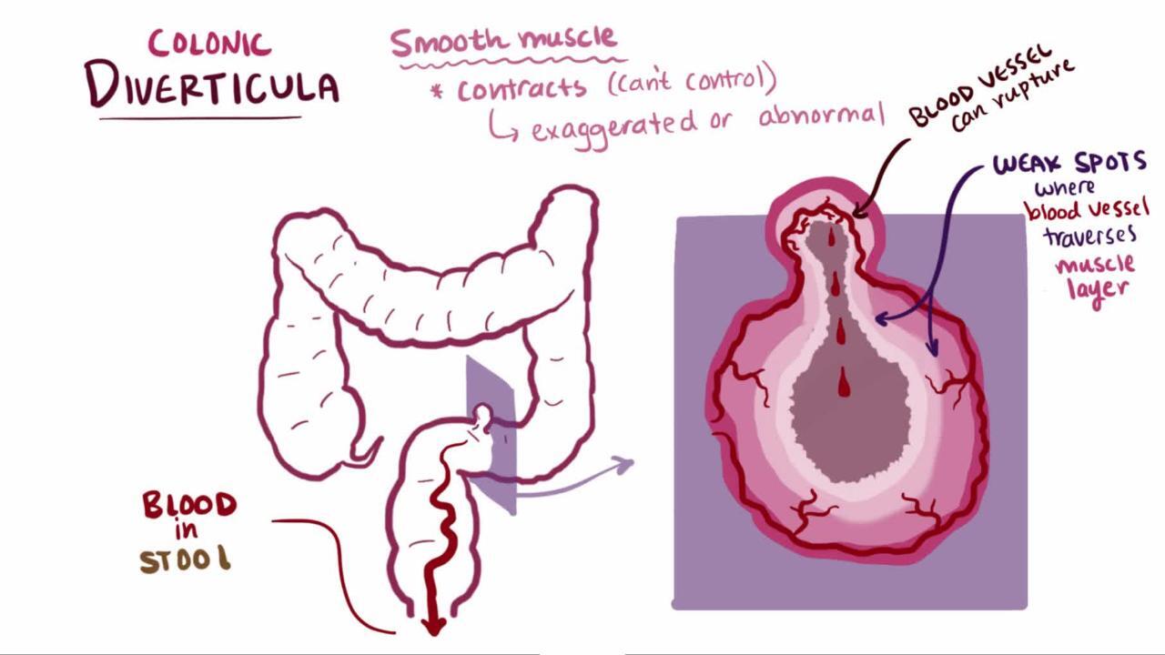
A colonic diverticulum is a saclike pouch of colonic mucosa and submucosa that protrudes through the muscular layer of the colon; because it does not contain all layers of the bowel, it is considered a pseudodiverticulum (see also Definition of Diverticular Disease).
Many people have multiple colonic diverticula (diverticulosis). In people > 50 years, acute diverticulitis is most common among women; in those < 50 years, it is most common among men (1). The predominant location of diverticular disease varies by ethnicity and geographic region; proximal (right-sided) disease is more common among patients of Asian and African descent, while distal (left-sided) diverticulitis is more common among European and United States populations. (2, 3).
Patients with HIV infection and those undergoing chemotherapy are at increased risk of developing acute diverticulitis (1).

Diverticula are usually asymptomatic but sometimes become inflamed (diverticulitis). Diverticulitis develops in approximately 1 to 5% of people with diverticulosis (4, 5, 6).
Diverticulitis that is managed nonoperatively can recur as either an acute or chronic process. The risk of a recurrent acute episode is up to 43%, although reported rates vary widely (7). A large population-based study found that after an episode of acute diverticulitis, the recurrence rate at 1 year was 8% and at 10 years was 22% (8). In some patients, however, recurrence manifests as chronic, ongoing abdominal pain; this may develop after 1 or more acute episodes.
General references
1. Francis NK, Sylla P, Abou-Khalil M, et al. EAES and SAGES 2018 consensus conference on acute diverticulitis management: Evidence-based recommendations for clinical practice. Surg Endosc. 2019;33(9):2726–2741. doi:10.1007/s00464-019-06882-z
2. Stollman N, Raskin JB. Diverticular disease of the colon. Lancet. 2004;363(9409):631-639. doi:10.1016/S0140-6736(04)15597-9
3. Fugazzola P, Ceresoli M, Coccolini F, et al. The WSES/SICG/ACOI/SICUT/AcEMC/SIFIPAC guidelines for diagnosis and treatment of acute left colonic diverticulitis in the elderly. World J Emerg Surg. 2022;17(1):5. Published 2022 Jan 21. doi:10.1186/s13017-022-00408-0
4. Shahedi K, Fuller G, Bolus R, et al. Long-term risk of acute diverticulitis among patients with incidental diverticulosis found during colonoscopy. Clin Gastroenterol Hepatol. 2013;11(12):1609–1613. doi:10.1016/j.cgh.2013.06.020
5. Brown RF, Lopez K, Smith CB, Charles A. Diverticulitis: A Review. JAMA. Published online July 24, 2025. doi:10.1001/jama.2025.10234
6. Long B, Werner J, Gottlieb M. Emergency medicine updates: Acute diverticulitis. Am J Emerg Med. 2024;76:1-6. doi:10.1016/j.ajem.2023.10.051
7. Sallinen V, Mali J, Leppäniemi A, Mentula P. Assessment of risk for recurrent diverticulitis: A proposal of risk score for complicated recurrence. Medicine (Baltimore). 2015;94(8):e557. doi:10.1097/MD.0000000000000557
8. Bharucha AE, Parthasarathy G, Ditah I, et al. Temporal trends in the incidence and natural history of diverticulitis: A population-based study. Am J Gastroenterol. 2015;110(11):1589–1596. doi:10.1038/ajg.2015.302
Etiology of Colonic Diverticulitis


The etiology and pathophysiology of diverticulitis are not fully understood and may vary among patients. It has long been thought that diverticulitis occurs when a micro or macro perforation develops in a diverticulum, resulting in the release of intestinal bacteria and triggering inflammation. However, emerging data suggest that in some patients, acute diverticulitis is more of an inflammatory than infectious process. Furthermore, cytomegalovirus may be a trigger of that inflammation. The virus disrupts the intestinal epithelium, and in one study, evidence of active viral replication was found in affected colon tissue in over two-thirds of patients with diverticulitis (1, 2).
Studies have suggested a direct correlation between red meat consumption per week, smoking, obesity, and the incidence of diverticulitis (3, 4). Nonsteroidal anti-inflammatory drugs (NSAIDs), aspirin, glucocorticoids, and opioids also increase the risk of diverticulosis and diverticulitis (3, 4). There is no association between consumption of nuts, seeds, corn, or popcorn and development of diverticulitis as was previously thought (5, 6).
Physical activity and dietary fiber have been shown to help prevent formation of diverticula and development of diverticulitis (7, 8, 9).
Etiology references
1. Hollink N, Dzabic M, Wolmer N, et al. High prevalence of an active human cytomegalovirus infection in patients with colonic diverticulitis. J Clin Virol. 2007;40(2):116-119. doi:10.1016/j.jcv.2007.07.008
2. Le-Trilling VTK, Ebel JF, Baier F, et al. Acute cytomegalovirus infection modulates the intestinal microbiota and targets intestinal epithelial cells. Eur J Immunol. 2023;53(2):e2249940. doi:10.1002/eji.202249940
3. Cao Y, Strate LL, Keeley BR, et al. Meat intake and risk of diverticulitis among men. Gut. 2018;67(3):466-472. doi: 10.1136/gutjnl-2016-313082
4. Strate LL, Keeley BR, Cao Y, et al. Western dietary pattern increases, and prudent dietary pattern decreases, risk of incident diverticulitis in a prospective cohort study. Gastroenterology. 2017;152(5):1023–1030.e2. doi: 10.1053/j.gastro.2016.12.038
5. Brown RF, Lopez K, Smith CB, Charles A. Diverticulitis: A Review. JAMA. Published online July 24, 2025. doi:10.1001/jama.2025.10234
6. Tursi A, Brandimarte G, Di Mario F, et al. Global guidelines on diverticular disease of the colon: the Fiesole Consensus report. Gut. Published online December 31, 2025. doi:10.1136/gutjnl-2025-336902
7. Bohm SK, Kruis W: Lifestyle and other risk factors for diverticulitis. Minerva Gastroenterol Dietol 63(2):110–118, 2017. doi: 10.23736/S1121-421X.17.02371-6
8. Schultz JK, Azhar N, Binda GA, et al: European Society of Coloproctology: Guidelines for the management of diverticular disease of the colon. Colorectal Dis 22 (supplement 2):S5–S28, 2020. doi: 10.1111/codi.15140
9. Veronese N, Gianfredi V, Solmi M, et al. The impact of dietary fiber consumption on human health: An umbrella review of evidence from 17,155,277 individuals. Clin Nutr. 2025;51:325-333. doi:10.1016/j.clnu.2025.06.021
Classification of Colonic Diverticulitis
Acute diverticulitis can be classified as (1, 2):
Acute uncomplicated diverticulitis: This is the most common manifestation of acute diverticulitis.
Acute complicated diverticulitis: This manifestation is defined by the presence of abscess, fistula, obstruction, or free perforation. Approximately 12% of patients with diverticulitis present with complicated disease (3).
Additional complications, such as peritonitis and sepsis, can develop after perforation of an inflamed diverticulum.
Approximately 15% of patients with acute complicated diverticulitis have a pericolic or intramesenteric abscess (4).
If acute diverticulitis does not heal completely, chronic diverticulitis will develop.
Chronic diverticulitis can be classified as:
Chronic uncomplicated diverticulitis: Defined as thickening of the colonic wall or chronic mucosal inflammation in the absence of stricture
Chronic complicated diverticulitis: Includes stenotic disease, which may cause acute colon obstruction and fistula formation (most commonly colovesical fistulas, from colon to bladder)
Colovesical fistulas are more common in women who have had a hysterectomy because the large intestine and bladder are no longer separated by the uterus (5).
Classification references
1.Schultz JK, Azhar N, Binda GA, et al. European Society of Coloproctology: Guidelines for the management of diverticular disease of the colon. Colorectal Dis. 2020;22(supplement 2):S5–S28. doi:10.1111/codi.15140
2. Brown RF, Lopez K, Smith CB, Charles A. Diverticulitis: A Review. JAMA. Published online July 24, 2025. doi:10.1001/jama.2025.10234
3. Gunby SA, Strate LL. Acute Colonic Diverticulitis. Ann Intern Med. 2024;177(3):ITC33-ITC48. doi:10.7326/AITC202403190
4. Lambrichts DPV, Bolkenstein HE, van der Does DCHE, et al. Multicentre study of non-surgical management of diverticulitis with abscess formation. Br J Surg. 2019;106(4):458-466. doi:10.1002/bjs.11129
5. Altman D, Forsgren C, Hjern F, et al. Influence of hysterectomy on fistula formation in women with diverticulitis. Br J Surg. 2010;97(2):251-257. doi:10.1002/bjs.6855
Symptoms and Signs of Colonic Diverticulitis
Patients have left lower quadrant abdominal pain and tenderness and often have a palpable sigmoid; pain is occasionally suprapubic. However, patients with right-sided diverticulitis often present with right-sided pain. The pain can be accompanied by nausea, vomiting, fever, and sometimes even urinary symptoms as a result of bladder irritation.
Peritoneal signs (eg, rebound or guarding) may be present, particularly with abscess or free perforation.
Fistulas may manifest as pneumaturia, fecaluria (feces in the urine), feculent vaginal discharge, or a cutaneous or myofascial infection of the abdominal wall, perineum, or upper leg. Patients with bowel obstruction have nausea, vomiting, and abdominal distention. Bleeding is uncommon.
Recurrent episodes of acute diverticulitis manifest similarly to initial episodes; they are not necessarily more severe (1).

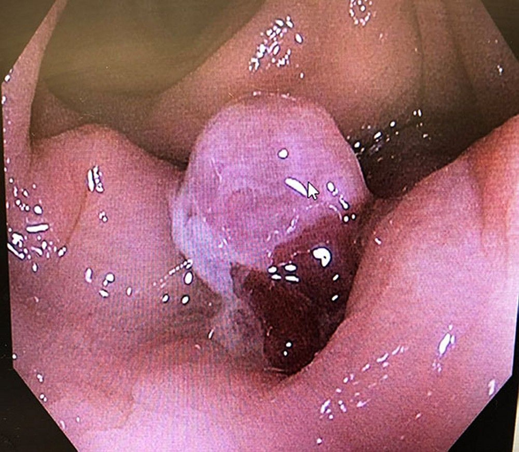
This image shows granulation tissue (arrowhead) protruding from a single inflamed diverticulum.
Photo courtesy of Drs. Joel A. Baum and Rafael A. Ching Companioni.
Symptoms and signs reference
1. Regenbogen SE, Hardiman KM, Hendren S, et al. Surgery for diverticulitis in the 21st century: a systematic review. JAMA Surg. 2014;149(3):292-303. doi:10.1001/jamasurg.2013.5477
Diagnosis of Colonic Diverticulitis
Abdominal and pelvic CT
Sometimes ultrasound or MRI
C-reactive protein (CRP) and sometimes other blood tests
Colonoscopy after resolution
Clinical suspicion is high in patients with known diverticulosis who present with characteristic abdominal symptoms. However, because other disorders (eg, appendicitis, colon or ovarian cancer, inflammatory bowel disease) may cause similar symptoms (and because many patients with diverticulosis are not known to have it), diagnostic testing is required.

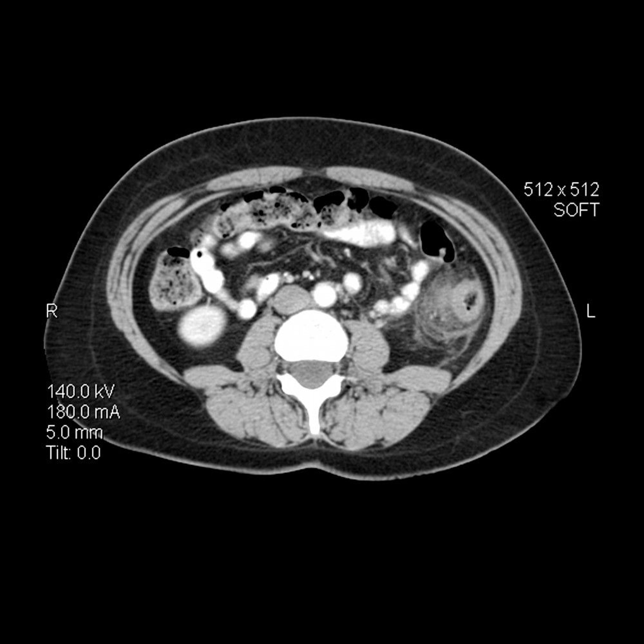
In this axial (cross-sectional) CT image of the abdomen in a person with diverticulitis, the wall of the descending colon is thickened. There is also inflammatory streaking with increased density in the adjacent pericolonic fat.
LIVING ART ENTERPRISES, LLC/SCIENCE PHOTO LIBRARY
Diverticulitis is evaluated preferentially with CT of the abdomen and pelvis with water-soluble contrast given orally and rectally; IV contrast also is given when not contraindicated (1, 2, 3). CT has a high sensitivity (95 to 99%) and specificity (95 to 100%) for acute diverticulitis (3). However, findings in approximately 10% of patients cannot distinguish diverticulitis from colon cancer. MRI is an alternative for pregnant and young patients (4).
Ultrasound is another alternative for people with diverticulitis, particularly in the ambulatory setting. Although ultrasound can achieve high accuracy, reported sensitivity (77 to 98%) and specificity (80 to 99%) vary widely and are operator-dependent. Ultrasound is also less sensitive (55%) in distinguishing complicated from simple diverticulitis (5). Ultrasound may be considered as a first-line approach in selected low-risk patients to reduce radiation exposure and emergency department length of stay, with CT reserved for equivocal cases or concern for complications (6).
CRP is recommended to guide decisions about antibiotic use and predict progression to complicated disease (1, 7, 8). Other laboratory testing, such as a complete blood count, play a role in the evaluation for sepsis.
Colonoscopy is recommended 1 to 3 months after an episode of complicated diverticulitis (and by some after any first-time episode of diverticulitis) and for patients not current with colon cancer screening (7, 9). However, in the absence of high-risk signs (eg, complicated diverticulitis, uncomplicated diverticulitis with imaging abnormalities or atypical course, family history of colorectal cancer, anemia, weight loss), the likelihood of malignant lesions or advanced adenomas after an episode of acute uncomplicated diverticulitis is low (1).
Diagnosis references
1. Francis NK, Sylla P, Abou-Khalil M, et al. EAES and SAGES 2018 consensus conference on acute diverticulitis management: Evidence-based recommendations for clinical practice. Surg Endosc. 2019;33(9):2726–2741. doi:10.1007/s00464-019-06882-z
2. Schultz JK, Azhar N, Binda GA, et al. European Society of Coloproctology: Guidelines for the management of diverticular disease of the colon. Colorectal Dis 22 (supplement 2):S5–S28, 2020. doi: 10.1111/codi.15140
3. Tursi A, Brandimarte G, Di Mario F, et al. Global guidelines on diverticular disease of the colon: the Fiesole Consensus report. Gut. Published online December 31, 2025. doi:10.1136/gutjnl-2025-336902
4. Stewart DB. Review of the American Society of Colon and Rectal Surgeons clinical practice guidelines for the treatment of left-sided colonic diverticulitis. JAMA Surg. 2021;156(1):94–95. doi:10.1001/jamasurg.2020.5019
5. Shokoohi H, Selame LA, Loesche MA, et al. Accuracy of "TICS" ultrasound protocol in detecting simple and complicated diverticulitis: A prospective cohort study. Acad Emerg Med. 2023;30(3):172-179. doi:10.1111/acem.14628
6. Barton MF, Barton KM, Goldsmith AJ, et al. POCUS-first in acute diverticulitis: Quantifying cost savings, length-of-stay reduction, and radiation risk mitigation in the ED. Am J Emerg Med. 2025;88:204-212. doi:10.1016/j.ajem.2024.12.079
7. Peery AF, Shaukat A, Strate LL. AGA Clinical Practice Update on Medical Management of Colonic Diverticulitis: Expert Review. Gastroenterology. 2021;160(3):906-911.e1. doi:10.1053/j.gastro.2020.09.059
8. Qaseem A, Etxeandia-Ikobaltzeta I, Lin JS, et al. Diagnosis and Management of Acute Left-Sided Colonic Diverticulitis: A Clinical Guideline From the American College of Physicians. Ann Intern Med. 2022;175(3):399-415. doi:10.7326/M21-2710
9. Redd WD, Holub JL, Nichols HB, et al. Follow-Up Colonoscopy for Detection of Missed Colorectal Cancer After Diverticulitis. Clin Gastroenterol Hepatol. 2024;22(10):2125-2133. doi:10.1016/j.cgh.2024.03.036
Treatment of Colonic Diverticulitis
Varies with severity
Sometimes antibiotics
CT-guided percutaneous drainage of abscess
Sometimes surgery
A patient with uncomplicated diverticulitis who is not very ill may be treated at home with rest (1, 2, 3). Symptoms usually subside rapidly.
For patients with uncomplicated diverticulitis, a clear liquid diet for 2 to 3 days is recommended with advancement to a low-fiber diet when pain abates (4, 5).
Patients with more severe symptoms (eg, moderate to severe pain, fever, marked leukocytosis, other signs of sepsis) should be hospitalized, as should patients taking prednisone (who are at higher risk of perforation and general peritonitis) (1, 5). Treatment is bed rest, nothing by mouth (for patients with acute complicated diverticulitis), and IV fluids.
Antibiotics
Data suggest that antibiotics may not improve outcomes in uncomplicated diverticulitis; therefore, patients who are immunocompetent, otherwise healthy, and have uncomplicated sigmoid diverticulitis without evidence of significant systemic inflammation or sepsis can often be safely managed without antibiotics (1, 2, 45, 6). Antibiotic therapy should be used for patients with acute complicated diverticulitis, immunosuppression, sepsis, or significant comorbidities. Some guidelines use C-reactive protein (CRP) specifically to guide decisions about antibiotic therapy in uncomplicated diverticulitis.
If antibiotics are used, they should cover gram-negative bacilli and anaerobic bacteria.
Oral antibiotic regimens that can be given to outpatients for whom treatment is elected include 7 to 10 days of one of the following (3, 7):
Metronidazole plus a fluoroquinolone (eg, ciprofloxacin)
Metronidazole plus a cephalosporin (eg, cephalexin)
Metronidazole plus trimethoprim/sulfamethoxazole
Amoxicillin plus clavulanic acid
Moxifloxacin (for patients unable to take penicillins or metronidazole)
IV antibiotic regimens for hospitalized patients are selected based on many factors, including severity of illness, risk of adverse outcome (eg, due to other illnesses, older age, immunosuppression), and likelihood of resistant organisms. Many regimens exist, such as a combination of metronidazole plus a cephalosporin or a fluoroquinolone. Single-antibiotic regimens include ertapenem, moxifloxacin, ticarcillin/clavulanic acid, imipenem/cilastatin, meropenem, doripenem, and piperacillin/tazobactam (3, 7).
If response is satisfactory, the patient should remain hospitalized until symptoms are relieved; a soft diet is resumed as tolerated. After the episode resolves, patients should consume a high-fiber diet and avoid routine analgesic use of nonsteroidal anti-inflammatory drugs (NSAIDs), opioids, or aspirin to prevent recurrence.
Percutaneous drainage or endoscopic ultrasound-guided drainage
CT-guided percutaneous or endoscopic ultrasound-guided drainage is becoming the standard of care for larger abscesses (over 3 to 4 cm in diameter), for abscesses that do not resolve with antibiotics, and/or for clinical deterioration (1, 6). However, abscesses that are multiloculated, inaccessible, or not improving with drainage require surgical intervention.
Surgery
Surgery is required immediately for patients with free perforation or when feculent peritonitis is suspected based on clinical examination or imaging (1). Other indications for surgery include severe symptoms that do not respond to nonsurgical treatment within 3 to 5 days and increasing pain, tenderness, and fever. Approximately 15 to 20% of patients admitted with acute diverticulitis require surgery during that admission (8).
For uncomplicated diverticulitis, surgical resection is recommended on a case-by-case basis rather than based solely on the number of episodes or the patient's age (1, 5, 9, 10). Patients for whom recurrent attacks pose a higher risk of death or complications are typically considered candidates for surgery.
For complicated diverticulitis, surgical recommendations vary. An elective segmental colectomy is not routinely recommended for patients after a conservatively managed episode of acute complicated diverticulitis (1). Surgery is recommended for patients with chronic complicated diverticulitis where fistulas, strictures, or persisting abscesses are present (10).
The involved section of the colon is resected. The ends can be reanastomosed immediately in healthy patients without perforation, abscess, or significant inflammation. Other patients have a temporary colostomy with anastomosis carried out in a subsequent operation after inflammation resolves and their general condition improves.
Treatment references
1. Schultz JK, Azhar N, Binda GA, et al. European Society of Coloproctology: Guidelines for the management of diverticular disease of the colon. Colorectal Dis. 2020;22(supplement 2):S5–S28. doi:10.1111/codi.15140
2. Qaseem A, Etxeandia-Ikobaltzeta I, Lin JS, et al. Diagnosis and Management of Acute Left-Sided Colonic Diverticulitis: A Clinical Guideline From the American College of Physicians. Ann Intern Med. 2022;175(3):399-415. doi:10.7326/M21-2710
3. Brown RF, Lopez K, Smith CB, Charles A. Diverticulitis: A Review. JAMA. Published online July 24, 2025. doi:10.1001/jama.2025.10234
4. Gunby SA, Strate LL. Acute Colonic Diverticulitis. Ann Intern Med. 2024;177(3):ITC33-ITC48. doi:10.7326/AITC202403190
5. Peery AF, Shaukat A, Strate LL. AGA Clinical Practice Update on Medical Management of Colonic Diverticulitis: Expert Review. Gastroenterology. 2021;160(3):906-911.e1. doi:10.1053/j.gastro.2020.09.059
6. Tursi A, Brandimarte G, Di Mario F, et al. Global guidelines on diverticular disease of the colon: the Fiesole Consensus report. Gut. Published online December 31, 2025. doi:10.1136/gutjnl-2025-336902
7. Bailey J, Dattani S, Jennings A. Diverticular Disease: Rapid Evidence Review. Am Fam Physician. 2022;106(2):150-156.
8. Wieghard N, Geltzeiler CB, Tsikitis VL. Trends in the surgical management of diverticulitis. Ann Gastroenterol. 2015;28(1):25–30.
9. Regenbogen SE, Hardiman KM, Hendren S, Morris AM. Surgery for diverticulitis in the 21st century: A systematic review. JAMA Surg. 2014;149(3):292–303. doi:10.1001/jamasurg.2013.5477
10. Hall J, Hardiman K, Lee S, et al. The American Society of Colon and Rectal Surgeons Clinical Practice Guidelines for the Treatment of Left-Sided Colonic Diverticulitis. Dis Colon Rectum. 2020;63(6):728-747. doi:10.1097/DCR.0000000000001679
Key Points
Diverticulitis is inflammation and/or infection of a diverticulum.
Inflammation remains uncomplicated in most patients; the remainder develop abscesses, peritonitis, bowel obstruction, or fistulas.
Diagnose using abdominal and pelvic CT with oral, rectal, and IV contrast, or sometimes with ultrasound or MRI; do colonoscopy 1 to 3 months in selected patients after the episode to look for cancer.
Management depends on severity but typically includes conservative management, often antibiotics, and sometimes percutaneous or endoscopic ultrasound-guided drainage or surgical resection.
Segmental Colitis Associated With Diverticulosis (SCAD)
Segmental colitis associated with diverticular disease refers to chronic colonic inflammation affecting the interdiverticular mucosa. Diagnosis is by endoscopy. Treatment is symptomatic.
(See also Colonic Diverticulosis.)
Segmental colitis associated with diverticulosis (SCAD) and chronic recurrent diverticulitis are terms used to describe chronic colonic inflammation attributed to diverticulosis. SCAD is an underrecognized inflammatory disorder occurring in segments of colon with diverticulosis (often left-sided). It can share features with inflammatory bowel disease and may be considered within the spectrum of diverticular disease–associated inflammation; careful clinicopathologic and radiographic evaluation is important to distinguish SCAD from Crohn disease and other colitides.
The cause of SCAD is unknown and may be multifactorial. Mucosal prolapse, fecal stasis, localized ischemia, alterations in the gut microbiota, and/or chronic inflammation may play a role. The prevalence of SCAD in people with diverticulosis is low (1 to 11%) (1, 2, 3). SCAD usually affects males > 60 years of age.
References
1. Sbarigia C, Ritieni C, Annibale B, Carabotti M. Common Diagnostic Challenges and Pitfalls in Segmental Colitis Associated with Diverticulosis (SCAD). J Clin Med. 2023;12(18):6084. Published 2023 Sep 20. doi:10.3390/jcm12186084
2. Urquhart SA, Ewy MW, Flicek KT, et al. Clinical and Radiographic Characteristics in Segmental Colitis Associated With Diverticulosis, Diverticulitis, and Crohn’s Disease. Gastro Hep Adv. 2024;3(7):901-909. doi:10.1016/j.gastha.2024.06.002
3. Tursi A, Brandimarte G, Di Mario F, et al. Global guidelines on diverticular disease of the colon: the Fiesole Consensus report. Gut. Published online December 31, 2025. doi:10.1136/gutjnl-2025-336902
Symptoms and Signs
Symptoms of SCAD include hematochezia, lower abdominal pain, change in bowel habits, and diarrhea.
Fever, nausea, vomiting, and weight loss are infrequent symptoms.
Diagnosis
Endoscopy
Sometimes CT or MR enterography.
The diagnosis of SCAD is suggested when endoscopy reveals erythematous, friable, and granular mucosa with either a diffuse or patchy distribution involving the interdiverticular mucosa.
CT or MR enterography may also show inflammation and lead to the diagnosis (1).
Diagnosis reference
1. Urquhart SA, Ewy MW, Flicek KT, et al. Clinical and Radiographic Characteristics in Segmental Colitis Associated With Diverticulosis, Diverticulitis, and Crohn’s Disease. Gastro Hep Adv. 2024;3(7):901-909. doi:10.1016/j.gastha.2024.06.002
Treatment
Symptom relief
Treatment of segmental colitis associated with diverticulosis (SCAD) is symptomatic; to date, high-quality randomized clinical trials have not been done.
Initial treatment with the oral antibiotics ciprofloxacin and metronidazole is recommended. In patients who do not improve with antibiotics, oral preparations of 5-aminosalicylic acid (5-ASA) can be used. Glucocorticoids (eg, prednisone) are used for refractory cases (1). Surgery (segmental colectomy) is an option for patients with refractory or glucocorticoid-dependent SCAD.
Treatment reference
1. Freeman HJ. Segmental colitis associated diverticulosis syndrome. World J Gastroenterol. 2016;22(36):8067-8069. doi:10.3748/wjg.v22.i36.8067
Drug Information for the Topic


