


Ankle fractures occur in the medial or posterior malleolus of the tibia and/or lateral malleolus of the fibula. These fractures may be stable or unstable. Diagnosis is with radiographs and sometimes MRI. Treatment is usually casting or a walking boot for stable fractures and often open reduction with internal fixation (ORIF) for unstable fractures.
(See also Overview of Fractures.)
Ankle fractures are common and can result from multiple injury mechanisms, but inversion injury while running or jumping is most common.
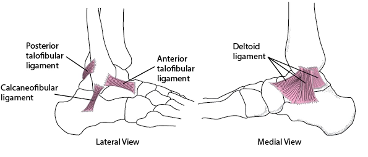
The ankle bones and ligaments form a ring that connects the tibia and fibula to the talus and calcaneus. Within the ring, stability is provided by
2 bones: Medial malleolus of the tibia and lateral malleolus of the fibula
2 ligament complexes: Medially, the deltoid ligament; laterally, mainly the anterior and posterior talofibular ligaments and calcaneofibular ligament (see figure )
Fractures that disrupt the ring in one place often disrupt it in another (eg, if only one bone is fractured, a ligament is often simultaneously and severely torn). If fractures disrupt ≥ 2 of the structures that stabilize the ankle ring, the ankle is unstable. Disruption of the medial deltoid ligament also causes instability.
The proximal fibula may also be fractured (called a Maisonneuve fracture) when the medial malleolus is fractured, the ankle mortise (the joint between the tibia and the talus) is open, and the distal fibula is not fractured. The peroneal nerve can be damaged with a Maisonneuve fracture. Without a fracture of the distal fibula, the joint can be disrupted only if the interosseous ligament between the tibia and fibula tears, as sometimes occurs when the proximal fibula is fractured.
Ligaments of the Ankle

Pain and swelling occur first at the injury site, then often extend diffusely around the ankle.


Diagnosis of Ankle Fractures
Radiographs
Sometimes stress radiographs and/or MRI
Ankle radiographs are taken in anteroposterior, lateral, and oblique (mortise) views. Specific criteria (eg, Ottawa ankle rules) are often used to avoid radiographs in patients unlikely to have a fracture. Based on the Ottawa ankle rules, ankle radiograph is required only if patients have ankle pain and one of the following:
Age >55
Inability to bear weight without assistance immediately after the injury and in the emergency department (for 4 steps), with or without limping
Bone tenderness within 6 cm of the posterior edge or tip of either malleolus
Ankle fractures are usually evident on radiographs.

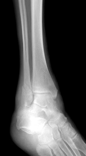
In this lateral view, a fracture of the distal fibula is visible as a small lucency and discontinuity (step-off) of the cortex.
In this lateral view, a fracture of the distal fibula is visible as a small lucency and discontinuity (step-off) of the
DR P. MARAZZI/SCIENCE PHOTO LIBRARY

This lateral view of the ankle shows a fracture of the distal fibula.
This lateral view of the ankle shows a fracture of the distal fibula.
GUSTOIMAGES/SCIENCE PHOTO LIBRARY

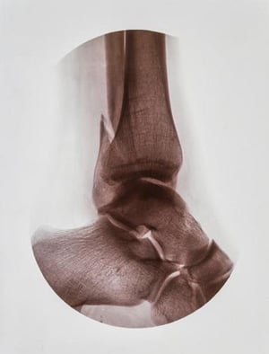
This ankle fracture involves the medial malleolus (of the tibia) and lateral malleolus (of the fibula).
This ankle fracture involves the medial malleolus (of the tibia) and lateral malleolus (of the fibula).
SCIENCE PHOTO LIBRARY

This ankle fracture affects the lateral malleolus but spares the medial malleolus.
This ankle fracture affects the lateral malleolus but spares the medial malleolus.
LIVING ART ENTERPRISES, LLC/SCIENCE PHOTO LIBRARY

The proximal fibula may be fractured (called a Maisonneuve fracture—shown here), when the medial malleolus is fractured, the ankle mortise (the joint between the tibia and the talus) is open, and the distal fibula is not fractured.
The proximal fibula may be fractured (called a Maisonneuve fracture—shown here), when the medial malleolus is fractured
EDWARD KINSMAN/SCIENCE PHOTO LIBRARY

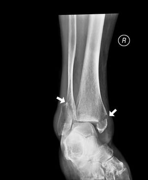
This radiograph shows a disruption (widening) of the medial mortise joint (blue arrow). Normally, the space of the mortise joint should be symmetric around the talus. With a concomitant proximal fibular fracture, this is known as a Maisonneuve fracture.
This radiograph shows a disruption (widening) of the medial mortise joint (blue arrow). Normally, the space of the mort
Image courtesy of Danielle Campagne, MD.

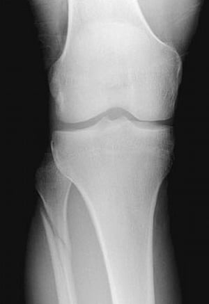
This radiograph shows a proximal fibula fracture. With a concomitant disruption (widening) of the medial mortise joint, this is known as a Maisonneuve fracture.
This radiograph shows a proximal fibula fracture. With a concomitant disruption (widening) of the medial mortise joint,
Image courtesy of Danielle Campagne, MD.
In this lateral view, a fracture of the distal fibula is visible as a small lucency and discontinuity (step-off) of the cortex.
In this lateral view, a fracture of the distal fibula is visible as a small lucency and discontinuity (step-off) of the
DR P. MARAZZI/SCIENCE PHOTO LIBRARY
This lateral view of the ankle shows a fracture of the distal fibula.
This lateral view of the ankle shows a fracture of the distal fibula.
GUSTOIMAGES/SCIENCE PHOTO LIBRARY
This ankle fracture involves the medial malleolus (of the tibia) and lateral malleolus (of the fibula).
This ankle fracture involves the medial malleolus (of the tibia) and lateral malleolus (of the fibula).
SCIENCE PHOTO LIBRARY
This ankle fracture affects the lateral malleolus but spares the medial malleolus.
This ankle fracture affects the lateral malleolus but spares the medial malleolus.
LIVING ART ENTERPRISES, LLC/SCIENCE PHOTO LIBRARY
The proximal fibula may be fractured (called a Maisonneuve fracture—shown here), when the medial malleolus is fractured, the ankle mortise (the joint between the tibia and the talus) is open, and the distal fibula is not fractured.
The proximal fibula may be fractured (called a Maisonneuve fracture—shown here), when the medial malleolus is fractured
EDWARD KINSMAN/SCIENCE PHOTO LIBRARY
This radiograph shows a disruption (widening) of the medial mortise joint (blue arrow). Normally, the space of the mortise joint should be symmetric around the talus. With a concomitant proximal fibular fracture, this is known as a Maisonneuve fracture.
This radiograph shows a disruption (widening) of the medial mortise joint (blue arrow). Normally, the space of the mort
Image courtesy of Danielle Campagne, MD.
This radiograph shows a proximal fibula fracture. With a concomitant disruption (widening) of the medial mortise joint, this is known as a Maisonneuve fracture.
This radiograph shows a proximal fibula fracture. With a concomitant disruption (widening) of the medial mortise joint,
Image courtesy of Danielle Campagne, MD.
Determining stability helps guide treatment. Instability may be obvious when the ankle is inspected or gently palpated. The knee, particularly the proximal fibula, should also be examined to look for a concomitant Maisonneuve fracture. Peroneal nerve function should be tested in patients with a Maisonneuve fracture by asking the patient to dorsiflex at the ankle.
If both the medial and lateral malleoli are fractured, the injury is probably unstable.
If only the fibula is fractured and the tibiotalar joint appears normal, an external rotation stress radiograph can be done; it may detect tibiotalar subluxation, which suggests deltoid ligament and thus ankle joint instability.
Pearls & Pitfalls
|
If a proximal fibula fracture seems possible, radiographs of the knee should also be taken.
Treatment of Ankle Fractures
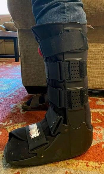
Walking boot or casting
Sometimes open reduction with internal fixation (ORIF)
Most stable ankle fractures can be treated nonsurgically with a walking boot or cast.

For unstable ankle injuries, ORIF is often done to align the bone fragments correctly and to better maintain alignment during fracture healing.
The prognosis is usually good if the ankle is stable and if treatment results in correct alignment. If bone fragments do not remain correctly aligned, arthritis may develop and fractures may recur.
Maisonneuve fractures are significant syndesmosis injuries that require ORIF.
Key Points
If an ankle fracture disrupts the ankle ring (formed by the ankle bones and ligaments) in one place, it often disrupts it in another; if ≥ 2 of the structures that stabilize the ankle ring are disrupted, the ankle is unstable.
Use the Ottawa ankle rules to try to limit radiographs to patients more likely to have a fracture.
Evaluate ankle stability (which determines treatment) by physical examination and, if needed, radiographs.
Treat most stable ankle fractures with a walking boot or cast and many unstable fractures with ORIF.


