


Paget disease of bone is a chronic disorder of the adult skeleton in which bone turnover is accelerated in localized areas. Normal matrix is replaced with softened and enlarged bone. The disease may be asymptomatic or cause gradual onset of bone pain or deformity. Diagnosis is by x-ray. Treatment includes symptomatic measures and often medications, usually bisphosphonates.
Approximately 2 to 3% of adults > 55 years old in the United States have Paget disease, with a slight male predominance (1). Prevalence increases with age; however, overall prevalence seems to be decreasing. The disease is most common in the United Kingdom and European countries including Spain, France, and Italy, as well as in countries settled by European immigrants (eg, Australia, New Zealand, the United States, and Canada) (2, 3). Paget disease is rare in Scandinavian countries and Asia.
General references
1. Ralston SH, Corral-Gudino L, Cooper C, et al: Diagnosis and management of Paget's disease of bone in adults: a clinical guideline. J Bone Miner Res 34(4):579-604, 2019. doi: 10.1002/jbmr.3657
2. Reid IR: Recent advances in understanding and managing Paget's disease. F1000Res. 2019;8:F1000 Faculty Rev-1485. Published 2019 Aug 22. doi:10.12688/f1000research.19676.1
3. Singer FR, Bone HG 3rd, Hosking DJ, et al: Paget's disease of bone: an Endocrine Society clinical practice guideline. J Clin Endocrinol Metab 99(12):4408-22, 2014. doi: 10.1210/jc.2014-2910
Etiology of Paget Disease of Bone
About 10% of patients with Paget disease have mutations of the SQSTM1 (sequestosome-1) gene, resulting in increased nuclear factor kappa-B activity, which increases osteoclast activity (1, 2). Several other mutations associated with Paget disease have been identified, many affecting the RANK (receptor activator of nuclear factor kappa-B) signaling pathway that is critical for osteoclast generation and activity.
A viral etiology, such as measles, has been proposed because nuclear inclusions in diseased osteoclasts that are similar to those seen in paramyxovirus-infected cells have been seen on electron microscopy. Although a viral cause remains controversial, it is hypothesized that in genetically predisposed patients an as yet unidentified virus triggers abnormal osteoclast activity.
Etiology references
1. Laurin N, Brown JP, Morissette J, Raymond V: Recurrent mutation of the gene encoding sequestosome 1 (SQSTM1/p62) in Paget disease of bone. Am J Hum Genet 70:1582–1588, 2002. doi: 10.1086/340731
2. Rea SL, Walsh JP, Layfield R, et al: New insights into the role of sequestosome 1/p62 mutant proteins in the pathogenesis of Paget's disease of bone. Endocr Rev 34(4):501-24, 2013. doi: 10.1210/er.2012-1034
Pathophysiology of Paget Disease of Bone
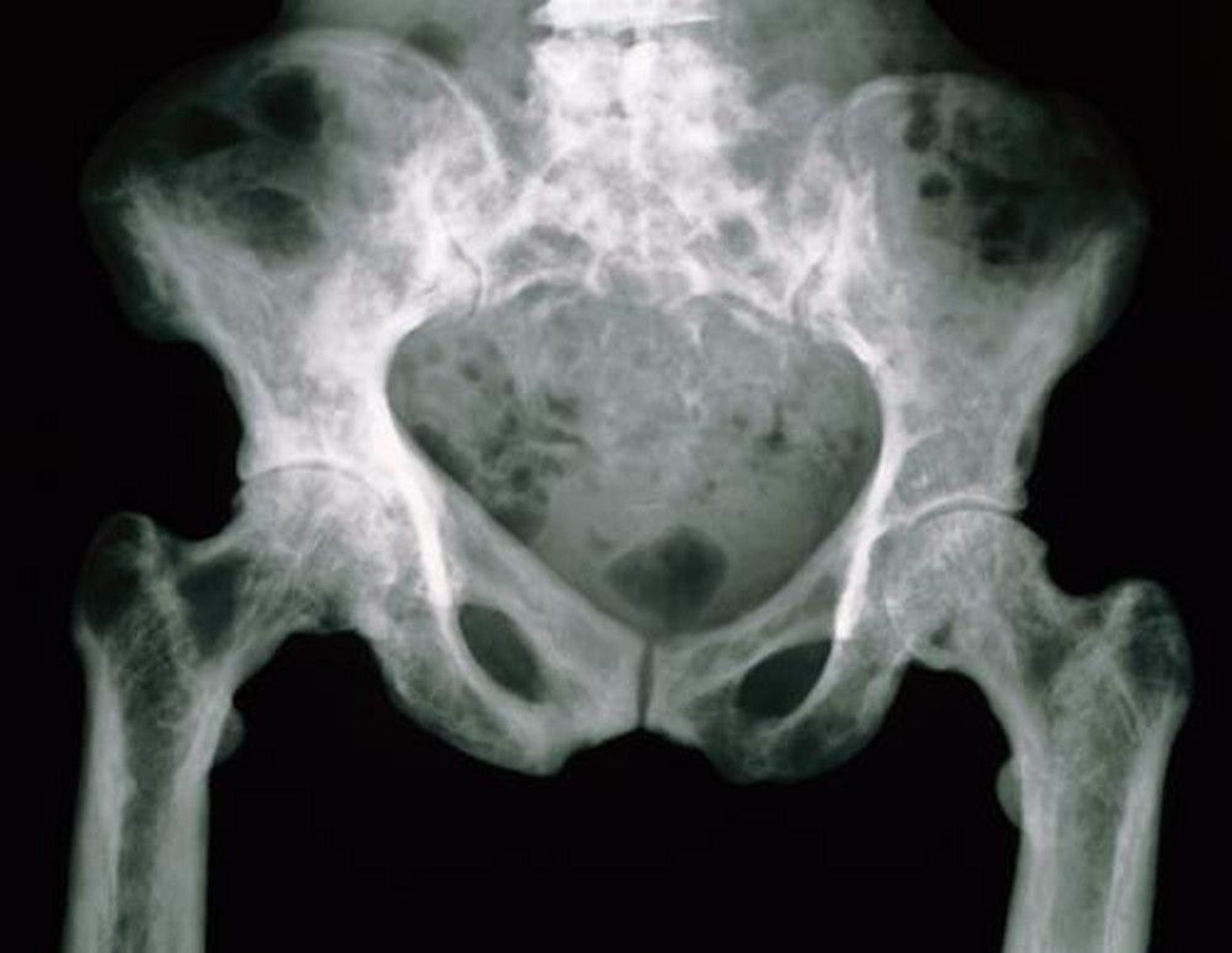
Any bone can be involved in Paget disease. The bones most commonly affected are the pelvis, femur, and skull. Other less commonly involved bones are the tibia, vertebrae, clavicle, and humerus.
Bone turnover is accelerated at involved sites. Pagetic lesions are metabolically active and highly vascular. Excessively active osteoclasts are often large and contain many nuclei. Osteoblastic repair is also hyperactive, causing coarsely woven, thickened lamellae and trabeculae. This abnormal structure weakens the bone, despite bone enlargement and areas of bone sclerosis.
Symptoms and Signs of Paget Disease of Bone
Paget disease of the bone is usually asymptomatic. If symptoms occur, they develop insidiously, with pain, stiffness, fatigue, and bone deformity. Bone pain is aching, deep, and occasionally severe, sometimes worse at night. Pain also may arise from compression neuropathy or osteoarthritis.
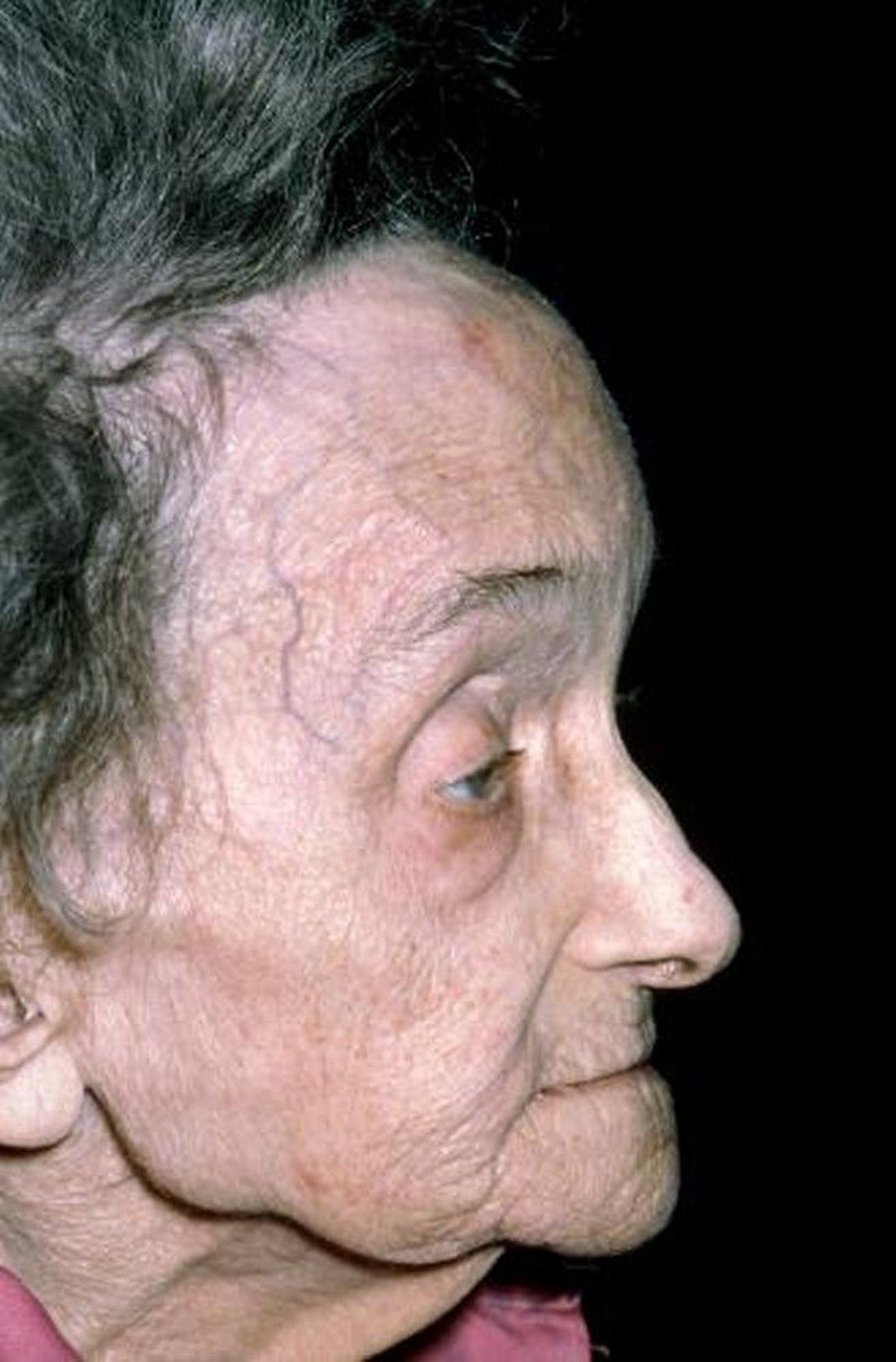
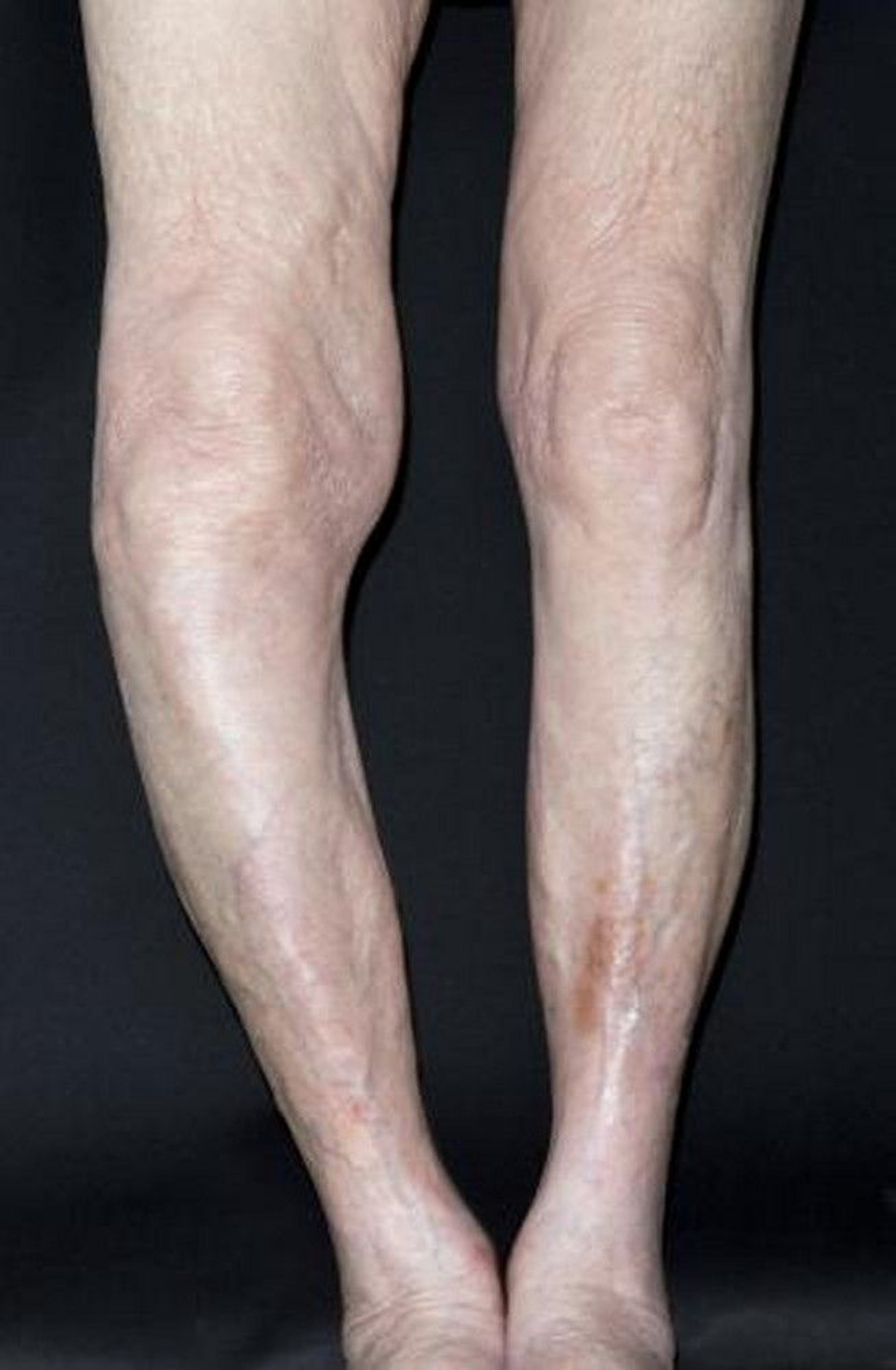
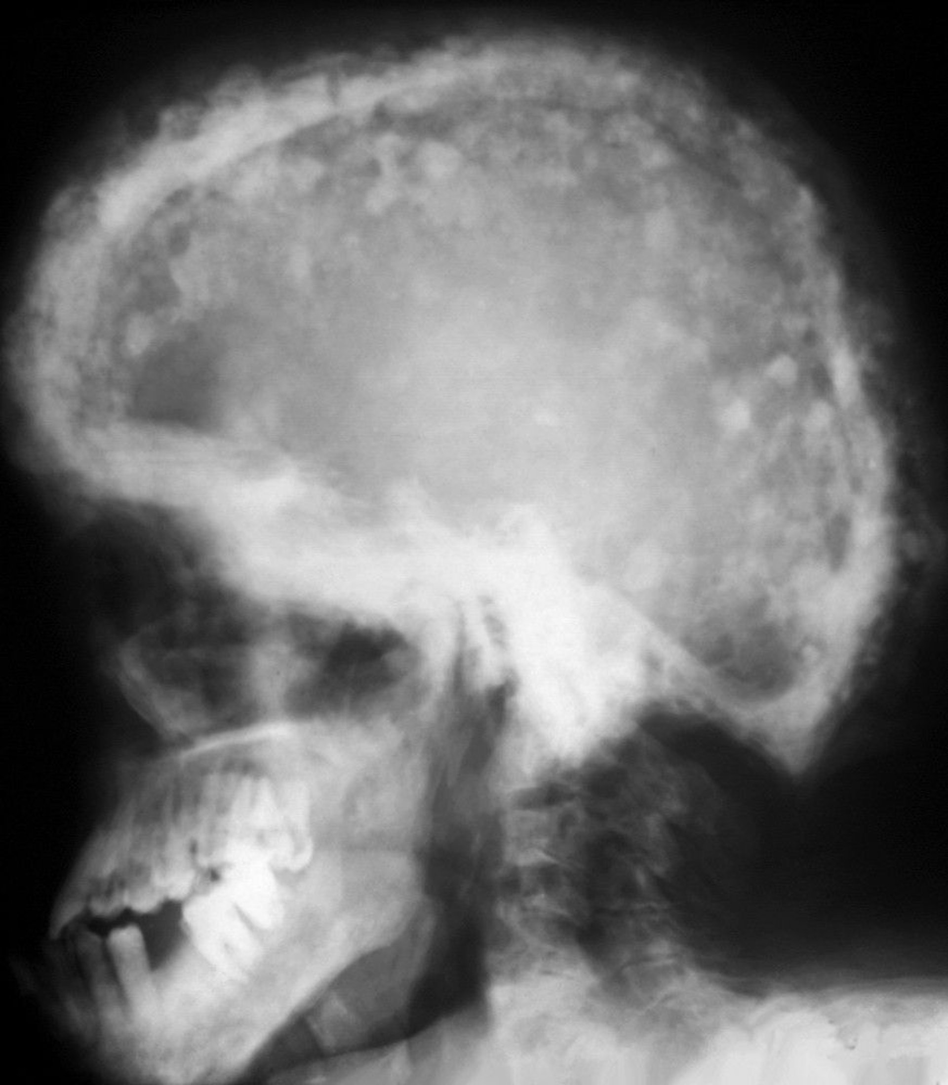
If the skull is involved, signs may include skull enlargement bitemporally and frontally (frontal bossing), dilated scalp veins, and nerve deafness in one or both ears. Symptoms may include vertigo, headaches, and hearing impairment. Deformities may develop from bowing of the long bones or osteoarthritis. Pathologic fractures may be the presenting manifestation. New onset or severe pain at a site of pagetic bone should prompt consideration of osteosarcoma.

DR P. MARAZZI/SCIENCE PHOTO LIBRARY

DR P. MARAZZI/SCIENCE PHOTO LIBRARY
Complications
The most common complication of Paget disease of bone is
Osteoarthritis occurs in up to 50% of patients and develops in joints adjacent to involved bone. Pathologic fracture is also common due to focal areas of weakened bone.
Overgrown bone may compress nerves and other structures passing through small foramina. Spinal stenosis or spinal cord compression may develop.
Rare complications include transformation to osteosarcoma in < 1% of patients. Highly vascular bones may bleed excessively during orthopedic surgery. Very rarely, hypercalcemia develops in patients who are immobile; however, hypercalcemia in ambulatory patients suggests the coexistence of hyperparathyroidism. High-output heart failure due to large or numerous hypervascular lesions has been reported.
Diagnosis of Paget Disease of Bone
Plain x-rays
Elevated serum alkaline phosphatase or other markers of increased bone turnover
Bone scan to establish the extent and location of disease
Paget disease should be suspected in patients with the following:
Unexplained bone pain or deformity
Suggestive findings on x-ray
Unexplained elevation of serum alkaline phosphatase on laboratory tests done for other reasons, particularly if liver-sourced 5'-nucleotidase or gamma-glutamyl-transpeptidase (GGT) is normal
Confirmation on x-ray is required to establish the diagnosis. Characteristic x-ray findings include abnormal architecture with coarse trabeculae and cortical thickening and sclerosis. Involved bone may be enlarged, and pagetic lesions in the tibia or femur may result in bowing. Once a diagnosis of Paget's disease of bone is made on x-ray, a radionuclide bone scan using technetium-labeled phosphonates should be done to determine the extent of bone involvement. However, in cases of diagnostic uncertainty, a biopsy should be performed to exclude possible metastatic disease. A biopsy is also indicated for patients with known pagetic bone involvement who have worsening symptoms at the affected site to evaluate for osteosarcoma or other tumors (eg, giant cell tumor of bone).

ZEPHYR/SCIENCE PHOTO LIBRARY

SCIENCE PHOTO LIBRARY
Pearls & Pitfalls
|
Treatment of Paget Disease of Bone
Supportive care for symptoms and complications
Bisphosphonates if bone disease is symptomatic or active in bones at risk of complications
Supportive treatment of Paget disease of bone includes analgesics or nonsteroidal anti-inflammatory drugs (NSAIDs) for pain. Orthotics help correct abnormal gait caused by bowed lower extremities. Some patients require orthopedic surgery (eg, hip or knee replacement, decompression of the spinal cord). Weight bearing should be encouraged, and bed rest should be avoided.
Localized, asymptomatic disease may not require treatment.
Pharmacologic therapy
Pharmacologic therapy is indicated to treat pain clearly related to the pagetic process and not to another source (eg, osteoarthritis). In addition, some guidelines (1) recommend treatment to
Prevent or retard progression of complications (eg, hearing loss, deformity, osteoarthritis, paraparesis or paraplegia related to vertebral Paget disease, or other neurologic deficits), particularly in a poor surgical candidate
Prevent or minimize bleeding if orthopedic surgery is planned at a site with pagetic involvement
Suppress excessive osteoclast activity when serum alkaline phosphatase (of bony origin) is > 2 times the normal level, even in the absence of symptoms
Although disease progression can be retarded by medications, existing deficits (eg, deformity, osteoarthritis, hearing loss, neural impingement) are not reversed.
Several antiresorptive agents that suppress osteoclast activity and decrease bone pain are available for treating Paget disease.
3); however, there are insufficient data to support its routine use.
Treatment references
1. Singer FR, Bone HG 3rd, Hosking DJ, et al: Paget's disease of bone: an Endocrine Society clinical practice guideline. J Clin Endocrinol Metab 99(12):4408-22, 2014. doi: 10.1210/jc.2014-2910
2. Reid IR, Lyles K, Su G, et al: A single infusion of zoledronic acid produces sustained remissions in Paget disease: Data to 6.5 years. J Bone Miner Res 26(9):2261–2270, 2011. doi: 10.1002/jbmr.438
3. Reid IR, Sharma S, Kalluru R, Eagleton C: Treatment of Paget's disease of bone with denosumab: Case report and literature review. Calcif Tissue Int 99(3):322–325, 2016. doi: 10.1007/s00223-016-0150-6
Key Points
Paget disease of bone is an often asymptomatic abnormality of bone metabolism, particularly among older adults.
Complications can include osteoarthritis, fractures, neural compression, osteosarcoma, and rarely hypercalcemia.
Confirmation is usually by x-rays showing findings such as bone sclerosis, coarse cortical trabeculation or cortical thickening, and bone bowing or enlargement.


